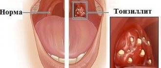
Causes of inflammation
As a rule, inflammation of the root part of the teeth is provoked by two factors - infection and trauma to the jaw and dental units.
In turn, the development of infections leads to:
- Untimely elimination of advanced caries.
- Incorrect treatment of pulpitis.
- A poorly made and then installed crown, under which food debris penetrates. When they decompose, they lead to the growth and reproduction of bacteria.
- Injuries to the gums due to an incorrectly installed crown or prosthesis. More often, these areas are visible as bleeding, scarlet-red, and swollen.
- Frequent diseases of bacterial sore throat.
- Chronic sinusitis, provoking the growth and reproduction of pathogenic flora.
In all of these cases, the root also becomes inflamed, since pathological processes are transferred to it.
As for injuries, as causes of pathology, these are:
- An incorrectly filled tooth, on which the load increases when chewing.
- Fractures that occur after injuries to the jaw, incorrectly installed crowns.
- Increased tooth mobility that occurs due to injuries to the nerves or vessels of the root canal.
- Excessively high crown restored from composite materials.
- The use of “old” antiseptics to treat canals that are inflamed (we are talking about arsenic).
Regardless of the cause of the development of the inflammatory root process, the patient experiences obvious discomfort and constant pain.
Scientific rationale for the modeling sequence of aesthetic restorations
The anatomy of human teeth, their classical features and forms are considered to be well studied for a long time [2, 4]. When using dental materials from previous generations, this knowledge was absolutely satisfactory to dentists. However, the appearance on the market of dental services of ceramics and composites, providing high aesthetic qualities of structures, showed limited information about the fine details of the tooth structure [3, 5]. On the other hand, the insufficiency and lack of demand for medical knowledge in the field of tooth morphology leads to the fact that even classical signs are not always reproduced in the design [7]. The result is more or less pronounced differences in the shape and relief of the restoration from the natural appearance of the tooth.
We studied the aesthetic characteristics of permanent teeth in 350 people using odontoscopy and odontometry.
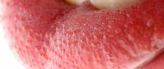
The need to bite, tear and grind food contributed to the formation of the main groups of teeth: incisors, canines, molars, premolars differ in shape, size, number of roots and are located in the dental arches. The upper arch is usually more rounded, the lower one is slightly compressed in the transverse direction. The anatomical crown, the border of which runs along the neck of the tooth, and the clinical crown, which is located above the gingival edge immediately after the tooth erupts, are identical in height. With age, the anatomical crown shortens as a result of tooth wear. The clinical crown also decreases, but may also lengthen due to gum recession (Fig. 1).
Rice. 1a. Changing the size of the central incisor. Rice. 1b. Changing the size of the central incisor.
Based on general characteristics, teeth were distinguished according to the signs of belonging to the right or left side, which relate to the curvature of the crowns, the ratio of the distal and mesial angles of the crown, and the inclination of the roots.
A sign of crown curvature is a greater convexity of the vestibular part of the crown, located near its mesial edge, and a gentle slope of the distal one. It is more clearly defined when examining the tooth from the occlusal surface and is expressed in 71% of central incisors (Fig. 2).
Rice. 2a. The sign of crown curvature is determined. Rice. 2b. The sign of crown curvature is determined.
In 23% of cases, the convexity is shifted to the distal side, in 6% of teeth the sign of curvature is not detected. The sign of the crown angle characterizes the situation when the mesial angles, composed of the mesial surface and the cutting edge (masticatory surface), are sharper than the distal ones (Fig. 3).
Rice. 3a. There is a pronounced sign of distal deviation of the periodontal dome. Rice. 3b. There is a pronounced sign of distal deviation of the periodontal dome.
This inequality of angles is observed in 85% of central incisors. In molars, the sign of angle is due to more massive mesial cusps. The sign of root inclination means that the root or its apex is curved in the distal direction in relation to the longitudinal axis of the tooth. In the oral cavity, the sign manifests itself as a distal displacement of the apex of the dentogingival dome.
Based on their appearance, teeth were classified into separate geometric shapes (rectangle, triangle, oval), which are characterized by their own characteristics (Fig. 4).
Rice. 4. Basic geometric shapes of permanent teeth.
Rectangular incisors in the absence of abrasion are detected in 55% of cases: the transverse dimensions of the vestibular surface in the gingival, equatorial region and at the cutting edge are close in value, as a result of which the proximal surfaces are almost parallel (Fig. 5).
Rice. 5a. Rectangular cutters. Rice. 5 B. Rectangular cutters.
The angle sign may be weakly expressed. The length of contact with adjacent teeth is significant: in 72% of cases, contact between teeth begins at the apex of the gingival papilla and ends at the cutting edge. With a triangular crown shape, which is characteristic of 39% of intact incisors, the transverse dimensions of the vestibular surface increase from the neck to the cutting edge (Fig. 6).
Rice. 6. Triangular shaped teeth.
The crown angle sign is well expressed in 88% of cases. The length of contact with adjacent teeth is insignificant. The oval shape of the crown occurs in 7% of cases, having similar values of transverse dimensions in the cervical region and near the cutting edge. The largest diameter is at the equator. The lateral surfaces are presented in the form of convex arcs. The corners at the cutting edge are smoothed. Contacts with neighboring teeth are point-like.
The vestibular surfaces of canines, premolars and molars have a more complex geometric shape due to protruding cusps.
According to a survey of young people, in 53% of cases the upper dental arch has an oval shape, in 42% - round, and in these arches there are teeth of any geometric shape: rectangular, triangular, oval. However, the rare rectangular and triangular dental arches (2.6% of cases) are characterized by rectangular and triangular teeth, respectively.
The visual perception of the geometric shape of a tooth can be influenced by the individual characteristics of the dentogingival contour. The dome-shaped gingival margin, characteristic of 56% of incisors, resembles a wedge or triangle in shape and gives the tooth a triangular shape. 39% of incisors have a rounded periodontal edge, 5% of teeth have a flattened, almost straight gingival contour. The shape of the incisal edge also reflects the individual characteristics of the tooth (Fig. 7).
Rice. 7a. The relief of the vestibular surface and scalloped cutting edge are pronounced. Rice. 7b. The relief of the vestibular surface and scalloped cutting edge are pronounced.
Immediately after eruption, the cutting edge is serrated. Then abrasion facets appear. An uneven (convex or concave) surface can be explained by the peculiarity of contact with antagonist teeth.
The relief of the vestibular surface of the tooth is of great aesthetic importance: vertical ridges, bridges, convexities, depressions, and areas. A single ridge is usually characteristic of the middle part of the vestibular surface. If there are two ridges, the most common ones are mesial and distal. The three enamel ridges are usually located mesially, medially and distally (Fig. 8).
Rice. 8a. Vertical ridges on the vestibular surface of the incisors. Rice. 8b. Vertical ridges on the vestibular surface of the incisors. Rice. 8th century Vertical ridges on the vestibular surface of the incisors.
Physiological wear of teeth leads to the formation of a smooth vestibular surface.
Modeling of aesthetic restorations should pursue the goal of reproducing in detail the natural classical, as well as individual characteristics of permanent teeth. However, an analysis of the quality of 145 restorations that were created by reproducing the characteristics of a symmetrical tooth showed differences between the artificial structure and the intact tooth in almost half of the cases [1, 6]. In most cases, this concerns the restoration of rectangular-shaped features where triangular or oval ones are required. In some cases, there is a recreation of oval (rectangular) tooth shapes in a triangular jaw.
The absence of signs of tooth affiliation was found in an average of 30% of restorations. Of these, 77.14% were modeled without taking into account the sign of crown curvature. Many patients had differences in height, width, or asymmetry in the size of individual sections of aesthetic structures (Fig. 9).
Rice. 9a. A veneer differs in size from an intact symmetrical tooth. Rice. 9b. A veneer differs in size from an intact symmetrical tooth. Rice. 9th century A veneer differs in size from an intact symmetrical tooth.
Among the age-related features characterizing the surface relief, there was practically no re-creation of perikymatia on structures in adolescents. The lengthening of the clinical crown of the tooth due to gum recession was not taken into account: the height increased significantly due to the filling material. No gum-mimicking polymers were used.
Individual features of the relief were absent in most restorations (vertical ridges, relief cutting edge).
At the filling stage, insufficient or excessive use of material changed the shape, size, and characteristics of the tooth. Thus, excess filling material on the vestibular surface caused an unnatural convexity of the crown (Fig. 9).
The lack of composite material led to the loss of proper anatomical volume, a decrease in the overall height and proximal slopes, and flattening of the crown. Single structures stood out as a result of asymmetry - differences from a similar tooth on the opposite side. Paired or multiple restorations looked unnatural if a single geometric type of crowns was not maintained, signs of tooth affiliation, and individual characteristics were not observed.
The results of scientific research, including analysis of the aesthetic characteristics of teeth and the quality of existing restorations, made it possible to develop recommendations for step-by-step planning of the sizes and shapes of aesthetic restorations.
The algorithm for planning the size and shape of the structure represents a certain sequence of measuring and describing specific anatomical structures of the tooth (Scheme 1).
Scheme No. 1. Restoration sequence algorithm.
First, it is necessary to conduct a comparative assessment of the size of the clinical and anatomical crown of the tooth. The presence of wear areas in the area of the cutting edge indicates a decrease in the height of the clinical crown. Gingival recession with exposure of the neck and root of the tooth is a sign of an increase in the vertical size of the clinical crown (Fig. 1).
The measurement (odontometry) is made with a micrometer (Fig. 10).
Rice. 10a. Measuring the transverse dimensions of a tooth. Rice. 10b. Measuring the transverse dimensions of a tooth. Rice. 10th century Measuring the transverse dimensions of a tooth.
The height of the clinical crown of the central and lateral incisors is assessed by the distance from the cutting edge to the marginal level of the gums along the vertical midline. Similar dimensions of the canine and premolars are measured along the midline from the apex of the cusp to the marginal level of the gingiva. The height of the molar crown is the distance from the gum level to the top of the most prominent cusp.
The shape of the tooth crown is considered rectangular if the lateral edges of the vestibular surface are parallel, and square if the height and width are equal. Mesiodistal dimensions in the neck of any tooth are measured by the distance between two points of opposite proximal surfaces at the level of the apexes of the interdental papillae. The horizontal parameters of the incisors in the equatorial region are determined at the level of the middle third of the crown height. Similar values of the central and lateral incisors in the area of the cutting edge are assessed by the distance between the protruding points of the mesial and distal edges of the crown. The mesiodistal dimensions of the canines and premolars are measured between the lateral sections of the crown located at the greatest distance. The transverse parameters of molars in the equatorial region are assessed as the distance between the most convex sections of the proximal surfaces.
Visual assessment and measurement results make it possible to describe the geometric shape of the tooth crown based on the relative position of the lateral surfaces.
The shape of the tooth crown is considered rectangular when the lateral edges of the vestibular surface are parallel (square when the height and width are equal). The triangular shape of the tooth is characterized by its maximum horizontal size at the cutting edge. A crown is considered oval in shape when the lateral surfaces have a rounded outline with the largest horizontal dimension in the area of the middle third of the tooth.
Next, the severity of the signs of belonging to the side of the teeth is assessed. The sign of the crown angle is recorded in the case of a predominance of the distal angle of the vestibular surface over the mesial one (Fig. 11).
Rice. 11. No signs of crown angle.
The sign of crown curvature is considered positive if the convexity of the vestibular surface is located closer to the mesial edge. In some cases, there is no sign of crown curvature (Fig. 12).
Rice. 12. Smooth vestibular surface of the central incisors.
A sign of tooth root deviation is noted in the map when there is a distal displacement of the apex of the dentogingival contour. The description of the individual characteristics of the tooth includes the surface topography, the shape of the gingival contour of the tooth, the shape of the cutting edge, and the length of contact with neighboring teeth. The type of relief of the vestibular surface of the incisors is determined by the presence or absence of vertical enamel ridges (Fig. 8).
The shape of the dentogingival contour is assessed by the upper border of the tooth crown, which starts from the top of one interdental papilla, then goes along the edge of the gums and ends at the top of the other interdental papilla (Fig. 13).
Rice. 13. Contours of the gingival dome.
The dentogingival contour can be round, dome-shaped or flat. It is necessary to plan the length of proximal contacts between teeth in such a way that there is enough space for the interdental papilla. The planning stage is completed by choosing the shape of the cutting edge of the teeth (Fig. 14).
Rice. 14. Planning the area of the cutting edge of the central incisors.
The anatomical features of chewing teeth require a careful assessment of the relationship of the cusps on the occlusal surface, and their shape can change significantly due to abrasion. Modeling restorations is a complex and demanding process.
The first stage is the formation of the base of the restoration, which includes the contours of the geometric shape of the dentin and mamelons at the cutting edge, with a clear designation of the lateral and lower boundaries of the dentinal layer (Fig. 15).
Rice. 15. Opaque base of restoration (diagram).
The second stage involves modeling the signs of teeth belonging to the side (signs of curvature, crown angle, deviation of the periodontal dome). The third stage is the reproduction of the individual characteristics of the tooth, including macro- and microrelief, the shape of the cutting edge and the gingival dome. The sequence of restoration of morphological elements corresponds to the sequence of odontoscopic examination and restoration planning. There is a gradual transition from recreating large details (the geometric shape of the vestibular surface) to reproducing medium-sized ones (signs of the angle and curvature of the crown), and then to modeling smaller elements (enamel ridges, teeth in the area of the cutting edge).
Using imaginary lines (two vertical and two horizontal), it is necessary to divide the vestibular surface of the tooth into segments, which will make it possible to more clearly determine the topographic position of a particular morphological element (Fig. 16).
Rice. 16. Conventional division of the vestibular surfaces of teeth into segments.
For example, in the upper tier the dentogingival contour is modeled, as well as a sign of tooth root deviation in the form of a displacement of the top of the gingival dome to the distal side. Predominantly in the middle part of the tooth, a sign of crown curvature is created through the formation of the largest convexity in the mesial region. Individual features of the cutting edge, expressed by varying degrees of transparency and mamelons, are located in the lower tier (Fig. 17).
Rice. 17a. Modeling the basis of the restoration. Rice. 17b. Modeling the incisal edge of the restoration.
Contacts between teeth, proximal slopes, as well as vertical enamel ridges are modeled, respectively, in the mesial and distal parts throughout the middle and lower tier. In the mesial region there may be a median enamel ridge (Fig. 18).
Rice. 18. Modeling of the individual relief of the vestibular surface.
Restoration of large morphological details must be carried out using opaque shades of the composite. The main guideline when working with opaque filling material is the border of the transparent tooth enamel. So, if the enamel evenly covers the entire surface, then for the corresponding layer of filling material it is necessary to leave no more than 0.5 mm of free space around the entire perimeter of the tooth. If the transparent strip is located in the area of the cutting edge, then the opaque is not brought to the lower border by 1.0 mm. In the case where a layer of transparent enamel is observed on the proximal edges, 1.0-1.5 mm should be left for the enamel layer. With severe abrasion, the dentinal layer is formed over the entire height of the tooth crown up to the cutting edge.
During the work, it is necessary to ensure that the thickness of the opaque layers does not exceed the amount of lost dentin. It is important that the opaque layer is not longer or shorter than on a symmetrical tooth and does not interfere with the type of transparency.
In accordance with the algorithm for planning the anatomical shape and relief, developed by the staff of the Department of Therapeutic Dentistry of BelMAPO based on their own odontometric and odontoscopic observations, 166 structures were made.
The results of visual examination of restorations modeled using the technique of preliminary planning of their morphology showed a significant improvement in quality compared to the previous period (see table).
In most cases, the optimal anatomical shape of the teeth was recreated, which corresponded to group affiliation, age and individual characteristics. The frequency of occurrence of signs of angle, crown curvature, and deviation of the gingival dome on restorations has increased almost 10 times. The vertical and horizontal dimensions of the created structures did not differ significantly from intact symmetrical teeth. Thanks to this, a single geometric type was maintained, characteristic of this group of teeth. Age-related features were simulated, including root exposure and age-related abrasion. In the vast majority of young people, enamel ridges and grooves on the vestibular surface were modeled. The alternation of highlights and shadows, due to the presence of macrorelief, emphasized the naturalness of the created restoration (Fig. 19).
Rice. 19a. Planning of aesthetic restorations. Rice. 19b. Modeling of aesthetic restorations.
Thus, the results obtained made it possible to identify errors made in the manufacture of composite restorations regarding sizes, shapes, and surface topography. Inaccuracies were allowed at the planning stage if signs associated with the group affiliation of the tooth, its age or individual characteristics were not recorded.
The lack of a strict sequence of actions, and often the exclusion of the very stage of planning the anatomical shape, caused a significant violation of the aesthetics of restorations. In turn, the use of the proposed algorithm of actions for assessing and reproducing the characteristics of tooth morphology in conjunction with the skills of odontoscopic examination helps to significantly reduce the number of errors and complications during the restoration of the anatomical shape of the restoration.
LITERATURE
- Danilova D.V. // Sat. Proceedings of the 5th Congress of Dentists of the Republic of Belarus. - Brest, 2004. - pp. 138-140.
- Dmitrienko S.V., Ivanov L.P., Krayushkin A.I., Pozharitskaya M.M. Practical guide to dental modeling. - M., 2001. - 239 p.
- Leonova L. E., Zheleznitskikh M. V., Maksimovskaya L. N. // Klin. dentistry. - 2002, No. 1. - P. 8-11.
- Lomiashvili L.M. // Institute of Dentistry. - 2003, No. 2. - P. 6-31.
- Lutskaya I.K. // Modern. dentistry. - 2003, No. 1. - P. 30-37.
- Lutskaya I.K., Danilova D.V. // Sovrem. dentistry. - 2004, No. 1. - P. 22-25.
- Novak I.V., Danilova D.V. // Sat. works of young scientists. - Mn., 2005. - P. 49-51.
Symptoms, signs of the disease
Inflammation of tooth roots can be acute or chronic. The first variant of pathology is more pronounced. Here the unfavorable symptoms look like this:
- Almost constant aching pain in the affected area. Pain syndrome manifests itself regardless of the time of day. It intensifies if the affected area is impacted (cleaning, chewing, touching).
- Sensation of tooth enlargement. The patient feels the jaw not closing tightly.
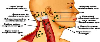
- Enlarged lymph nodes under the jaw from the side of the inflamed part of the dental unit.
- Throbbing pain, accompanied by chills and increased body temperature.
- Pain transmitted to the trigeminal nerve.
Acute processes are often accompanied by the accumulation of pus in the affected area. Here the doctor faces the main task - removing secretions, eliminating the painful focus, and channel therapy according to modern protocols.
Signs of chronic pathology are:
- Pain occurs when pressing on the crown or chewing. If the tooth is not touched, it will not be painful.
- Bad breath.
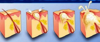
- Formation of a fistula and exit of exudate through it. This symptom is especially characteristic of granulating periodontitis. This zone is often visible as a section of gum that has changed in color and appearance.
Chronic inflammation is often treated conservatively. Only in the most difficult situations is surgical intervention indicated - removal of the root tip or the entire unit at once.
Braces
pros
- Classic proven method
leading to a positive result.
- Possible design variations makes metal braces invisible
(lingual installation, sapphire vestibular).
- When using the simplest design, complete the cost will be relatively inexpensive
.
- Coping with different types of pathologies
, up to serious ones.
- Many doctors can successfully work with braces
.
Minuses
- Metal structure visible
In most cases.
- Takes some time to get used to
to the device in the oral cavity.
- Possible mucosal injury
design details.
- Careful hygiene is required.
- Diet required
, exclusion of hard and viscous foods.
- Wearing the structure for 1-2 years
.
- Painful sensations during placement
and detail adjustments.
- In case of insufficient hygiene there is a risk of caries
.
- Arsenal of care products
behind the teeth and structure.
- In the vast majority of cases eights need to be removed
.
Professional treatment of the inflammatory process
If any of the symptoms of inflammation in the roots occur, you should immediately contact your dentist. Depending on the clinical picture, the doctor selects one of the treatment tactics:
- Conservative help. It involves eliminating the cause of the inflammatory process, cleaning the canals, re-treating them, disinfecting the affected area, filling the canals, installing new temporary fillings with anti-inflammatory drugs. After complete neutralization of the inflammatory process, the doctor restores the anatomy of the unit with composite material or a crown. With conservative treatment, the patient must be prescribed a course of antibiotics. The drugs neutralize inflammation well. The doctor selects the type of drug therapy for internal administration and rinsing according to the age, as well as the medical indications of the patient.
- Surgical intervention (surgical therapy). It all depends on the area of root damage. The doctor decides to remove 1/3 of its area (apex), ½ part or the entire unit plus the dental alveoli. Today, modern dentistry practices maximum tooth preservation. In approximately 80-90% of cases, a diseased tooth can be saved and treated using a conservative method using properly selected antibiotics and without even removing the top of the root.
Is it possible to straighten crooked teeth as an adult?
Dentistry today offers many ways to correct malocclusion in adults. When a person, year after year, day after day, sees in the reflection a smile with uneven teeth, it will be very interesting for him to look at the prospect, to see how his smile will look with a corrected bite. It will be interesting to know how this will affect the external image. Finding out this became possible with the 3D virtual setup technique, when the patient can see the step-by-step change in the position of his teeth.
The technique of correcting a bite with aligners involves preliminary drawing up a virtual treatment plan. The dentist takes photographs of the patient: his face, teeth. X-rays and dental impressions are taken. The data is processed by a computer program, resulting in a three-dimensional model in which the simulated dentofacial system is visible from all sides.
The orthodontist has been trained in imaging software and can create a treatment plan by moving the curved units along the desired path to create an even row on the arch. The computer calculates the duration of treatment until the desired result is achieved, as well as the required number of droppers for therapy. The technique is almost 100% accurate. Computer modeling is programmed in such a way as to calculate all stages of change on the path to correcting the pathology.
Treatment at home
In case of inflammation of tooth roots, home remedies or your own efforts are not enough. Since the process occurs hidden, only a doctor can determine the intensity of the lesion and later select treatment tactics. Self-administration of any antibiotics, anti-inflammatory drugs, and even more so the use of traditional therapy is strictly prohibited. Ignoring the problem and trying to treat root inflammation at home can lead to the formation of a purulent cyst, its further rupture and, as a result, sepsis.
It is worth considering that the roots of the teeth are located as close as possible to the brain. Therefore, any infection, pus, or serous contents that enter the bloodstream here quickly reach the brain vessels. No folk remedies (compresses, rinses) used at home will help here.
The only acceptable drug for home use is a local analgesic. To eliminate pain, you can take medications such as Ketanov, Ketalong, Analgin. After this, you should consult a doctor as soon as possible.
Aligners
pros
- Invisible guards, unnoticeable during communication.
You can only see them from a distance of less than a meter. Transparent material does not spoil the patient's appearance.
- The removable design allows you to adhere to absolutely no diet.
There are no restrictions on the consumption of different types of products.
- Easy hygiene.
The tray is removable and can be easily cleaned with a soft brush. Clean your mouth as usual.
- Short period of defect correction.
Over a period of six months to a year, with minor pathology, you can see a real effect.
- Creating a 3D setup
, where the future result is modeled, gives the patient an excellent visual picture of a beautiful smile, which motivates and inspires treatment.
- No long period of adaptation
, convenient use of the trays, do not cause severe discomfort. The patient gets used to the design for a couple of days, and then stops noticing the aligners.
- The enamel does not deteriorate
, since hygiene is carried out in the usual format, no additional hard-to-reach places are created.
- It is possible to do without removing the eights
.
- Visits to the doctor every one and a half to two months
.
Minuses
High price.
Depending on the choice of a foreign or domestic manufacturer, the cost changes dramatically. The technologies are the same, so Russian companies do not spend money on transportation. The result is the same, so you can choose a more economical option.
They cannot cope with complex bite pathologies.
To find out if you are included in this number, you need to consult with a specialist. The percentage of patients who are not candidates for aligners is small.
Aimed Center - professional dental care
All types of dental services are provided by the Aimed clinic for residents of Moscow, Northern, Southern Butovo. The center employs doctors with many years of experience. Specialists provide therapeutic, surgical, endodontic, orthopedic, orthodontic treatment according to new European protocols. When treating tooth root inflammation and providing other services, we guarantee our patients:
- Complete painlessness of the procedure, use of proven, certified anesthetics;
- A comfortable clinic environment that is incomparable to hospital conditions;
- Use of new Japanese, American, European equipment for accurate diagnosis and treatment;
- Use of modern consumables, drugs from Israel, USA, Japan;
- Possibility of visiting the center from 08:00 to 21:00, seven days a week, holidays;
- Reception of patients of any age from the youngest to the elderly;
- Formation of favorable prices for all services without compromising the quality of the work performed.
- To make an appointment with a specialist, call +7 (495) 120-66-43. Clinic operators select a convenient time for your visit. And in the clinic’s parking lot there is always a free space for your car. We located at the metro station st. Gorchakova.
Remember, you should never ignore toothache. A minute of delay or self-therapy with folk remedies (rinses, compresses) threatens with serious complications!
What is the best way to correct crooked teeth?
Sensible adults do not need to be reminded that home methods for correcting bites, praised on the Internet, do not actually work, but have the opposite effect. From home experiments, the enamel, the teeth themselves, and also the periodontium may suffer.
Among the leading effective ways to correct crooked teeth in adulthood are veneers, braces and aligners.
. The difference between them is that veneers correct the defect only cosmetically, externally. Beautiful overlays create the illusion of even units. But in the jaws, the teeth remain in their original places, the load remains the same, and the pathology can progress. Veneers are effective for minor defects.
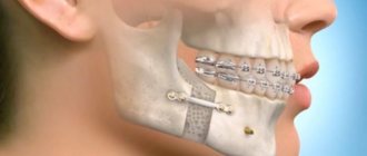
Aligners and braces radically correct curvature, making the bite and dentition straight. Among braces, there are many options for setting them, differing in material and installation location - lingual or vestibular. Braces are a permanent structure, while aligners are removable.
The cost of orthodontic treatment with braces and aligners differs slightly. The price depends on many factors: the severity of the pathology, the manufacturer of the aligners (domestic or foreign), the fastening of braces (lingual or vestibular), as well as the material from which they are made. The prestige of the clinic and the doctor’s academic degree will also be reflected in the final cost.
For children, orthodontic treatment may include the use of plates, trainers, functional appliances and 2x4 braces, where brackets are attached only to the frontal units. Treatment is selected by the doctor together with the patient.
How to prevent pathology
prevent your child from developing crooked teeth by regularly visiting the orthodontist's office. Even if your baby has a genetic predisposition to certain diseases, dealing with them at an early stage is much easier.
Adults can avoid the appearance of upper and lower crooked teeth if they do not delay the installation of implants after the loss of one of the molars, and also remove their wisdom teeth in a timely manner. In addition, it is necessary to get rid of the habit of gnawing on durable objects, not to knock your teeth against each other and not to clench your jaw during emotional stress.
Why is it worth correcting crooked teeth?
Statistics note that 80% of people have malocclusion to one degree or another. In most cases, malocclusion develops from childhood. From this time on, some people develop complexes and are embarrassed to smile. In addition to aesthetic problems, health difficulties arise.
In the case of crooked teeth, carious lesions occur more often due to crowding of the teeth. Places that are difficult to reach for hygiene accumulate bacteria, plaque, tartar, caries develop, and periodontal disease occurs. The consequence of this is digestive problems. When your teeth and gums hurt, it is more difficult to chew food to the required state.
There is no need to attribute everything to a genetic factor. Genetics is something that is very difficult to prevent in advance. You can only deal with the consequences. And other factors that arise during life and affect the curvature of teeth can be prevented. There are many reasons for curvature. They begin to develop in childhood and also arise over time. Here are several reasons why this pathology appears:
- Food quality
. Our food in big cities is often easy to chew. Soft food does not develop the jaw muscles, the dental system is reduced. Refined food contributes to the deterioration of the quality of dental tissue, caries forms more often, which causes premature loss of milk units.
- Bad habits
, originating from childhood. The habit of holding foreign objects (pacifier, finger) in the mouth creates incorrect position of the teeth. Gradually the jaw becomes deformed. If a child constantly pushes his teeth with his tongue, they begin to change their position in the jaw.
- The lower jaw is larger than the upper jaw
. This pathology can be corrected surgically.
- Loss of teeth.
When a primary or permanent unit is lost, an empty space appears into which the adjacent teeth gradually tilt under the pressure exerted by the chewing load. In primary dentition, this affects the growth of the erupting molar. In permanent cases, not only the neighboring ones suffer, but also the antagonist tooth. Bone tissue also has a negative impact; it decreases over time if an implant is not placed. The temporomandibular joints are affected.
- Bruxism
(involuntary contraction of the muscles responsible for chewing, occurring in paroxysms). The pathology is common in children. The cause is said to be stress. In adulthood, it can occur after the installation of crowns or large-scale restoration work.
If you or your loved ones notice any of the above reasons, you should consult a doctor. The sooner treatment begins, the easier it will be and the fewer health consequences there will be. Many clinics offer a free consultation where you can learn more about the condition of your teeth, the causes of their crookedness and treatment options. On our portal you will find a suitable clinic, choose an experienced specialist and receive competent advice.