Quick transition Treatment of acute external otitis
Acute otitis externa is an inflammatory disease of the external auditory canal.
Acute external otitis can be caused by infectious, allergic and dermatological diseases, but its most common cause is an acute bacterial infection.
Acute external otitis can occur at any age, but the peak of the disease occurs in childhood and adolescence (5-14 years).
The incidence is more common in the summer and is apparently associated with increased environmental humidity, heat and swimming.
Sulfur is one of the main protective mechanisms that prevent the development of acute external otitis (AEO)
When it decreases, the environment in the external auditory canal (EA) becomes alkaline instead of acidic, moisture accumulates in the ear and, as a result, the ear canal becomes an ideal breeding ground for microorganisms, which leads to an inflammatory process.
Risk factors
- Swimming or other exposure to water. Excess moisture leads to skin irritation and destruction of the skin-sulfur barrier.
- Injury. This includes excessive or aggressive ear cleaning. Not only leads to a decrease in sulfur, but can also lead to injury to the skin, allowing microorganisms to gain access to its deeper layers.
- Obstruction of the NSP. Earwax, foreign body, less often - devices that close the ear canal: hearing aids, in-ear headphones.
- Allergic contact dermatitis. Exposure to shampoo, cosmetics, allergic reaction to metal earrings.
- Dermatological diseases. Psoriasis, atopic dermatitis.
- Radiation therapy. It can lead to ischemia (local decrease in blood supply) of the NSP, alter the production of sulfur and its normal excretion.
Normally, NSP is populated by aerobic and anaerobic bacteria. Staphylococci are the most common bacteria (63%) found in the ear canal, most commonly Staphylococcus auricularis and Staphylococcus epidermidis.
The most common pathogenic organisms causing inflammation of the outer ear are Pseudomonas aeruginosa (38%). Less commonly - Staphylococcus epidermidis (9%) and Staphylococcus aureus (8%). Anaerobic bacteria are detected in 4-25% of cases. Fungal infections account for 2-10% of cases of ANO.
Tsiprolet, 10 pcs., 500 mg, film-coated tablets
Drugs that cause prolongation of the OT interval
Caution should be exercised when ciprofloxacin, like other fluoroquinolones, is used concomitantly in patients receiving drugs known to prolong the QT interval (for example, class IA and III antiarrhythmic drugs, tricyclic antidepressants, macrolides, antipsychotics).
Chelation formation
Concomitant use of tablet forms of ciprofloxacin and cation-containing drugs, mineral supplements containing calcium, magnesium, aluminum, iron, sucralfate, antacids, polymeric phosphate compounds (sevelamer, lanthanum carbonate) and drugs with a large buffer capacity (such as didanosine tablets) containing magnesium, aluminum or calcium reduces the absorption of ciprofloxacin. In such cases, ciprofloxacin should be taken either 1-2 hours before or 4 hours after taking these drugs.
This restriction does not apply to drugs belonging to the class of histamine H2 receptor blockers.
Intake of food and dairy products
The simultaneous use of ciprofloxacin and dairy products or drinks fortified with minerals (milk, yogurt, calcium-fortified orange juice) should be avoided as the absorption of ciprofloxacin may be reduced. However, calcium contained in other foods does not significantly affect the absorption of ciprofloxacin.
Omeprazole
With the combined use of ciprofloxacin and omeprazole, a slight decrease in plasma Cmax and a decrease in the area under the concentration-time pharmacokinetic curve (AUC) may be observed.
Theophylline
The simultaneous use of ciprofloxacin and drugs containing theophylline may cause an undesirable increase in the concentration of theophylline in the blood plasma and, accordingly, the occurrence of theophylline-induced adverse events; in very rare cases, these adverse events can be life-threatening for the patient. If the simultaneous use of these two drugs is necessary, it is recommended to constantly monitor the concentration of theophylline in the blood plasma and, if necessary, reduce the dose of theophylline.
Other xanthine derivatives
The simultaneous use of ciprofloxacin and caffeine or pentoxifylline (oxpentifylline) may lead to an increase in the concentration of xanthine derivatives in the blood serum.
Nonsteroidal anti-inflammatory drugs
The combination of very high doses of quinolones and some non-steroidal anti-inflammatory drugs (excluding acetylsalicylic acid) can provoke seizures.
Cyclosporine
With the simultaneous use of ciprofloxacin and drugs containing cyclosporine, it was observed
short-term transient increase in plasma creatinine concentration. In such cases, it is necessary to determine the concentration of creatinine in the blood twice a week.
Oral hypoglycemic agents
With the simultaneous use of ciprofloxacin and oral hypoglycemic agents, mainly sulfonylureas (for example, glibenclamide, glimepiride), the development of hypoglycemia may be due to an increase in the effect of oral hypoglycemic agents.
Probenecid
Probenecid slows down the rate of excretion of ciprofloxacin by the kidneys. The simultaneous use of ciprofloxacin and drugs containing probenecid leads to an increase in the concentration of ciprofloxacin in the blood serum.
Phenytoin
With the simultaneous use of ciprofloxacin and phenytoin, a change (increase or decrease) in the content of phenytoin in the blood plasma was observed. It is recommended to monitor phenytoin therapy in patients taking both drugs, including determination of phenytoin plasma levels.
Methotrexate
With the simultaneous use of methotrexate and ciprofloxacin, the renal tubular transport of methotrexate may slow down, which may be accompanied by an increase in the concentration of methotrexate in the blood plasma. This may increase the likelihood of developing side effects of methotrexate. In this regard, patients receiving concomitant therapy with methotrexate and ciprofloxacin should be closely monitored.
Tizanidine
As a result of a clinical study involving healthy volunteers with the simultaneous use of ciprofloxacin and drugs containing tizanidine, an increase in the concentration of tizanidine in the blood plasma was revealed: an increase in Cmax by 7 times (from 4 to 21 times), an increase in AUC by 10 times (from 6 to 24 times ). Hypotensive and sedative side effects are associated with increased serum concentrations of tizanidine. Therefore, the simultaneous use of ciprofloxacin and drugs containing tizanidine is contraindicated.
Duloxetine
Clinical studies have shown that the simultaneous use of duloxetine and potent inhibitors of the CYP450 1A2 isoenzyme (such as fluvoxamine) may lead to an increase in the AUC and Cmax of duloxetine. Although there is no clinical data on possible interactions with ciprofloxacin, the likelihood of such an interaction can be anticipated when ciprofloxacin and duloxetine are used concomitantly.
Ropinirole
The simultaneous use of ropinirole and ciprofloxacin, a moderate inhibitor of the CYP450 1A2 isoenzyme, leads to an increase in the Cmax and AUC of ropinirole by 60 and 84%, respectively. Monitor for adverse effects of ropinirole during coadministration with ciprofloxacin and for a short time after completion of combination therapy.
Lidocaine
In a study on healthy volunteers, it was found that the simultaneous use of drugs containing lidocaine and ciprofloxacin, a moderate inhibitor of the CYP450 1A2 isoenzyme, leads to a decrease in the clearance of lidocaine by 22% when administered intravenously. Despite the good tolerability of lidocaine, when used simultaneously with ciprofloxacin, side effects may increase due to interaction.
Clozapine
With the simultaneous use of clozapine and ciprofloxacin at a dose of 250 mg for 7 days, an increase in serum concentrations of clozapine and N-desmethylclozapine was observed by 29% and 31%, respectively. The patient's condition should be monitored and, if necessary, the dosage regimen of clozann should be adjusted during its combined use with ciprofloxacin and for a short time after completion of combination therapy.
Sildenafil
With simultaneous use of ciprofloxacin at a dose of 500 mg and sildenafil at a dose of 50 mg in healthy volunteers, a 2-fold increase in Cmax and AUC of sildenafil was observed. In this regard, the use of this combination is possible only after assessing the benefit/risk ratio.
Vitamin K antagonists
The combined use of ciprofloxacin and vitamin K antagonists (for example, warfarin, acenocoumarol, phenprocoumon, fluindone) may lead to an increase in their anticoagulant effect. The magnitude of this effect may vary depending on concomitant infections, age and general condition of the patient, so it is difficult to assess the effect of ciprofloxacin on increasing the international normalized ratio (INR). The INR should be monitored fairly frequently during co-administration of ciprofloxacin and vitamin K antagonists, as well as for a short time after completion of combination therapy.
Diagnostics
Common symptoms of ENO include otalgia (ear pain), which may be triggered or worsened by touching the pinna of the ear; itching; discharge from the ear and hearing loss (including due to swelling of the LES).
There are mild, moderate and severe forms of the disease, with symptoms ranging from mild discomfort and itching in the ear, otalgia, to lymphadenopathy and fever.
During otoscopy (examination of the ear), swelling and hyperemia (redness) of the NSP is noted, discharge in the lumen is yellow, brown, white or gray; the eardrum can be only partially visible, sometimes hyperemic.
With otomycosis, the otoscopic picture may vary depending on the type of fungus - dark, black dotted plaque with aspergillosis (Aspergillus niger); white, curdled with candidiasis (Candida albicans).
The diagnosis of ANO should always be questioned if the patient has a perforated eardrum, in which case inflammation of the outer ear may be secondary.
To make a diagnosis and prescribe treatment, in the vast majority of cases, it is enough to assess symptoms, collect anamnesis and otoscopy results.
Ear culture is performed in cases of severe disease, frequent relapses, and ineffective empirical therapy. Microbiological testing is also indicated for patients with chronic external otitis, after ear surgery, and in immunodeficient conditions (after transplantation, patients with HIV, patients undergoing chemotherapy or radiation therapy).
Differential diagnosis
- Contact dermatitis - can be caused by medications (including ear drops), cosmetics or shampoos. Itching in this case is the dominant symptom.
- Chronic suppurative otitis media—symptoms include ear discharge, possibly otalgia, decreased hearing, ringing in the ears, or dizziness.
- Carcinoma is cancer of the NSP. It is rare, but in the early stages is often indistinguishable from IT. The final diagnosis is made by biopsy of the affected area.
- Psoriasis - there is redness and flaking of the skin of the NSP, the process often extends to the auricle.
Publications in the media
Secretory otitis media (SOM) is a catarrhal inflammation of the auditory tube, accompanied by a violation of its functions, which leads to a change in the condition of the tympanic cavity and the appearance of transudate in it; occurs with inflammatory diseases of the nose and nasopharynx. The predominant age is children (from 4 to 10 years), the most common cause of hearing loss (up to 55%).
Etiology • Unknown • According to one theory, the disease is caused by hypovirulent pathogens, according to another - the impact of a viral infection • Constitutional characteristics of the body - predisposition of the mucous membrane of the middle ear to allergic edema, inflammation, hypersecretion. Pathogenesis • When the functions of the auditory tube are impaired, the pressure in the tympanic cavity decreases, retraction of the tympanic membrane occurs • This in turn leads to an increase in the blood supply to the vessels of the mucous membrane of the middle ear and the sweating of serous exudate.
Risk factors • Frequent inflammatory diseases of the nasal cavity and paranasal sinuses • Presence of adenoid growths • Rhinosinusopathy of an allergic nature • Sudden changes in atmospheric pressure (when flying on an airplane, etc.) • Irrational treatment of acute purulent otitis media, refusal of myringotomy.
Classification • Acute CVS • Chronic CVS •• Stage I - the contents of the tympanic cavity are represented by transudate mixed with mucus (serous otitis), the surface epithelium proliferates, the number of goblet cells and mucous glands increases •• Stage II - the entire surface of the mucous membrane of the tympanic cavity secretes mucus . The latter, mixing with the products of cellular decay, becomes viscous and creates the picture of a “sticky ear” •• Stage III - the amount of mucus decreases, the accumulated viscous exudate is organized, conditions arise for the formation of an adhesive process, ultimately leading to adhesive otitis media or cicatricial obliteration of the tympanic cavity • Since CVS occurs more often in children, who are not always able to give a correct assessment of their condition, this disease often becomes chronic in them. • In adults, CVS becomes chronic when treated untimely or incorrectly.
Clinical picture • The general condition is usually not affected • There is no pain in the ear • Noise in the ear • Decreased hearing with its fluctuation when the head is tilted • A feeling of fullness in the ears • Autophony (increased perception of one’s own voice in one ear) - in acute CVS.
Otoscopy • Acute CVS: retraction of the tympanic membrane, injection of its vessels, the presence of fluid in the tympanic cavity (bubbles or the border of exudate in the form of an arc, concave upward: the tympanic membrane in the lower parts has a yellowish or yellowish-greenish color, with influenza - bluish) • Chronic CVD •• Stretching of the eardrum due to its thinning, it becomes so flabby and thin that it appears as if it is missing •• When the auditory tube is blown, the eardrum is again partially or completely displaced into the lumen of the external auditory canal. In this case, sometimes calcareous plaques are clearly visible, shining through the fibrous and epidermal layers of the tympanic membrane (myringosclerosis).
Diagnostics • Tuning fork tests (Rinne's, Federici's experiments were negative) • Audiometry - increased airborne sound conduction thresholds • Impedance measurement - flattened curve.
TREATMENT Therapeutic tactics • Most doctors in the treatment of CVS give preference to conservative therapy and only if it is ineffective perform surgical intervention • Etiotropic therapy (conservatively - diseases of the nasal cavity and paranasal sinuses, surgically - adenoids) • Catheterization of the auditory tubes with the introduction of proteolytic enzymes into them, GK, antibiotics • Anemization of the nasal mucosa with vasoconstrictors • Blowing of the auditory tube after the subsidence of acute processes in the nasal cavity and nasopharynx • Pneumotube massage, electrical stimulation of the auditory tubes • Physiotherapy.
Drug therapy • Local •• Administration through a catheter of proteolytic enzymes (trypsin, chymotrypsin), hydrocortisone, penicillin antibiotics •• Vasoconstrictors - 0.1% naphazoline solution, 0.05–0.1% xylometazoline solution 2– 3 times a day • Resorptive •• NSAIDs •• Antihistamines (chloropyramine, clemastine, mebhydroline, hifenadine).
Physiotherapy • Electrophoresis with 0.5% diphenhydramine solution and 2% calcium chloride solution, lidase • Helium-neon laser therapy through the pharyngeal mouth of the auditory tube - 5-6 sessions of 5-7 minutes each, total dose - 120– 148 J/cm2.
Surgical treatment • Tympanopuncture (low-frequency ultrasound needle or carbon dioxide laser) • Myringotomy • Tympanotomy - for dissection of scars and adhesions in the tympanic cavity, revision of the windows of the labyrinth, the chain of auditory ossicles and the tympanic opening of the auditory tube • Bypass of the tympanic membrane using the classical method or according to Soldatov - through a tunnel , created by cutting the skin of the external auditory canal in a small area of the posterior wall and separating it along with the tympanic membrane • Transmastoid drainage of the mastoid cave, the entrance to the cave and the tympanic cavity.
Prevention • Systematic examinations of children and adolescents in kindergartens and schools • Hardening, physical education • Periodic audiological examination of patients with a history of cardiovascular system • In the presence of dysfunction of the auditory tubes, work associated with changes in atmospheric pressure is not recommended.
Synonyms • Qatar of the middle ear • Secretory otitis • Serous otitis • Tubootitis. Reduction. SSO - secretory serous
ICD-10 • H65.0 Acute serous otitis media
Complications of IT
- Periauricular cellulitis is manifested by hyperemia, edema, and local hyperthermia of the skin around the auricle. Pain is usually mild and systemic symptoms are usually absent, which helps distinguish cellulitis from malignant otitis externa.
- Malignant otitis externa (necrotizing) is a severe, potentially fatal complication of bacterial Otitis. It is most common in older patients with diabetes or people with weakened immune systems. In this case, the infection spreads to the base of the skull and surrounding soft tissue. There is severe otalgia and discharge from the ear. In the NSP - granulation, edema, erythema, necrosis (death) of the NSP.
Treatment of acute external otitis
To reduce pain, including before being examined by a doctor, it is recommended to use systemic painkillers, such as ibuprofen or paracetamol.
If the eardrum is intact, drops containing painkillers (for example, lidocaine) can be used. However, the advantage still remains with systemic anesthesia, and the issue of using drops with anesthetics is still controversial.
- For mild cases of ON, local agents containing acetic acid and/or glucocorticoid are indicated. Treatment is aimed at changing the environment in the NSP to a more acidic one, as well as reducing inflammation.
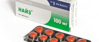
- For moderate cases, local agents are prescribed that contain an antibiotic or an antibiotic in combination with a glucocorticoid. The antibiotic must have activity against the most common pathogens of ONO - the drugs of choice are ciprofloxacin, neomycin, polymyxin B. If perforation of the eardrum is suspected, the use of drops containing aminoglycosides should be avoided.
- In severe cases, in addition to local therapy, the option of additional systemic antibacterial therapy is considered - most often oral forms of quinolones (ciprofloxacin or ofloxacin).
Drugs for local use remain the treatment of choice for ANO, as they allow creating a high concentration of the drug at the site of inflammation.
There are no randomized studies comparing topical and oral antibiotic therapy. But according to available data, the addition of an oral antibiotic to local antibacterial therapy for uncomplicated ANO does not have a significant effect on the course of the disease and recovery time.
The basis of treatment for otomycosis is NSP toilet and local antifungal therapy - the drugs of choice are clotrimazole and miconazole. Systemic antifungals are indicated for patients with suspected invasive fungal disease.
General recommendations
- During treatment, the ear should be protected from water - while bathing, a cotton swab soaked in petroleum jelly or glycerin should be placed in the ear.
- It is necessary to refrain from water sports for 7-10 days.
- You should limit the wearing of hearing aids and headphones as much as possible until the pain completely subsides and there is no discharge from the ear.
How is acute external otitis treated at the Rassvet Clinic?
For ONO, we give preference to local empirical therapy. An important stage of treatment is the toilet of the NSP, performed by an otorhinolaryngologist. In the clinic, we perform it under a microscope, which makes the procedure less traumatic and more effective. During the procedure, pathological discharge and granulations are removed from the ear canal, which in turn increases the effectiveness of local treatment.
Our otorhinolaryngologists select medications for local treatment based on otoscopy and medical history. Not only the effectiveness of the antibiotic against the most common pathogens is taken into account, but also their side effects and possible drug resistance.
We prescribe systemic antibiotics only when indicated. First, compared with local therapy, systemic antibiotics may increase the likelihood of drug resistance. Secondly, the use of only oral agents without local antibacterial therapy may lead to relapse of the disease, which is likely due to lower concentrations of antimicrobial drugs at the site of inflammation.
Acute external otitis media resolves within 7-14 days. With a longer course or repeated changes of tactics during the treatment period, frequent relapses, we always exclude other diseases, including immunodeficiency states and oncology.
Antibiotic therapy for otitis media
This topic can be presented in several sections, since we should talk not only about external and otitis media, but also about acute and chronic processes in these parts of the ear, since each of these diseases requires an independent medical approach.
Unfortunately, at present, along with the successes of pharmacotherapy, social changes have occurred in society that largely reduce the effectiveness of the use of new drugs that can not only shorten the duration of acute ear disease, but also prevent its transition to a subacute and chronic process. This refers, first of all, to the lack of the previous possibility of carrying out a general medical examination of patients with ear diseases.
The decline in the level of well-being of the population, the quality of life, and the increase in infectious diseases significantly complicate the provision of appropriate care to patients with inflammation of the middle ear, especially chronic ones.
The problems of providing effective care to patients with ear diseases cannot be reduced only to the choice of a drug that has a pronounced anti-inflammatory effect, and one cannot rely only on the “power” of the newest antibiotic. Therefore, the use of antibiotics should be under the control of otoscopy with an analysis of the effectiveness of the treatment, otherwise the opposite can be achieved - allowing the acute process to become chronic or unsuccessfully influencing chronic purulent otitis media, which may require surgical intervention. It is noted that one of the reasons that indirectly contribute to the occurrence, persistent and long-term course of the so-called exudative otitis media, the incidence of which is growing, is acute respiratory viral diseases, often against the background of an enlarged pharyngeal tonsil and allergies. Irrational use of antibiotics, for example, at the first signs of catarrh of the upper respiratory tract, otitis media, leads to the suppression of the active purulent process in the middle ear, when some of its symptoms (for example, pain) disappear and the process turns into sluggish inflammation. This ultimately leads to disruption of the basic function of the ear - permanent hearing loss. Elimination of ear pain, the most troubling symptom for a patient, does not mean the elimination of inflammation, but only helps to reduce the patient’s motivation for further treatment.
As practice shows, 1–2 days after the prescription of potent antibiotics, ear pain disappears, and the patient rarely continues to take them at the full course (50% stop taking them on the 3rd day, 70% on the 6th day). Therefore, you should not trust only antibacterial therapy for acute otitis media and exacerbation of a chronic disease, since in addition to fighting an active infection, it is necessary to create all the conditions for the evacuation of inflammatory exudate, including purulent, from the cavities of the middle ear, otherwise the inflammatory process will be delayed. In addition, the opinion that flora plays a decisive role in the development of inflammation in a closed cavity, for example, in the middle ear, and especially in the chronicity of inflammation, is currently debatable, since the state of immunity, both general and local, has an undoubted influence on the nature of the inflammatory process. The microorganism gives impetus to the emergence of an acute process in the ear, and then the complex of opposing forces of the body determines the transition of the process to chronic or recovery occurs. Therefore, antibacterial therapy for various inflammatory diseases of the middle ear should be carried out, presenting the nature of the inflammatory process (catarrhal, purulent, destructive, allergic), taking into account otoscopy data and always after obtaining information about the nature of the microbial flora in the discharge from the ear, its sensitivity to this or that antibiotic. One thing can be said for sure: for a patient with an inflammatory process in the middle ear, and especially in the inner ear, the use of “ototoxic” antibiotics of the aminoglycoside group is contraindicated
, since this can, even after a single instillation of the solution into the ear, not to mention parenteral administration, lead to a severe outcome - irreversible hearing loss and even deafness due to damage to the receptor part of the cochlea.
Based on the above, first of all, it must be emphasized that antibiotic therapy cannot be a panacea, but is only part of a complex of therapeutic measures that provide relief from acute and exacerbation of chronic processes in the cavities of the outer and middle ear
. The methods of using antibiotic therapy for ear diseases can be varied - from topical application in the form of drops introduced into the ear through the external auditory canal, to taking the antibiotic orally and parenterally.
At the same time, it can be noted that since the infection penetrates into the middle ear, mainly through the auditory tube from the nasopharynx, its sanitation, including with the help of antibiotics applied locally, is entirely justified. For example, fusafungin, used in the form of a spray, provides sanitation of the nasal cavity, nasopharynx, nasopharyngeal mouth of the auditory tube, and has an effect on gram-positive and gram-negative cocci and bacilli, anaerobes, and fungi.
Acute otitis media
It is generally accepted that until a natural perforation of the eardrum has occurred and exudate has not appeared, which can be characterized as purulent, acute otitis can be classified as “catarrhal”, and treatment does not require antibiotic therapy, with the exception of the previously mentioned fusafungin for irrigation of the nasal mucosa , nasopharynx. This prevents further infection of the middle ear and suppresses the flora in the nasal cavity, pharynx, and trachea. Antibiotics for acute otitis media must be prescribed in cases where the measures taken (anemization of the mucous membrane of the nasal cavity, nasopharynx; warming compress on the mastoid area; analgesics, etc.) do not stop the process
– ear pain continues, hearing decreases, body temperature persists, and the patient’s general condition worsens. Typically, acute catarrhal otitis media with a favorable course ends by the 4th–5th day. After perforation occurs or is created artificially (tympanopuncture, paracentesis), it becomes possible to identify the flora and determine its sensitivity to antibiotics. But even before the eardrum is perforated, it is sometimes necessary to prescribe broad-spectrum antibiotics, taking into account the most common flora in acute otitis media:
• Amoxicillin
1 capsule 3 times a day with plenty of drink 2 hours after meals, course 7-10 days.
• Ampicillin trihydrate
orally, 1 tablet or capsule (adults 250–1000 mg, children 125–500 mg) 4 times a day for 5 days.
• Phenoxymethylpenicillin
orally, 3 tablets per day, for children under 14 years of age 50–60 thousand units per 1 kg of body weight per day.
• Spiramycin
6–9 million IU, i.e. 2–3 tablets.
• Azithromycin
orally 1 hour before meals, 500 mg once a day for 3 days. Children: 1 tablet 125 mg 1 time per day.
• Cefazolin
by injection intramuscularly in a solution of procaine. The bottle contains 0.25–0.5 or 1 g of powder for dilution. For 7 days, 1–4 times a day.
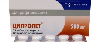
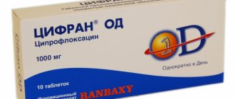
• Ciprofloxacin
1 tablet 2 times a day for 7 days.
Thus, the arsenal of antibiotics, which have a very effective effect in the treatment of patients with acute purulent otitis media, is sufficient, and by the end of the week after the start of treatment, almost complete remission occurs - ear pain stops, there is no discharge from the ear canal. However, the criterion for complete recovery is the restoration of hearing, for which a wide range of measures should be used that are not related to the fight against inflammation in the ear.
Chronic otitis media
Treatment of patients with chronic suppurative otitis media necessarily involves the use of various antibiotics
, since with chronic inflammation of the middle ear, polyflora or even flora that changes during long-term treatment can be isolated.
The doctor encounters particular difficulties in the presence of Proteus and Pseudomonas aeruginosa in the purulent discharge .
In these cases, treatment can last for months or even years. For chronic otitis, the use of antibiotics orally or parenterally is indicated only in cases of exacerbation of the process in the middle ear or in the postoperative cavity, if the patient has undergone sanitary surgery on the ear. Basically, antibiotics for chronic otitis media have to be used in the form of drops, washing liquids, and ointments. It is necessary not only to eliminate the identified flora, but also to achieve reparation of the mucous membrane damaged by inflammation, which is sometimes associated with the need for immunocorrection. It has been proven that, along with anti-inflammatory therapy, detoxification therapy is necessary, since the level of average molecular peptides in the blood plasma correlates with the severity of intoxication. From the arsenal of antibiotics used for chronic purulent otitis media and its exacerbations, you can use drugs from the list above. Adding to it may be a list of antibiotics that affect Proteus, Pseudomonas aeruginosa and Escherichia coli:
• Ciprofloxacin
orally 125–500 mg, 1 tablet 2 times a day.
• Chloramphenicol
(alcohol solution: 100 ml contains 0.25 chloramphenicol) for instillation into the ear for detected Proteus, E. coli, salmonella, bacteria resistant to penicillin, streptomycin.
• Netilmicin
used for injection in the process in the ear, supported by gram-negative microorganisms. Use 4 mg per day in equal portions 2 times a day.
Otitis externa
Skin diseases of the external auditory canal can also have an acute and chronic course with exacerbations, accompanied by ear pain, congestion, itching and purulent discharge. Depending on the nature of the pathogen, the clinical picture may indicate a fungal or bacterial-coccal flora
. Based on the clinical manifestations, the doctor can guess the nature of the pathogen, but only a microbiological examination allows you to accurately select the type of medication that is appropriate in each case.
As a rule, local treatment for fungal external otitis
is quite successful. In cases of protracted otitis externa caused by a fungus such as candida, the antibiotic nystatin is used (for adults, 500 thousand units orally 3-4 times a day for 10-14 days). Among ointments containing antibiotics, many doctors still prefer to prescribe the well-proven Oxycort ointment (it contains hydrocortisone and oxytetracycline).
For a boil of the external auditory canal and for diffuse inflammation of the skin of the external auditory canal, the use of gramicidin
in the form of alcohol drops (2% alcohol solution, 2–3 drops). The very active sofradex also contains gramicidin (the bottle contains 5–8 ml of solution, apply 2–3 drops 3 times a day for 1 week). It must be remembered that the local use of some antibiotics can have an ototoxic effect in the presence of perforation in the eardrum. It is necessary to emphasize once again that antibiotics for inflammation of the skin of the external auditory canal cannot be monotherapy and in the vast majority of cases should be used in combination with measures to activate the body's defenses and additional effects on the tissue (physiotherapeutic effects, opening the boil, etc.).
| Applications to the article |
| Antibacterial therapy for otitis media should be carried out after obtaining information about the sensitivity of the microbial flora to antibiotics. |
Prevention
Prevention of ONO is carried out in case of frequently recurring episodes of the disease, concomitant dermatological diseases, as well as with regular contact with water - in athletes.
When engaging in water sports or frequently visiting the pool, it is recommended to use swimming caps or special earplugs, and dry your ears after swimming. The outer ear can be gently wiped with a towel. To remove residual water, shake your head vigorously in different directions.
Author:
Chekaldina Elena Vladimirovna otorhinolaryngologist, Ph.D.