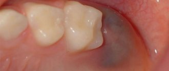
If not treated promptly, caries under the gum can lead to severe tooth decay and even loss. The enamel at the base of the tooth darkens noticeably, and it begins to react painfully to cold and hot food and mechanical stress. Read the article about the causes of subgingival caries and methods of treating the disease.
In this article
- Causes of caries in the gums
- Additional reasons for the development of caries under the gum
- Features of the disease
- Symptoms of root caries
- Treatment of cervical caries
- Consequences of cervical caries if left untreated
- Features of filling caries near the gums
- Prevention of subgingival caries
Causes of caries in the gums
The main cause of the disease is cariogenic microbes that cause damage to enamel and dentin. They exist in everyone, but they begin to develop only when favorable conditions are created: consumption of large amounts of carbohydrate foods, poor oral hygiene and due to some other reasons. The development of caries at the base of the tooth is associated with its localization and is caused by the following factors:
- Due to the characteristic structure of the tooth, plaque constantly accumulates in the root area, which cannot always be removed even with a toothbrush. If you do not make an effort when cleaning, then gradually the neck of the tooth will begin to collapse under the influence of cariogenic bacteria contained in plaque.
- In the depressions near the gums, the so-called “pockets,” food debris accumulates. If oral hygiene is not sufficiently thorough, food decomposes and lactic acid is formed, which destroys tooth enamel.
A risk factor that provokes the development of cervical caries may be the consumption of large quantities of too acidic foods that contain quickly fermentable carbohydrates. As a result of their fermentation, organic acid is released, which corrodes tooth enamel and washes away calcium.
Why can baby teeth turn black?
Baby teeth are much more susceptible to caries than molars, since they do not have such a dense structure. Enamel can change color due to the development of various pathological processes that can be provoked in a child:
- fragility of tooth enamel;
- calcium deficiency;
- improper oral care;
- chips and cracks in the enamel;
- vitamin deficiency;
- endemic fluorosis;
- chronic gastrointestinal diseases;
- genetics.
Sometimes the cause of darkening of the teeth of very young patients is artificial feeding of the baby at night. Consuming formula or milk at night reduces saliva production. The acid that accumulates on the teeth during feeding is not washed away by saliva and the process of destruction of tooth enamel begins.
Additional reasons for the development of caries under the gum
In addition to factors directly related to the impact on the teeth, there are also third-party causes that can provoke the occurrence of a carious process between the gum and tooth.
Let's name the most common ones:
- hormonal or endocrine disorders in the body;
- use of medications that increase enamel porosity;
- lack of certain vitamins, especially group B;
- poor quality of drinking water;
- heredity;
- increased acidity of the body;
- age factor and others.
Taking into account these additional reasons, the treatment of gum caries should not be limited only to the work of the dentist. In this case, it should be comprehensive with the involvement of several specialists, for example, an endocrinologist and others. If the cause is not eliminated, the pathology will return again and again.
Prevention of the development of gingivitis and periodontitis
- The first place is always hygienic cleaning of the oral cavity, including regular brushing of teeth using the products that are most suitable in each particular case.
- Regular visits to the dentist for preventive examination.
- If you have crowns and dentures, it is recommended to use an irrigator so that you can thoroughly clean them at home. The Waterpik WP-100 E2 irrigator has proven itself well.
- The use of medicinal toothpastes containing the latest generation of antiseptics.
- The use of effective rinses that prevent the growth of bacteria and, as a result, the formation of pathogenic plaque on the gums and teeth.
Features of the disease
This type of caries is especially dangerous and unpleasant, as it has some peculiarities in its course and location. Its manifestations are associated with the following factors:
- Carious lesions are localized in the weakest area near the neck of the tooth (this is the part covered by the gum). In this zone, the enamel is also weakly mineralized, and this factor enhances the development of caries.
- Circular distribution. Cervical caries affects the tooth in a circle, covering increasingly larger areas. As a result, part of it may break off, since the integrity will be broken in many places.
- Damage to the front teeth. An unpleasant feature of gingival caries is its frequent location on the incisors. A person experiences psychological discomfort when he has to talk, smile, laugh.
Under the influence of the bacteria Streptococcus Mutans, which causes the development of the disease, the enamel and dentin underneath are actively destroyed. Inflammation can spread to the pulp and ultimately lead to pulpitis or periodontitis.
Symptoms of root caries
Pathology has the same symptoms as caries of other classifications, but its impact is more destructive, and the consequences are more unpleasant and more dangerous for dental health. Carious lesions can spread deep inside, gradually destroying all the canals. Here is what the main signs of cervical caries look like:
- increased reaction of teeth to cold, hot, sour, sweet foods;
- the appearance of dark spots on gums with carious lesions;
- pain when chewing food and while brushing teeth.
All of these symptoms indicate the development of a disease that consists of several stages.
- Chalk stain. At this stage, a slight dark spot appears on the surface of the enamel without changing its shape or size. Particular sensitivity to acidic foods may occur. At the matte spot stage, caries under the gum is eliminated without the use of a drill.
- Superficial stage. The spot becomes rough, and gradual destruction of the enamel begins. A diseased tooth reacts to various irritants, in addition to sour: hot, cold, including cold air, mechanical stress. The pathology begins to actively progress.
- Middle stage. Cavities form in teeth as dentin is destroyed. The defect is already becoming clearly visually noticeable.
- Deep stage. The process spreads deep into the tooth, reaching the roots and nerve endings. The pain can be very severe, especially at night, and discomfort occurs when inhaling cold air.
There is no clearly defined boundary between the listed stages; the transition from one to another is so smooth that it is almost impossible to track it. In an advanced stage, serious consequences can develop: for example, circular caries, when the process already covers the entire gingival area of the crown in a circle. Teeth at this stage break off easily.
Treatment of loose gums (gingivitis or periodontitis)
- In dentistry, a periodontist will perform a complete cleaning of the teeth using a mechanical method or using ultrasound.
- Be sure to carry out an anti-inflammatory course of treatment of the whole body according to the recommendations of a dentist.
- Agree to undergo a curettage procedure, during which the cavity formed due to inflammation is completely cleaned and treated with antiseptics. It is better to use ultrasonic cleaning.
- In advanced forms of the disease, when treatment is already overdue, perform a splinting procedure. In such cases, the teeth are fixed together using fiberglass.
Treatment of cervical caries
It is very important to contact your dentist for help while the process has not yet become global. A competent specialist knows how to effectively treat this disease in order to save teeth and eliminate the consequences. At the initial stage, you can still do without the use of a drill and filling, which allows you to significantly save your budget. The doctor determines treatment methods depending on the degree of pathology and the general clinical picture. Each stage requires its own approach to therapy.
Treatment at the spot stage
In this case, remineralizing therapy and additional means of protection are used. The doctor removes tartar and plaque from the teeth, then applies fluoride strips to the affected areas. This element helps regenerate enamel layers and stop the carious process. At home, you can use herbal decoctions, solutions of fluoridated salt and water to rinse the mouth, use pastes and threads containing fluoride.
How to treat cervical caries of the superficial and middle stages
At these stages, the doctor removes the damaged tooth tissue and polishes the carious area. The procedure is completed with remineralizing therapy. If necessary, a filling is placed.
Method for treating deep caries
At this stage of the disease, coping with it is not an easy task. The treatment consists of several sessions. Here are the procedures the dentist performs:
- Anesthetic injection. When treating cervical caries, the doctor must provide anesthesia, since the area near the gums is very sensitive to action.
- Cleaning enamel from plaque and tartar. This is necessary to prevent infection from reaching the affected area.
- Selecting the color of the filling material.
- Removing carious tissue using a drill.
- Treatment of the cavity with an antiseptic and adhesive material.
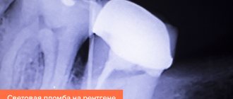
- Filling. The composite material is applied in layers, each of them is treated with a photopolymerization lamp for hardening.
- Grinding and polishing the tooth surface. The dentist gives the surface the correct shape and makes sure that he has achieved the correct bite.
Thus, each carious tooth is treated. The process can take a long time and cost a lot of money - another argument in favor of regular prevention.
Now about the production of metal ceramics in our clinic
Temporary crowns are made for you before preparing your teeth so that you don’t walk around for a single day with ground-down “stumps” that scratch your tongue and lips and frighten those around you. The doctor carefully, and therefore slowly, fits these crowns to your teeth and gums. Yes, yes, even temporary crowns should under no circumstances irritate the gums. And this is achieved by high-quality, which again means quite a long work, by preparing the teeth, with their obligatory polishing and smoothing of the ledge, designed to protect the gums from the pressure of the crown.
Impressions for permanent crowns are taken using modern, absolutely painless gum retraction technology of the 21st century - the gums are moved back not with a traumatic retraction thread, but with a special composition that does not cause the slightest pain. The quality of the impression material plays an important role at this stage.
When your new teeth are ready, they must be tried on, taking into account your wishes; the fit of the crowns to the ledge and gum is very carefully checked in order to exclude any pressure on it, as well as contacts with neighboring teeth so that they are tight and pieces of food do not fall between the teeth ( a very common mistake when making crowns). The crowns are then fixed to temporary material so that the color, shape and ease of use can be checked in natural conditions. If necessary, the crowns are adjusted, achieving absolute harmony and comfort. And only after some time the metal ceramics are fixed with permanent cement.
Of course, all these manipulations take time, and high-precision technologies that guarantee quality, by definition, cannot be cheap.
You wouldn’t believe it if they offered you to buy “a car for the price of a bicycle”? What is the difference between metal-ceramic crowns?
Do you want to buy something inexpensively that you simply won’t be able to use in the future?
After all, as more than 20 years of practice and simple mathematics show, the production of cheap metal ceramics, which is promoted on every corner, is much more expensive. And not in the distant future, but in the quite foreseeable.
Consequences of cervical caries if left untreated
Advanced caries near the gums is very dangerous, it can progress quite quickly and cause complications such as:
- periodontitis;
- pulpitis;
- inflammation of the gums;
- an abscess or a well-known flux.
In addition, the development of caries in the gums is often associated with pathologies of the thyroid gland and diabetes mellitus. It is necessary to conduct a comprehensive diagnosis to determine the exact cause of the disease. After the therapy prescribed by the endocrinologist, the disease may recede.
Features of filling caries near the gums
Placing fillings in the area near the gums is much more difficult than with other types of caries - approximal or fissure. This is due to some factors that arise during the work process:
- the area near the gums is inconvenient for filling, especially on the upper teeth;
- Saliva gets on the affected area all the time, even placed cotton rolls do not help;
- During the treatment process, blood is always released, which also enters the working area.
The closer to the root a tooth is damaged, the more difficult it is to treat. Due to the fact that cervical caries most often affects the front teeth, you need to choose the color of the material with special care - it should ideally match the color of the enamel, since the aesthetic visual effect plays a role here.
Prevention of subgingival caries
The development of the disease is provoked by many external and internal factors. And even if caries has never bothered you, preventive measures will help prevent its occurrence and development. Here are some simple rules doctors recommend following:
- Brush your teeth with toothpastes high in minerals.
- After each meal, use a mouthwash and dental floss - it will help effectively remove pieces of food stuck between your teeth. Oral irrigators can also be used.
- Half an hour after eating, brush your teeth if possible. This should be done twice a day for a couple of minutes.
- Include in your mandatory daily diet foods rich in fluoride, calcium and other beneficial microelements to strengthen enamel, eat more solid vegetables and fruits.
- Avoid excessive consumption of sweets, flour and sour foods.
- Periodically massage the gums: it improves blood circulation in the gum tissues, protects against the formation of deep periodontal pockets and the accumulation of deposits in them, which reduces the risk of caries formation at the neck of the tooth.
If the enamel is thin and susceptible to the rapid formation of tartar on it, regular remotherapy helps a lot - this is the name for applying fluoridating compounds to the teeth. The procedure reduces the risk of enamel destruction and improves the condition of hard tissues. To further strengthen the enamel, you can periodically take special vitamin complexes containing useful minerals. In addition, it is necessary to visit the dentist’s office at least once every six months for a dental examination. The doctor will make sure that everything is fine with them, or will prescribe additional diagnostics using x-rays. Once every 6-9 months, it is recommended to carry out professional enamel cleaning using ultrasound or other methods. During the procedure, plaque and tartar are removed from the surface of the teeth. It polishes and becomes smoother, and less deposits accumulate on the enamel. During cleaning, the doctor can also clean periodontal pockets if plaque and deposits accumulate under the edge of the gums.
People who are at risk for endocrine diseases need to be especially attentive to pain in their teeth. With them, teeth are often affected by cervical caries. Treatment must be comprehensive and systemic to prevent caries from damaging the tooth root. Gingival caries is a dangerous pathological process if it is started, but it can be successfully treated in the early stages.
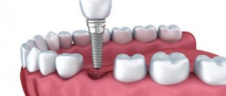
Modern dentistry eliminates caries of various types, but it also happens that if the stage of the disease is too deep, some teeth cannot be saved. We have to remove them and resort to various methods of restoring the dentition. You just need to be attentive to your oral health, taking simple preventive measures, so as not to have to deal with expensive treatment later. With regular dental examinations, you will be able to detect the problem in time and solve it.
Clinical researches
ASEPTA products have clinically proven effectiveness. Repeated clinical studies have proven that the use of ASEPTA adhesive gum balm for a week can reduce gum bleeding by 51% and reduce inflammation by 50%.
As part of the research, it was also proven that the two-component mouth rinse ASEPTA ACTIVE more effectively combats the causes of inflammation and bleeding compared to single-component rinses - it reduces inflammation by 41% and reduces bleeding gums by 43%.
Sources:
- The use of adhesive balm "Asepta®" in the treatment of inflammatory periodontal diseases L.Yu. OREKHOVA*, Dr. med. Sciences, Professor, Head of Department V.V. CHPP**, Dr. med. Sciences, Professor, Head of Department S.B. ULITOVSKY*, Dr. med. Sciences, Professor A.A. LEONTIEV*, dentist A.A. DOMORAD**, O.M. YAKOVLEV** SPbSMU named after. acad. I.P. Pavlova, St. Petersburg - *Department of Therapeutic Dentistry, **Department of Microbiology
- The effectiveness of the use of Asept “adhesive balm” and Asept “gel with propolis” in the treatment of chronic generalized periodontitis and gingivitis in the acute stage (Municipal Dental Clinic No. 4, Bryansk, Kaminskaya T. M. Head of the therapeutic department Kaminskaya Tatyana Mikhailovna MUZ City Dental Clinic No. 4, Bryansk
- Study of the clinical effectiveness of treatment and prophylactic agents of the Asepta line in the treatment of inflammatory periodontal diseases (A.I. Grudyanov, I.Yu. Aleksandrovskaya, V.Yu. Korzunina) A.I. GRUDYANOV, Doctor of Medical Sciences, Prof., Head of Department I.Yu. ALEXANDROVSKAYA, Ph.D. V.Yu. KORZUNINA, asp. Department of Periodontology, Central Research Institute of Dentistry and Maxillofacial Surgery, Rosmedtekhnologii, Moscow
