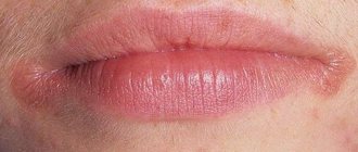
Cheilitis is a disease and inflammation that is expressed by damage to the lips (“ cheilitis on the lips ”), more precisely, inflammation of the red border, the mucous membrane of the lips and (in some cases) the skin of the lips. According to medical classification, there are different types of cheilitis. For example, simple cheilitis (Cheilitis simplex, the most common form), angular cheilitis (Cheilitis angularis, or angular cheilitis , or seizure, or inflammation of the corners of the mouth). Cheilitis can be either an independent disease (for example, exfoliative cheilitis, glandular cheilitis, actinic cheilitis), or a symptom of other diseases (for example, atopic cheilitis , eczematous cheilitis ). There are more than twenty types or types of cheilitis.
What it is
The most common condition of the lower lip, which foreshadows a cancerous tumor, is diagnosed in most cases in males of retirement age. This disease is called abrasive pre-cancerous cheilitis of Manganotti, and it is most often provoked by various types of injuries.
A characteristic feature of precancrosis Manganotti cheilitis is the formation of small oval or round erosions on the lips without infiltration, which do not heal for a very long time and often appear over a long period of time.
This disease got its name in honor of the Italian dermatologist Manganotti, who practiced in the first half of the twentieth century and described his observations regarding the manifestations of this type of precancerous inflammation of the red border of the lips.
1.General information
The collective diagnosis of “cheilitis” covers a group of etiologically heterogeneous inflammatory diseases affecting the lips, their border and, in some cases, the surrounding skin. Like any other inflammation, especially those occurring in a chronic form, cheilitis is associated with the risk of malignancy - the appearance of mutated atypical cells that can trigger the growth of a malignant tumor.
It is known that for different types of inflammatory pathology this risk is not the same: some processes are relatively safe, others are interpreted as an obligate precancerous condition. It is precisely this intermediate position that is occupied by abrasive precancerous (precancerous) Manganotti cheilitis - a special form of ulcerative inflammation localized on the red labial border and a very high risk of further malignancy.
A detailed clinical description of this form of cheilitis was given in 1933 by the Italian dermatologist Giuseppe Manganotti, after whom the disease is named.
Manganotti cheilitis develops predominantly on the lower lip, usually in elderly patients (over 60 years of age). Men get sick several times more often than women.
A must read! Help with treatment and hospitalization!
Causes
The main reason for the development of this disease is old age. It is in old age that the immune defense weakens, pathological processes begin to form in the body, internal organs fail, and metabolism is disrupted. Because of these changes, the external defense also suffers and the epithelial layer weakens.
In old age, a person has few healthy teeth left; many have to be removed, which inevitably leads to the need to wear dentures. And since the majority of elderly people in our country are not well-off enough for treatment in paid clinics, they have to turn to state-owned clinics for the manufacture of prostheses, where the quality of the source material and the final product suffers significantly.
As a result, a complex of influencing factors, namely malnutrition of the skin of the lips, vitamin deficiencies, constant traumatic effects of low-quality prosthesis, as well as natural factors and injuries, lead to the formation of erosions on the lips.
2. Reasons
First of all, it should be noted that elderly, pre-senile, and senile age in itself is a factor of high cancer risk - due to the natural wear and tear of tissues, changes in the processes of cellular nutrition and general metabolism. The etiopathogenetic basis of Manganotti cheilitis is considered to be degenerative-dystrophic processes, which then transform into a local focus of atrophy. Much of these patterns, however, remains unclear today.
Provoking factors are dental problems - incomplete dentition, poorly fitting dentures, sharp chips on the teeth - and, in general, any trauma, mechanical or chemical irritation of the lip, especially if such effects are repeated or permanent (including smoking, the habit of biting lip, etc.).
The triggering factor may be intense insolation, i.e. ultraviolet part of the spectrum of direct sunlight.
Visit our Dermatology page
Symptoms
The following symptoms are characteristic of abrasive precancrosis cheilitis of Manganotti:
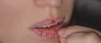
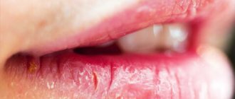
- Formation on the red border of the lower lip of one or two or three small erosions of a round, oval or irregular shape.
- The erosion surface is most often smooth, bright red or yellow-red in color.
- Sometimes a serous or bloody crust may form on the surface of the erosion, and when removed surgically, the opened wound will bleed a little.
- Patients never complain of severe pain with abrasive cheilitis of Manganotti; on the contrary, most often the erosions are completely painless.
- The resulting erosions may not heal for several weeks or even months, and new ones soon appear in place of the healed ones or nearby.
- The disease is chronic and can last for years if the inflammation does not develop into a malignant tumor. The onset of the cancer process can be determined by the compaction of the base of the erosion and the appearance of blood at the slightest damage to the upper layer.
Precancerous diseases of the red border of the lips (RBL) and oral mucosa (ORM)
I. K. Lutskaya
Doctor of Medical Sciences, Professor, Head of the Department of Therapeutic Dentistry BelMAPO (Minsk)
Diseases characterized by a high tendency to malignancy (obligate precancers of the oral mucosa and red border of the lips) require special consideration in dental practice. They are characterized by the absence of objective signs of a cancerous tumor, but in the presence of pathogenic factors they become malignant. The clinical manifestations of this group of diseases are quite diverse, which makes their diagnosis difficult. On the other hand, the prognosis depends on a number of factors, primarily the nature of the carcinogens, as well as the local status and general condition of the body. If adverse effects are excluded, reverse development of the lesion elements, stabilization of the process without significant changes, or further development without a tendency to degeneration are possible. The persistence of an unfavorable background leads to malignancy of the lesion.
The main signs of malignant degeneration can be the following symptoms: a sharp change in the clinical picture, namely acceleration of the development of a tumor or ulcer, exophytic growth or ulceration of the tumor (Fig. 1).
Rice. 1 a. Signs of malignancy.
Rice. 1 b. Signs of malignancy.
The next signal points are bleeding of the lesion, the appearance of hyperkeratosis, infiltration and compaction at the base (Fig. 2). The lack of effect of conservative treatment within 7-10 days is the basis for referring the patient for consultation to an oncologist or maxillofacial surgeon. Malignancy is confirmed by the results of morphological studies, namely the identification of atypical cells in the biopsy material.
Rice. 2. Bowen's disease.
Systematics of keratoses as precancerous conditions
I. Keratoses without a tendency to malignancy (initial form of leukoplakia, soft leukoplakia, geographic tongue, etc.).
II. Optional precancer with the possibility of malignancy up to 6% (flat form of leukoplakia, hyperkeratotic form of lichen planus; pemphigoid form of lichen planus, etc.).
III. Optional precancer with a tendency to malignancy (malignancy tolerance) from 6 to 15% (elevating form of leukoplakia; warty form of leukoplakia; erosive form of leukoplakia; warty form of lichen planus; erosive form of lichen planus; rhomboid glossitis - hyperplastic form, etc.).
IV. Obligate precancerosis with the possibility of malignancy over 16% (ulcerative form of leukoplakia; keloid form of leukoplakia; ulcerative form of lichen planus; follicular dyskeratosis; Bowen's syndrome; atrophic keratosis; xeroderma pigmentosa, ichthyosis vulgaris, etc.).
Bowen's disease
Bowen's disease has the highest potential risk of malignancy (Bowen, 1912), since its histological picture is cancer in situ (intraepithelial cancer without invasive growth). Clinical manifestations of Bowen's disease vary depending on the location, stage of the disease, and associated factors. The patient's complaints can be reduced to discomfort, roughness of the corresponding parts of the mucous membrane, more or less pronounced itching. In some cases, there are no subjective sensations, so the lesion will be detected during a routine examination of the oral cavity.
The favorite localization of lesion elements in the form of spots, papules, scales, erosions, and areas of keratinization are the posterior sections of the mucous membrane: the palatine arches, the root of the tongue. Clinical manifestations of the disease on the cheeks, lateral surface of the tongue, and soft palate are described. More often one, less often two or three areas of altered mucosa are found.
Most typical for the initial stages of the disease is the appearance of a limited area of hyperemia, which has a nodular-spotted or smooth appearance (Figure 3). A distinctive feature may be the peculiar velvety surface as a result of small papillary growths. The further clinical picture resembles leukoplakia or lichen planus due to the formation of areas of hyperkeratosis. There is a tendency to erosion. The lesion will rise above the level of surrounding tissues if nodules form and merge into plaques. With a long course, atrophy of the mucous membrane develops, and in such cases the affected area seems to sink.
Diagnosis is difficult when a small focus of hyperemia subsequently becomes covered with scales, resembling leukoplakia or lichen planus in appearance. The diagnosis of Bowen's disease is made on the basis of a histological picture: giant cells with an accumulation of nuclei in the form of lumps, the so-called monstrous cells, are found in the spinous layer.
The prognosis of the disease is unfavorable: the development of lesions within 2-4 months ends with invasive growth without a tendency to regression (reverse development).
Treatment for Bowen's disease involves complete excision of the lesion, which may include surrounding healthy tissue. In some cases, close-focus X-ray therapy is used.
Warty precancer
Warty precancer is an independent clinical form (A. L. Mashkilleyson). It is characterized by a pronounced tendency to malignancy: already 1-2 months after the onset of the disease. The patient's complaints boil down to the presence of a cosmetic defect and discomfort. The favorite localization of the lesion (usually single) on the red border of the lower lip allows for a detailed study of the clinical picture. The main element of the lesion is a nodule with a diameter of up to 10 mm, protruding above the level of the mucosa and having the usual color of the red border of the lips or a persistent red color (Fig. 4). The surface of the nodule may be covered with thin, tightly attached scales that cannot be removed when scraped. The tissue surrounding the lesion is not changed. Upon palpation, the compacted consistency of the nodule is determined, there is no pain.
Differential diagnosis with a wart, keratoacanthoma, papilloma is carried out on the basis of the clinical picture with mandatory pathological confirmation. Clinically, a common wart is characterized by a lobulated structure or papillary growths with a rim of stratum corneum along the periphery. Keratoacanthoma is characterized by a large number of keratinized cells filling the depression in the form of a funnel formed by a dense ridge of surrounding hyperemic mucosa. The papilloma has a stalk of soft consistency.
Treatment of warty precancer is only surgical with complete excision of the lesion and histological examination of the tissue. Confirmation of warty precancer is the proliferation of the epithelium due to the spinous layer both towards the surface and deep into the mucosa.
Rice. 3. Warty precancer.
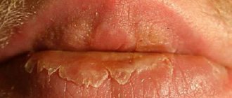
Limited precancerous hyperkeratosis
As an independent disease, limited precancerous hyperkeratosis has a less pronounced degree of malignancy than warty precancer: the lesion can remain in a stable phase for months, even years. However, clinical signs of malignant degeneration are very unreliable, since increased keratinization processes, the appearance of erosion and compaction can be detected some time after the onset of malignancy. Therefore, the basis is histological examination. The patient makes no complaints or indicates a cosmetic defect. The characteristic localization of an area of altered polygonal mucosa on the red border of the lips makes it possible to diagnose limited hyperkeratosis. The grayish lesion may sink or rise above the surrounding unchanged red border without spreading to the skin or Klein's area (Fig. 5). The elevation of the lesion above the level of the lip is associated with the layering of scales, which cannot be removed when scraped. On palpation, the compaction at the base is not detected, but the dense consistency of the surface of the lesion is felt.
It is necessary to differentiate limited precancerous hyperkeratosis from leukoplakia, lichen planus, and lupus erythematosus. A diagnostic sign can be the lesion itself, which is small in size from a few mm
up to 1.5
cm
of polygonal shape with a tendency to form scales on the surface, which is not typical for leukoplakia. When localized on the lip, lichen planus is characterized by hyperemia, infiltration, whitish stripes, spots, and significant prevalence. With lupus erythematosus, inflammation, atrophic scars, and often erosion and diffuse damage to the red border are detected. Since histological examination is critical for the diagnosis of malignancy, biopsy should be performed as early as possible.
Treatment of limited hyperkeratosis consists of surgical removal of the lesion within healthy tissue; wedge-shaped excision is advisable. It is mandatory to exclude local irritants and bad habits: smoking, lip biting.
Rice. 4. Limited hyperkeratosis.
Abrasive precancrosis cheilitis Manganotti
The disease was first described by Manganotti (1913) and is considered in the group of obligate precancers due to its tendency to malignancy. It differs from other diseases of this group in its long course, tendency to regression (remissions), reappearance and development. Degeneration may occur after several months or many years.
A single lesion (rarely there are two) is localized on the red border of the lips in the form of oval or irregularly shaped erosion (Fig. 6). The surface of the erosion, having a bright red color, looks polished, can be covered with a thin layer of epithelium and does not tend to bleed. Drops of blood can be detected when crusts or crusts (serous, bloody) are separated, which can appear in the form of layers on the surface of erosions.
Rice. 5. Heilith Manganotti.
The element is localized on the unchanged red border of the lips. In some cases, congestive hyperemia and infiltration can be detected, but the background inflammation in Manganotti cheilitis is unstable.
Palpation does not detect changes in tissue consistency or pain. A feature of the clinical course of the disease is its intermittent nature: once it appears, erosion can spontaneously epithelialize, and then reappear in the same or another place, limited in size from 5 to 15 mm
.
It is necessary to differentiate Manganotti cheilitis from erosive-ulcerative forms of hyperkeratoses (leukoplakia, lichen planus, lupus erythematosus), herpetic lesions at the erosion stage, pemphigus, erythema multiforme. It is based on a characteristic picture of bright red erosion, often oval in shape, without bleeding or with the formation of bloody crusts on the surface. There are no lesions characteristic of hyperkeratosis in the form of spots, papules, stellate scars, or persistent hyperemia. In contrast to vesicular and vesicular lesions, fragments of blisters and specific cells are not identified (with pemphigus, herpetic stomatitis).
Erythema multiforme is characterized by an acute onset and pronounced prevalence of the process. Erosion in herpetic stomatitis is distinguished by scalloped edges as a result of the fusion of vesicles. If you have the habit of biting a portion of your lip, swelling and hyperemia are clearly visible, and signs of inflammation may transfer to the skin. Upon examination, individual point erosions or erosions merging into one are determined. Frequent lip biting confirms the traumatic nature of the disease. The history, as a rule, reveals a primary lesion in the form of a herpetic, mechanical, chemical, etc. lesion.
In doubtful cases, a histological examination is carried out, the diagnosis is confirmed by detecting epithelium infiltrated with histiocytes, lymphocytes, mast cells, as well as changes in the spinous layer and connective tissue.
Treatment of Manganotti cheilitis includes general and local treatments. Vitamins are prescribed internally to stimulate regeneration processes. Drugs that have an epithelializing effect are used locally: oil solutions of vitamins A, E, methyluracil, solcoseryl. In the presence of background inflammatory phenomena, it is possible to use corticosteroid ointments.
Conservative treatment of Manganotti cheilitis in the absence of signs of degeneration and a positive effect (the onset of stable remission) can last 2-3 months. Frequent recurrence, clinical growth, especially the appearance of the slightest signs of malignancy serve as an indication for surgical treatment (removal of the lesion within healthy tissue) with histological examination of the material.
Eliminating bad habits, irritating factors, and sanitation of the oral cavity are mandatory.
Tactics of a dentist
| Possible situations | Dentist tactics |
| Local manifestations in the presence of a local etiological factor: traumatic injuries, Vincent’s ulcerative necrotizing stomatitis | - diagnostics and treatment at the dentist — additional research is possible - therapy is usually local |
| Local manifestations in the presence of a common cause: chronic recurrent herpetic stomatitis, fungal infections, erythema multiforme | — diagnosis and treatment by a dentist after additional research and consultation with a specialist — local therapy, general therapy is possible |
| Local lesions against the background of general diseases: diseases of the gastrointestinal tract, cardiovascular system; allergic reactions, neurogenic and endocrine disorders; childhood infections, radiation sickness, myocardial infarction, blood diseases, tuberculosis, pemphigus | - dental examination — the final diagnosis is made by a doctor specializing in the field of this disease (hematologist, dermatovenerologist, allergist, etc.) - treatment by a specialist in the profile of pathology - general (etiotropic, pathogenetic), by a dentist - local, often symptomatic |
Particular vigilance should be exercised in cases of detection of a possible social risk or threat to the patient’s life. Depending on the specific clinical situation, the dentist bases his actions, including the following general rules.
- Each patient is admitted using personal protective equipment and sterile instruments.
- The initial examination of the oral mucosa is carried out only using instruments (mirror, probe, spatula, tweezers). Before special studies (serological, bacteriological, cytological) are carried out, the elements of the lesion are not palpated.
- In all doubtful cases, which include the primary detection of lesions on the mucous membrane or the lack of effect of treatment for a previously detected soft tissue change, the patient must be examined for syphilis and HIV infection. This recommendation is due to the fact that even a clinically clear local picture of the disease can accompany a general severe or infectious disease (for example, herpetic eruptions in AIDS, aphtha-like plaque in syphilis).
- Identification of positive serological blood reactions to syphilis or detection of Treponema pallidum in the lesion elements serves as an indication for treatment of the patient in a specialized institution. The situation is similar when identifying the causative agent of tuberculosis. HIV-infected people are served in AIDS centers, but they can also receive the necessary help from a dentist, which every outpatient doctor should remember.
- The initial detection of an ulcer with hardened edges or the lack of effect of treatment during an ulcerative process (7-10 days) requires consultation with an oncologist or maxillofacial surgeon, accompanied by a cytological or histological (biopsy) examination of the affected tissue. If signs of a tumor malignant process are detected, surgical treatment methods are used.
- A diagnosis confirmed by laboratory tests and excluding oncological or contagious diseases serves as the basis for prescribing conservative treatment, taking into account the etiology and associated factors.
General examination scheme
The first stage of interaction between a doctor and a patient is the collection of factual material, that is, the identification of symptoms of deviation from the norm. Next comes the clarification of the information received until the final diagnosis is made.
Research methods used in dentistry can be divided into the following groups: interviewing the patient (his relatives), examination, palpation, instrumental examination (probing, percussion of teeth, thermal diagnostics), assessment of gum and plaque condition indices, physical methods (electrical, radiological), laboratory studies (biochemical, bacteriological, cytological), special tests (blister test, histamine test, Kovetsky test, capillary resistance test), blood, urine, saliva tests.
Experts from the World Health Organization (WHO) recommend the following approach. The survey includes three parts: A
) extraoral region of the head and neck;
B
) perioral and intraoral soft tissues;
C
) teeth and periodontal tissues.
Settings, equipment and materials required (for all stages): adequate lighting, two dental mirrors and two gauze pads. We should not forget about gloves, a mask, and hygiene control.
The patient is in a sitting position.
First part of the examination - A
— requires a limited number of tools and takes no more than 5 minutes. The head, face, and neck are examined. The doctor evaluates changes in the size, color and shape of the parts of a given anatomical region.
When examining the skin, attention is paid to the presence of congenital changes (nevi, hemangiomas), as well as elements of damage in diseases. Notes the color, turgor, elasticity, moisture of the skin.
Some pathological changes, for example contractures, atrophy of facial muscles, are noticeable even during an external examination and must be recorded in the outpatient card (from a legal point of view, this is important in order to avoid a conflict situation when the patient is dissatisfied with the medical treatment provided).
It is necessary to pay attention to the shape and size of the pupils, which may reflect organic damage to the nervous system. Movement of the eyeballs is assessed, especially the presence of nystagmus (eye twitching). External examination of the facial muscles is insufficient. It is advisable to ask the patient to wrinkle his forehead, nose, open his mouth wide, and show his teeth. With facial nerve paralysis, tic-like twitching of the affected facial muscles, changes in the width of the palpebral fissure, and increased mechanical excitability of the muscles are observed. Peripheral paralysis of the lingual muscles causes fibrillary twitching with atrophy of the tongue (which may be a symptom of syringobulbia or amyotrophic lateral sclerosis). Bilateral paresis of the tongue causes a speech disorder such as dysarthria. Articulation defects and scanned speech are identified during conversation and questioning of the patient.
Part Two - B
- includes 7 steps: red lip border; mucous membrane and transitional fold of the lips; corners of the mouth, mucous membrane and transitional fold of the cheeks; gums and alveolar margin; language; floor of the mouth; hard and soft palate.
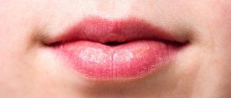
B1 - lips are examined with the mouth open and closed. The color, shine, and consistency of the red border are recorded.
B2 - examine the mucous membrane of the lips and transitional fold (color, consistency, moisture, etc.). Normally, small elevations are sometimes found on the inner surface of the lip due to small mucous glands, which is not a pathology.
B3 - using two mirrors, first examine the right, then the left cheek (mucous) from the corner of the mouth to the palatine tonsil (pigmentation, color change, etc.). Along the line where the teeth meet, there may be derivatives of the sebaceous glands, which should not be mistaken for a pathology. These pale yellow nodules with a diameter of 1-2 mm do not rise above the mucous membrane. It must be remembered that at the level of the 17th and 27th teeth there are papillae on which the excretory duct of the parotid gland opens, sometimes also mistaken for deviations.
B4 - gums: first examine the buccal and labial areas, starting from the right upper posterior area, then move along the arc to the left. They descend onto the lower jaw from the left back and move to the right along an arc. Then the lingual and palatal areas of the gums are examined: from right to left on the upper jaw and from left to right along the lower jaw. Discoloration, swelling and swelling of various shapes and consistencies may occur on the gums. Along the transitional fold there are fistulous tracts, which most often arise as a result of a chronic inflammatory process in the apical periodontium.
B5 - tongue: consistency, mobility, all types of papillae are assessed. The shape, size, color of the tongue, the nature of the location and development of the papillae are determined. A healthy tongue has a soft pink color, its back is velvety, clean, in the morning it is somewhat whitish due to keratinization of the filiform papillae. Deviations from the norm should be considered changes in the size of the tongue, the presence of plaque, areas of increased desquamation of the epithelium, swelling, erosion, excoriation, ulcers, changes in relief, various anomalies of shape, attachment of the frenulum, etc.
B6 - floor of the mouth: changes in color, vascular pattern, etc. are recorded.
B7 - palate: examined with the mouth wide open and the head tilted back; Use a wide spatula to carefully press the root of the tongue, and use a dental mirror to examine the hard and then the soft palate.
When characterizing lesions of the mucous membrane, they clarify the boundaries of the pathological focus (clear, fuzzy), the relationship of its edges with the surrounding tissues (at the same level, roller-shaped edges), color (white, gray, pink, yellowish, cyanotic), the nature of the surface of the lesion (moist, shiny , dull), surface relief (smooth, lumpy, covered with small papillary processes), vascular pattern (number, shape and diameter of capillaries, uniformity of vessels, their color, presence or absence of deformation, flask-shaped swelling, intermittency).
Third part - C
- includes examination of the dentition and periodontium.
C1 - first, all teeth are examined. Otherwise, the true cause may remain undetected if the pain radiates to a healthy tooth or there is a phenomenon of repercussion, complications or a combination of diseases. In addition, examination of the dentition during the first visit allows you to outline a general treatment plan, that is, sanitation, which is the main task of the dentist.
It is recommended to carry out the examination in the same order, according to a certain system, using a dental mirror and probe. The mirror allows you to examine poorly accessible areas and direct a beam of light to the desired area, and the probe allows you to check all fissures, depressions, pigmented areas, and defects. If the integrity of the enamel is not broken, then the probe glides freely over the surface of the tooth without getting caught in the grooves and folds of the enamel. If there is a carious cavity in the tooth, sometimes invisible to the eye, the probe gets stuck in it.
The occlusal and proximal surfaces are carefully examined and probed, on which it is quite difficult to detect a cavity, especially a small one.
The examination of the dentition ends with the registration of the dental formula in the outpatient card and the calculation of the CP with an analysis of its structure.
C2 - during periodontal examination, in addition to determining the condition of the gums ( see
. stage B4), describes the state of the dentogingival attachment.
Optimal determination of periodontal parameters is ensured by the study of special indices.
Diagnostics
The diagnosis of precancerous Manganotti cheilitis is usually made based on an external examination of the affected area and questioning the patient about the nature of the disease. In case of difficulties that may arise due to the similarity of the manifestations of this type of cheilitis with erosive and ulcerative forms of other diseases (herpes, leukoplakia, lupus erythematosus, lichen planus), analysis of scrapings from the damaged area of the lip will help. In addition, histological examination makes it possible to timely recognize the formation of cancer cells.
Reasons for the development of cheilitis
Factors that can lead to the development of cheilitis include:
- dermatoses (erythematoses, tuberculosis, psoriasis, syphilis and other skin diseases);
- unfavorable weather and climatic conditions (the influence of too hot or cold air, excessive insolation, strong wind);
- prolonged work outdoors;
- sensitization of epithelial tissues of the lips by UV radiation, chemicals and other allergens;
- neuritis of the facial nerve.
Treatment
In the advanced stage, Manganotti cheilitis is very difficult to treat with medications. However, if it is detected in the early stages, the prognosis for the patient is favorable. If you notice typical symptoms of this type of cheilitis, contact your dentist. Perhaps he will refer you for additional consultation with an oral and maxillofacial surgeon to eliminate factors traumatic to the lip.
Treatment of the disease begins with thorough sanitation of the oral cavity. In parallel with the treatment, the doctor prescribes the patient a gentle diet, consisting of fresh and lightly salted foods, rich in vitamins and minerals. Being in the open sun, much less smoking, is strictly contraindicated for such patients.
Treatment of Manganotti cheilitis consists of a course of taking multivitamins, theonicol and methyluracil, as well as an oil solution of vitamin A. The course of treatment usually lasts at least a month. It is recommended to constantly lubricate the lips themselves with retinol acetate or sea buckthorn oil. For more serious injuries, the doctor may prescribe corticosteroid ointments or even surgical removal of the inflammation. If cancer cells are detected in a biosample, surgery is an emergency measure to prevent further spread of the tumor.
1.3. Treatment
Inflammation of the lips - cheilitis, as mentioned earlier, can occur as a result of exposure to endogenous and exogenous factors. In this regard, treatment of any form of cheilitis should be aimed at eliminating them [47].
Exomatous cheilitis
Treated in the same way as eczema. Detoxification therapy is prescribed: prednisolone 40-50 mg (orally or intravenously), 10% calcium chloride, and courses of antihistamines (fenkarol, claritin, zyrtec) [23].
Since all patients, to one degree or another, suffer from functional disorders of the nervous system, sedative therapy (tenaten, afobazole, persen), and at the same time vitamins B and C are indicated. The prescription of group B drugs, in particular B1 and B6, is pathogenetic, since thiamine and pyridoxine influence the metabolism of amino acids and histamine, which normalizes the psycho-emotional state [24]. The prescription of vitamin C for eczematous cheilitis is explained by its participation in redox reactions and carbohydrate metabolism; vitamin C normalizes capillary permeability and eliminates tissue hypoxia [4, 67].
Also used are lotions for several days on the lips from a borax solution, as well as applications of 1% naphthalan oil [2].
Currently, creams containing 0.5% prednisolone and other corticosteroid drugs (advantan, celestoderm B, flucinar) are widely used [44].
If cracks appear with eczematous cheilitis (especially in the corners of the mouth), a microbiological examination is carried out. If streptococci or staphylococci are detected, ointments containing antibiotics and steroids (levomikol, tetracycline 1% eye ointment) are prescribed. But since long-term use of antibiotics can cause exacerbation of eczematous cheilitis, they are used for a short time [18]. In the contact form of eczematous cheilitis, eliminating the effect of a chemical agent quite quickly leads to a cure.
Treatment of exfoliative cheilitis
still remains a challenge, since the pathogenesis and etiology of the disease are unclear [25].
Patients with this disease experience more or less significant impairment of the nervous system. Therefore, general treatment should be carried out jointly with a neurologist and be accompanied by the prescription of drugs that calm the nervous system for a long time. Antihistamines are also indicated along with vitamins [27].
In case of persistent dermatosis, Bucca rays in an initial dose of 100 Gy once a week have an effective therapeutic effect. Treatment is carried out at intervals of 7-10 days until the pathological elements disappear (3-4 procedures) [34].
According to S.A. Rabinovich (2000), for exfoliative cheilitis, blockade of the red border of the lips with solutions of 1% novocaine or 1% lidocaine solution is effective.
For simple glandular cheilitis
, complicated by eczema, topical ointments based on corticosteroids are prescribed, and in case of secondary infection, creams and ointments containing corticosteroids and antibiotics, and in some cases, injections of histoglobulin [19, 60].
It has been clinically proven that the long-term course and chronicity of pathological changes in the red border of the lips can be facilitated by violations of the local immunity of the oral cavity [35]. The use of the drug Imudon stimulates the cells of the reticuloendothelial system and promotes the healing of the oral cavity (6-8 tablets per day for 2-3 weeks) [12].
Sometimes phonophoresis with interferon is prescribed (if PCR diagnostics reveal viral DNA). In this case, general antiviral drugs can also be prescribed [39, 40].
For adenomatous hyperplasia of the lip, electrocoagulation is prescribed [41], and surgical excision of the red border of the lip is extremely rarely used [38].
In the treatment of actinic cheilitis
The influence of insolation should first be excluded. The use of photoprotective creams and ointments (melascrine light, SPF 50+; bioderma, SPF 50; photoderm Ski SPF) helps stop the process. Among the means of general treatment, vitamin therapy is indicated, in particular vitamins B12, B6, A. Some authors believe that this, on the one hand, affects the normalization of proliferative processes, and on the other, affects the peripheral nervous system, normalizing psycho-emotional reactions and reducing the feeling of itching [69, 77].
In case of persistent actinic cheilitis, they resort to laser ablation of the lesion, electrodissection or chemical peeling. These methods are quite aggressive and cause cosmetic defects (scars) [22, 51, 58].
Treatment of abrasive precancerous cheilitis Manganotti
consists, first of all, in eliminating local traumatic factors; mandatory conditions - the patient’s refusal to smoke and eat hot and spicy foods; carry out sanitation of the oral cavity, including rational prosthetics; Vitamins A and E are prescribed orally, as well as applications of these vitamins on erosion and ointments containing corticosteroids (Advantan, hydrocortisone, Triderm, Flucinar) [6, 57].
If the erosion does not heal within 1-1.5 months, then it is surgically excised within healthy tissue, followed by histological examination. When using X-rays, the therapeutic effect is achieved only in large doses. All patients with Manganotti cheilitis require clinical observation [36].
Treatment of cheilitis must be improved, and a search for new effective methods of therapy is necessary.
Prevention
In order to avoid encountering this unpleasant disease, patients at risk are recommended to:
- be examined by a dentist at least once every six months and promptly remove sharp edges of teeth, tartar, and damaged dental crowns that injure the lip;
- dentures should be made only from high-quality materials to suit the individual characteristics of the patient’s jaw apparatus;
- give up bad habits that destroy your health;
- do not stay in the sun for a long time, use protective equipment, including oil-based lipstick;
- pay attention to and begin treatment of chronic diseases of the gastrointestinal tract, endocrine system, internal organs;
Follow a diet that excludes salty, spicy, sour and spicy foods, as well as very hot foods and drinks.
Prevention of cheilitis
Since the causes of the disease are extremely varied and often not reliably established, it is impossible to indicate a set of preventive measures to prevent lip damage. Prevention is possible only in certain cases, for example, excluding allergen-containing products from the diet if you are prone to atopic dermatitis. But in general, it is necessary to strengthen the body’s immune system to successfully suppress infections and treat diseases that manifest as lip diseases.
Request a consultation
Treatment of cheilitis
The main measures aimed at combating the manifestations of cheilitis are:
- obtaining consultation from a neurologist with subsequent prescription of tranquilizers and sedatives;
- physiotherapy (ultrasound, laser or radiation therapy);
- eliminating dry lips with moisturizing lipsticks;
- vitamin therapy;
- increasing the reactivity of the patient’s body using UVOC, autohemotransfusion and other methods;
- the use of anti-inflammatory creams and ointments;
- elimination of allergens (with atopic cheilitis);
- following a hypoallergenic diet;
- minimizing the harmful effects of meteorological factors.
If treatment for cheilitis is started in a timely manner and in the absence of symptoms of malignancy, the prognosis of the disease is favorable. And, on the contrary, a long course of the pathological process increases the risk of developing cancer and precancerous conditions.