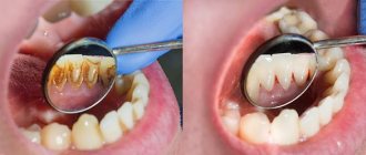
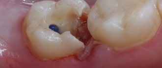
If caries is not cured in time, and there is a deep carious lesion, the only way out is to remove the dental nerve. As caries develops, microorganisms violate the integrity of the enamel, as a result of which the nerve is exposed and reacts to any influence with unbearable pain.
Depulpation (nerve removal) is a modern way to preserve a unit of dentition without implantation.
When nerve removal is indispensable
In modern dental practice, both complete and partial resection are used. Indications for the procedure:
- deep carious tissue damage;
- chronic pulpitis (including asymptomatic);
- bactericidal infection spread by the apex of the tooth root;
- the presence of an extensive pulp area, the threat of developing periodontitis or transition to periodontitis;
- trauma leading to nerve exposure and tooth destruction;
- the need to correct a medical error;
- the need for prosthetics with low crowns.
Unbearable pain can also be an indication for depulpation.
The dentist will never prescribe such a serious intervention as long as it is possible to save the tooth.
Important : problematic “eights” when affected by caries, as a rule, are removed. This is due to the location of the “wisdom” teeth at the end of the dentition, which makes cleaning them from plaque and stone and filling the canals very problematic. Indications for removal are also the wrong direction of eruption, displacement of the dentition, malocclusion, and the development of pulpitis.
Indications for depulpation
When should a dental nerve be removed?
- Advanced caries;
- Pulpitis – including asymptomatic;
- Severe tooth trauma – partial destruction of the tooth and exposure of the nerve;
- Preparation for prosthetic crowns (at the discretion of the orthopedic dentist);
- Periodontitis;
- Consequences of unsuccessful treatment;
- Unbearable pain.
What is the dental nerve called?
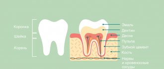
In dentistry, the dental nerve, or pulp, is a complex structure - an interweaving of nerves and vessels located inside the root and crown of the tooth and responding to external stimuli.
Depulpation allows you to save the damaged tooth, but has negative consequences:
- Since the pulp acts as a barrier to infections, removing the nerve deprives the tooth of the necessary level of blood supply and mineralization, which shortens its “life.”
- A tooth deprived of a nerve loses sensitivity, the enamel becomes more fragile and faded, and the strength of the tooth decreases.
Functions of nerves in teeth
The main function of the nerve is sensory. When bacterial damage (caries) spreads to the deep tissues of the tooth, a painful reaction occurs. Pain is the body’s signal that something is wrong and help is needed. The nerve is not located in the tooth cavity in isolation. It is part of the neurovascular bundle, a component of the dental pulp. The functions of the pulp are varied:
- tooth nutrition;
- growth and development;
- mineralization of enamel and dentin;
- immune protection.
The tooth pulp is treated and tried to be preserved in all cases where possible. It is especially important to do this in children and adolescents, since their teeth have not yet fully formed.
Stages of the depulpation process
- X-ray – necessary to assess the condition of the pulp, the number, length and branching of the canals to be filled.
- Anesthesia - local or complete anesthesia completely covers the issue of pain when removing a nerve (it doesn’t hurt!). At the same time, general anesthesia is used relatively rarely - when treating children, with true dental phobia, etc.
- Installation of a rubber dam - a latex film for insulating a tooth. A modern solution to improve the dentist’s working conditions and protect against the possible spread of microorganisms through saliva.
- The actual removal of the nerve is the excision of tissues affected by caries, opening the pulp chamber and extracting the nerve with a special instrument (pulpoextractor). In modern practice, this is a careful cutting of the pulp without affecting sensitive areas.
- Installation of a temporary filling (for a period of 1-2 weeks).
- Control x-ray.
If the x-ray and the condition of the tooth are satisfactory, after the control period a permanent filling is installed.
Modern method of depulpation
Today, depulpation is carried out using a pulp extractor - a thin metal rod with teeth that can penetrate even remote areas of the root canals. The apex locator device helps determine the depth of the canal to reduce the risk of injury. After the procedure, a temporary filling is usually installed, but a permanent one can also be installed if the doctor does not see the risks of complications and has high-precision instruments on hand, for example, a dental microscope.
Depulpation before prosthetic crowns
Depulpation is unconditional if there is serious tooth decay. It is preferable to remove the dental nerve during prosthetics in the following cases:
- The size of the teeth determines the low position of the crown (too small or short teeth);
- The inclination of the prosthetic tooth is from 15°;
- Increased sensitivity of teeth;
- According to aesthetic requirements.
The need to remove the dental nerve during prosthetics is dictated by deep removal of the top layer of dental tissue: if the doctor is not sufficiently qualified, heating or touching the nerve can cause an attack of unbearable pain. However, cases of nerve-sparing crown installation also occur in modern dental practice.
If over time the nerve under the crown becomes bothersome, it is possible to remove the pulp through the top of the crown and fill the hole after removal.
The danger of such a procedure is associated with the risk of complications in the future due to surgery on the exposed dental nerve.
About dental anesthesia
Standard dental practice is an injection of anesthetic; Taking tablet analgesics before going to the dentist is not recommended, as it leads to a decrease in the effectiveness of anesthesia (usually the effect of an anesthetic injection is 45 minutes). For patients suffering from a fear of injections, it is possible to use paste anesthesia, which has a similar effect.
The choice of anesthesia is up to the dentist; Taking into account the individual pain sensitivity threshold of each patient, the standard dose is sometimes doubled or even tripled. With proper selection of an anesthetic, discomfort during the procedure for the patient is excluded.
Why can a tooth hurt after pulp removal?
This often happens: the nerve has already been removed, but the pain remains. Such manifestations are acceptable in the first 3-4 days as a natural reaction of the body (for example, to clenching the jaws or to temperature stimuli).
Other causes of toothache after pulp removal:
- Careless cleaning of the canals, preservation and intensification of the inflammatory process due to incomplete removal of tissue damaged by caries.
- Using during the procedure a pulp extractor that is not suitable for the size of the canal or improper handling of the instrument.
- Broken dental instrument, retention of its remains in the upper part of the root (such a medical error leads to the need to remove the tooth).
- The occurrence of a secondary inflammatory process in the treated canals (residual pulpitis) occurs against the background of an incompletely removed nerve.
- Allergic reaction of the patient to the filling material.
Modern pulp removal
Today, it is enough to go to the dentist once to remove the pulp. The doctor gives an anesthetic injection and calmly works with the tooth, removing the destroyed layer of dentin, extracting the pulp, treating and filling the canal. Sometimes inspection using a microscope is possible. To be on the safe side, your doctor may place a filling temporarily to make sure he has completely removed the pulp. And only if everything turns out to be in order, the final filling is installed.
Incomplete removal of the pulp is also possible, which makes sense if only the outer part of the pulp is affected by inflammation. Dead tissue is removed from the opened tooth, the outermost part of the pulp is removed, and the root remains intact. This is especially important on children's teeth, since the pulp is responsible for the formation of roots. When the pulp is not completely removed, the tooth is kept alive, and further destruction occurs more slowly.
Change in enamel color after nerve removal
If the dental clinic follows international depulpation protocols, such phenomena are excluded. However, in budgetary institutions there are frequent cases of using outdated practices.
The color of the enamel after nerve removal can be affected by:
- Improper preparation of the tooth for installation of a filling;
- Poor quality instrumental processing of channels;
- Low quality filling materials.
Thus, when using resorcinol-formalin paste (the use of the material is practiced in the treatment of baby teeth), the enamel acquires a pinkish tint; When using endomethasone, the tooth may turn yellow after a few years.
Sequence of treatment
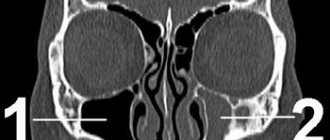
- Diagnostics . After the doctor has examined you and collected your medical history, an X-ray is taken. This diagnosis allows you to evaluate the structure of the tooth and plan treatment. To see the full anatomy of the root canals, a computed tomography (CT) scan is performed - a 3D diagnostic that allows you to see all the structures of the tooth.
- Pain relief . Local infiltration or conduction anesthesia is performed. Before the injection, the gums are lubricated with an anesthetic gel so that the injection is not felt.
- Setting up insulation . Root canal work should be done in clean, dry conditions. For this purpose, rubber dam (insulation) is used. It is a latex plate that is placed on the tooth. Advantages of isolation: saliva and plaque do not enter the root canals. Medicines and solutions also do not flow into the patient’s mouth.
- Preparation of hard dental tissues . The doctor uses a drill to remove the destroyed tissue. To prevent overheating of the tooth, water flows from the tip. The doctor creates direct access to the root canals.
- Nerve removal . It is performed with thin sterile instruments similar to long needles. The doctor places an instrument into the root canal and removes the nerve. Machine rotary tools may also be used. This stage is completely painless, since anesthesia was previously performed.
- Mechanical and medicinal treatment of canals . The canals are washed with special antiseptic solutions and passed through with instruments. This allows you to completely remove the diseased nerve and relieve inflammation.
- Temporary filling . Root canal work is complex, so it is not always possible to treat a tooth in one visit. An anti-inflammatory paste is placed into the canals, a temporary filling is placed, and a further stage of treatment is planned.
For permanent restoration of the crown part of the tooth, ]esthetic dentistry[/anchor] will be required.
Possible complications
A medical error associated with poor-quality disinfection of the canal when removing a nerve can trigger the process of suppuration with subsequent transition to a periodontal abscess (in the absence of adequate treatment). This complication leads to the need for tooth extraction.
Other possible complications:
- Pain for several days after nerve removal. The duration of discomfort is individual for each patient; if pain persists for a long time, it is necessary to consult a doctor to re-open the canals and carry out disinfection.
- Increased bleeding of the canal occurs when the pulp extractor is removed after the pulp has been torn off. To avoid this phenomenon, many specialists carry out the procedure in stages with copious rinsing of the tissues with an antiseptic. Bleeding control is carried out directly at the appointment.
- The appearance of granuloma, fistula, cyst, gumboil.
Specific problems can arise if the material is applied incorrectly: if the filling extends beyond the boundaries of the root apex, the jaw nerve may be pinched.
If you experience pain in the lips and chin, you should urgently consult a dentist: a possible complication is facial paralysis.
Removal with arsenic
Once upon a time there was only one way to remove it, it was terribly painful, especially since you had to visit the doctor several times. At first, the doctor drilled the tooth down to the pulp with a drill, which was extremely painful. Next, arsenic was placed in the hole and it was temporarily sealed with a filling. In a couple of days, the arsenic dealt with the nerve, and the tooth hurt so much that no medicine could help. Then I had to go to the doctor again, he opened the temporary filling, removed the nerve, and cleaned the root canals. Pulp is a tenacious substance, so this process was also extremely painful.
This technology poses a certain danger, because arsenic is a poison, and the doctor, when using it, must be especially careful, and the patient must be careful: if you are late with the second visit to the doctor and leave arsenic in the tooth, it will completely destroy it, which will end deletion. Dentists stopped using arsenic and developed new techniques for removing pulp.