Introduction
It is known that facial prostheses, such as ear prosthetics, made of wax, were used in ancient Egypt.
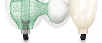
The first historical evidence of the use of facial prostheses dates back to the sixteenth century: the French surgeon Ambroise Paré describes the first artificial nose made of gold, silver and papier-mâché, which was attached to the face using a headband. In the second half of the 19th century, Claude Martin put forward the idea of creating an immediate denture using the tissues of the upper and lower jaws as a matrix to create complex structures. In the 20th century, with the advent of silicone materials, the quality and realism of craniofacial prostheses improved significantly, but the problem of fixation, which affects the appearance, function of the prosthesis, and patient comfort, was not completely solved. With increasing aesthetic demands, traditional fixation methods such as adhesives, pockets, loops and glasses have become unacceptable. In 1997, Brånemark installed the first bone-anchored extraoral hearing aid implant, and in 1979 the bone-anchored ear prosthetic implant. These events changed the entire concept of maxillofacial prosthetics. Since then, extraoral bone implants have been widely used to support prosthetic eyes, ears, and noses. Their use partially solves the problems of discoloration and deterioration of the prosthetic material, since these problems are often associated with adhesive materials. The skin and mucous membranes are less irritated by the mechanical effects of permanent fixatives and the chemical effects of adhesives and solvents. Implant-retained craniofacial prostheses have improved significantly from an aesthetic point of view due to their ease of installation, as well as thinner edges that blend in color with the skin. A large number of scientific and clinical studies confirm the success of their use and note an improvement in the quality of life of patients. Facial injuries and mutilations, especially after radical surgery, often lead not only to external physical disabilities, but also to functional and psychological disorders. In such cases, maxillofacial surgeons and dental prosthetists have to carry out a whole range of rehabilitation procedures. The options for reconstructive plastic surgery are often limited due to unfavorable conditions, such as damage to the vascular system of the surgical site due to radiation therapy, or insufficient volume of residual soft and hard tissue. In such cases, rehabilitation of patients with severe craniofacial defects is carried out with the help of prostheses, which represent an acceptable solution from an aesthetic point of view. A facial prosthesis is created by a craniofacial surgeon, maxillofacial orthopedist and prosthetic technologist as a good alternative to reconstructive surgery. To replace missing hard and soft facial tissues, prostheses using extraoral implants with a ball abutment, a bar design, or a choice of magnetic abutment are used. The shape of the prosthetic nose, eyes and ears, its color and texture should, as far as possible, be indistinguishable from the surrounding natural tissues. Rehabilitation can only be successful when patients are able to appear in public without attracting unwanted attention. Extraoral implants have many advantages in correcting facial defects over both adhesive prostheses and spectacle-mounted prostheses. They are characterized by ease of fixation of the prosthesis, which ensures the ability to properly install the prosthesis, thereby promoting comfort and confidence in its use. There is no skin irritation from the adhesive, and there is no need to wash off the adhesive every time after using the prosthesis. A thinner denture can be made, with thinner edges that blend into the skin, improving the overall appearance. During preoperative planning, when meeting with the patient, specialists from various fields not only show him the options for prostheses, but also teach him how to care for the abutment and prosthesis.
Implants have played a critical role in improving patient acceptance of facial prostheses. Patients enjoy the safety, comfort, and convenience of implant-supported dentures—benefits previously unattainable with older fixation methods. Over time, surgeons have noted a decrease in the number of patients requiring multiple complex surgical procedures. When correcting large defects, a combined approach combining tissue grafting and prosthetics using implants is recommended to achieve optimal results.
A three-implant facial prosthesis is the preferred method of replacing missing hard and soft orofacial tissue. Fixation on extraoral implants stimulates more confident behavior of the patient in public. Multidisciplinary treatment will require the combined efforts of several specialists. If we are talking about a defect in the eye socket, then the efforts of an ophthalmologist, a craniofacial surgeon, a maxillofacial orthopedist and a prosthetic technician are required.
Midface defects can be divided into two broad categories: midline midface defects, which include the nose and/or upper lip, and lateral facial defects, which include the cheeks and orbit.
The choice between plastic surgery and a prosthesis for serious facial injuries remains difficult and depends on the area and etiology of the damage, as well as on the wishes of the patient. The development and use of extraoral implants for the correction of facial defects has to some extent changed the attitude of patients towards facial prostheses. Implants provide comfortable donning and reliable fastening of the prosthesis, which improves the perception of the prosthesis by patients.
We will conduct a retrospective review of patients who underwent facial prosthetic reconstruction from 2005 to 2010. Clinical reports describe step by step the materials and methods used, including the implant procedure.
There are three main diagnoses that require surgical intervention: oncological, traumatological and genetic and/or acquired diseases. The goals of surgical and prosthetic rehabilitation are to replace missing hard and soft facial structures, restore physical impairments, alleviate social problems and improve quality of life.
Treatment protocol
Treatment of a patient with oral cancer or after trauma is a collaborative effort between craniofacial surgeons, oncologists, radiation therapists, and prosthetic technicians.
Typically, immediately after radical surgery, a free tissue graft is performed to restore both soft and bone tissue. A large percentage of patients receive 4 to 6 weeks of radiation therapy after surgery. The total radiation dose to the tumor bed depends on the presence or absence of cancer cells at the surgical margin of the tumor. The radiation dose usually does not exceed 55 Gy, for cases with a clear pathological margin, and from 65 to 70 Gy, with a narrow pathological margin. Radiation localizers are widely used, and radiation fields are adjusted in such a way as to minimize the possibility of large salivary glands entering the area of exposure to large doses of radiation; however, intensity-modulated radiation therapy was not used. Radiation dose to bone can often be minimized in areas where implants are to be placed. Approximately 6 weeks after radical surgery, the patient was expected to undergo radiation therapy for 5 to 7 weeks. Once the patient had recovered from plastic surgery and radiation therapy, new standard prostheses were made. The implants were planned to be placed 4-6 weeks after plastic surgery, depending on the need for post-operative radiation therapy. All implants were placed based on wound healing from osteotomy and recovery from radiation therapy. Three implants were installed. The implants were installed in the existing native bone tissue and/or in the free vascularized bone tissue of the reconstructed mandible. During the operation, before installing the implants, excess plate segments were removed to connect the bone fragments.
Prosthetic options include classically anchored dentures, then implants with ball-shaped attachments, bar structures and magnets.
The long-term prognosis is important, but having a high-quality prosthesis for even a year or two can significantly improve a patient's quality of life. The advantage of installing implants and facial prostheses is that the perfect stability of the prosthesis guarantees the patient's return to social life.
Nasal prosthesis
Clinical report
A 63-year-old woman diagnosed with recurrent basal cell carcinoma of the nose was treated.
After nasal amputation, external beam radiation therapy and chemotherapy were performed. Oncological treatment was completely completed in 2007. Surgical restoration and installation of the prosthesis began a year after the end of treatment (Fig. 1, 2). Three extraoral implants were inserted along the edge of the postoperative defect. The operation was performed under general anesthesia with nasopharyngeal intubation. The main implant bed for the nasal prosthesis was a pear-shaped ridge at the base of the nose (2 implants - left and right). The other bed was the glabella; we paid special attention to the degree of pneumatization of the frontal sinus and the amount of overlying bone tissue. The operation lasted 65 minutes and went without complications. The wound healed without inflammation. Lincomycin (600 mg orally) was used as an accompanying antibiotic. Four days after the operation, the patient was discharged in good condition. Six months after the installation of the implant and the end of the healing period, prosthetics began. A control tomogram was performed. Magnetic mounts were inserted. The prosthesis was prepared as follows. A one-stage prosthetic technology was used; a nasal prosthesis with separately located abutments and magnetic fixation was chosen. It was investigated in which direction the protruding parts of the abutments are directed. It was required that the fixation elements be oriented so that the prosthesis would extend beyond the anterior edge of the maxillary bone. To achieve this and position the fixation components in a position that is easy to manipulate, cantilever abutments were chosen. An additional set of magnetic abutments was ordered to select the appropriate cantilever angle.
The defect impression is recorded as a partial alginate impression. A plaster model of the prosthetic element was made. Base wax was used to sculpt the edges of the acrylic resin base. The wax-up model was prepared, evaluated on the patient and brought to its final form.
The plaster mold is cast. The wax is melted out, and the surface of the plaster model and casting mold is coated with a release agent and auto-polymerizing acrylic plastic. Acrylic plastic is subjected to pressure treatment in a polymerizer. The shape of the prosthesis, its color and texture are finalized using special plastic. The finished prosthesis is ground and polished. A magnet is attached to the top of the support. An impression is made of the contact surface between the abutment and the fixture using single-phase silicone. Magnets connect to the abutments.
Figure 1. Nasal prosthesis: a) wax cast; b) polymerization; c) coloring; d) installation of magnets.
Figure 2. Nasal prosthesis: a) CT scan after installation of 3 implants, b) magnetic attachments; c) patient after rehabilitation.
Types of maxillofacial devices and their manufacturing technology
Devices that are used in maxillofacial orthopedics are divided according to their purpose - fixing, repositioning (helping to move or extend fragments in the required direction), forming and replacing. Devices in orthopedics are classified according to the material used and wearing time. Orthopedic devices carry both a basic load and have auxiliary functions. Let's consider these varieties in detail.
Removable and non-removable
Maxillofacial orthopedics offers many methods for eliminating defects in the frontal part of the skull. Devices are divided according to the method of fixation - removable and non-removable. Removable ones include temporary ones, used as first aid to the patient, and those used before surgery. The production of fixed ones is carried out in dentofacial laboratories; they are attached to the teeth with dental cement. These include removable tires Port, Limberg, Weber, Vankevich.
Polymer, metal and combined
Maxillofacial prosthetics divides the devices used according to manufacturing technology, design and material. There are the following types:
Ocular prosthesis
The absence of an eye may be a birth defect. Eye loss may also result from trauma, tumor, ophthalmia migrans, removal of a painful blind eye, or the need for histological confirmation of a suspected diagnosis. Eye diseases are relatively rare, but given the anatomy of the surrounding structures, they are very serious diseases. Tumors in the eye area account for 0.2 to 0.5% of the total. Tumors of the eye and adjacent organs manifest themselves in the following symptoms: dystopia, impaired mobility of the eyeball, exophthalmos and diplopia. Depending on the severity of the disease, surgery may follow one of the following three approaches: evisceration, enucleation, or exenteration. Evisceration
is a surgical operation during which the internal contents of the eyeball are removed, the sclera, Tenon's capsule, conjunctiva, external muscles of the eyeball and the optic nerve are preserved; the cornea of the eye can be preserved or excised.
Enucleation
is a surgical operation to remove the eyeball and part of the optic nerve from the socket.
Exenteration of the orbit
– this is the complete removal of the eye socket, often accompanied by partial or complete removal of the eyelids; produced primarily for malignant ocular tumors.
In particular, the diagnosis is made by an ophthalmologist; An examination by an otolaryngologist and a dentist is recommended. Radiation diagnostic methods are an integral part of the diagnostic procedure. They include radiography of the skull in frontal, semiaxial and lateral projections. In addition, ultrasound, computed tomography, and very often nuclear magnetic resonance imaging are used. In accordance with the details of the diagnosis, it is possible to accurately determine the extent of damage to the eye and surrounding structures of the orbit and choose a treatment method. In the treatment of solid tumors, radical surgery is used in most cases, despite the risk of functional and appearance defects.
Losing an eye is a disability that can lead to serious physical and emotional problems. Replacing a lost eye as quickly as possible is essential for the patient's physical and psychological recovery and helps improve the patient's perception of others.
Restoring facial defects using prosthetics improves the capabilities and level of self-esteem of patients. However, when using facial prostheses, difficulties may arise due to the moving tissue bed, the quality of fixation of the prostheses and reactions when interacting with adhesives. The use of extraoral implants in the craniofacial region reduces the limitations associated with the use of adhesives and is a treatment method with high long-term success. A three-implant facial prosthesis is the preferred method of replacing missing hard and soft orofacial tissue. The shape of the prosthesis, color and texture should, as far as possible, be indistinguishable from the surrounding natural tissues. Rehabilitation can only be successful when patients are able to appear in public without attracting unwanted attention. An important prerequisite for successful treatment of patients with such physical defects is healthy bone tissue at the edges of the defect. The following clinical report follows the step-by-step process of making an ocular prosthesis.
Clinical report
The patient, aged 21 years, was referred to the Clinic of Dentistry and Maxillofacial Surgery in Prague.
At age 2, he underwent surgery to remove retinoblastoma in his left eye. (Enucleation was followed by external beam radiation therapy and six cycles of chemotherapy (etoposide and cisplatin (cDDP). Cancer treatment was completed in 1983. Most cases of retinoblastoma are discovered in early childhood (80%), the incidence rate is 1:18,000 newborns The age of onset of the disease is mainly from 1 to 2 years. The etiology is unknown. A genetic predisposition associated with the Rb 1 mutation is possible.
The patient had no other chronic illnesses, was in good health, had no serious injuries, had suffered no illnesses other than common childhood illnesses during childhood, and had no allergies. The patient did not use a prosthetic eyeball and was very distressed by his injury. Routine preoperative examination was performed, as well as semiaxial cranial radiography and magnetic resonance imaging. According to the treatment plan, 3 implants were installed. After engraftment, the implants were equipped with ball attachments, and an ocular prosthesis was made with a system of clip fixation of males on matrices. The operation was performed under general anesthesia with nasopharyngeal intubation. In March 2004, 2 implants were implanted in the upper edge and 1 implant in the lower edge of the left orbit. The operation lasted 65 minutes and went without complications. The wound healed without an inflammatory process. Lincomycin (600 mg orally) was used as an accompanying antibiotic. On the fourth day after surgery, the patient was discharged in good condition. Six months after the installation of the implant and the recovery period, the production of the prosthesis began. A control semiaxial X-ray and magnetic resonance imaging study was performed. Three ball attachments were fixed to the implants. An impression of the anophthalmic orbit was then made using a standard impression tray specifically designed for orbital impressions (tray material: acrylic plastic). A working model was made, and a prefabricated plate with 3 male clips was also prepared.
Figure 3. Ocular prosthesis: a) CT scan before treatment; b) eye socket; c) installation of implants.
Figure 4. Ocular prosthesis: a) installation of implants, b) taking an impression; c) ball attachments and ocular prosthesis; d) prosthesis; e) patient after rehabilitation.
The wax-up model was prepared, evaluated on the patient and brought to its final form. The shape of the model corresponded to the contours of the eye socket and the configuration of the eyelids. After individualization, the manufacturing process of the prosthetic eye with the eyeball was completed. The shape of the prosthesis, color and texture should, as far as possible, be indistinguishable from the surrounding natural tissues. The finished prosthesis was ground and polished and inserted into the orbit of the eye. Glasses help to achieve a symmetrical position relative to the second eye and the face as a whole (Fig. 3, 4).
Why are devices used in maxillofacial orthopedics needed?
Maxillofacial orthopedics uses devices to restore patients after trauma to the upper, lower jaw or as a result of surgery. The devices are used as an alternative to facial surgery, which is contraindicated in some cases. We are talking about blood diseases, cardiovascular diseases, and disorders of the central nervous system.
There are diseases whose treatment is more effective with orthopedic rather than surgical methods - all kinds of defects of the alveolar process, cleft palate. The main objectives of maxillofacial prosthetics are as follows:
- prosthetics for jaw and facial anomalies, congenital or resulting from trauma,
- restoration (prosthetics) of deformed areas of the dental system,
- treatment of facial fractures and their complications,
- treatment of disorders of the masticatory muscles and TMJ.
Auricular prosthesis
Indications for autoplastic reconstruction of the auricle in comparison with a prosthesis with fixation on an extraoral implant were described in the work “Plastic and Reconstructive Surgery” in 1994. The choice between the two existing methods, autoplasty and prosthesis, depends more on the surgeon's training and tradition than on an analysis of the indications for a particular procedure in a specific clinical situation. For example, most children with microtia in the United States undergo plastic surgery. In Sweden, on the contrary, the same defect is corrected with the help of a prosthesis. Patients with post-traumatic defects of the auricle or after radical operations are usually adults. There are some differences between physical disabilities in adults and those in children. First, skin loss and scarring from trauma or previous surgery can make standard plastic reconstruction difficult. Second, the auricular tragus is often preserved in postoperative and trauma patients, which significantly improves the impression of the prosthesis. The presence of a tragus allows the leading edge of the prosthesis to be hidden, a great advantage in terms of appearance. It has long been noted that the area of the auricle is the most reliable place for an implant. All potential patients undergo a preoperative tomographic scan with radiographic stent markers installed. This procedure allows you to evaluate the intended implantation site in order to be able to maximize the length of the implant. In particularly difficult cases, the mastoid cells of the temporal bone make it difficult to select a site for implantation, and sometimes the choice has to be made again. There is no evidence that opening the mastoid cells during implant placement has a negative effect. If the bone is sufficient to ensure reliable fixation, the implant is left in the same place, otherwise, it is necessary to look for another area. Thickening of the bone along the edge is a feature favorable for implant placement. The use of three implants in the ear area reduces the need for hanging mounting and creates a tripod effect with mechanical strengthening. All implants are connected using a tissue beam structure, and fixation is achieved using clips.
Clinical report
The patient, a 45-year-old man, lost his left ear during a car accident and was referred for prosthetic replacement (Fig. 5, 6).
The patient received three implants in a two-stage surgical procedure. Titanium implants were inserted into the temporal bone using a gentle surgical technique, and once the implants had healed, it was possible to go deeper into the skin to create a non-contact channel. During the first stage of the procedure, implants were inserted into the bone tissue in the area of the craniofacial defect. Previously, bone thickness in the mastoid region was measured (minimum 6 mm) using a frontal, axial and three-dimensional model based on computed tomography scanning. Implantation was performed under general anesthesia. A 4 mm longitudinal incision was made behind the external auditory canal and the temporal bone was exposed. The estimated time for osseointegration of implants inserted into the temporal bone is 6 months. The second stage consisted of thinning the subcutaneous tissue, opening the implant and attaching the abutments to the implants. This procedure involved removing some of the subcutaneous tissue to minimize movement of the skin around the implant. Healing caps were placed on the abutments and wrapped in gauze soaked in ointment to improve skin-bone contact and prevent hematoma and swelling. The postoperative treatment plan included instructions for caring for the operated site. The sutures were removed after 10 days, and during this period the patient did not complain of pain or complications. Four weeks after the end of the second stage, the prosthesis was manufactured and attached to the implants. Fabrication of the implant-retained prosthesis began three weeks after attachment of the abutments, following routine clinical and laboratory procedures. Fixation was carried out using a beam structure with a clamp.
The auricle prosthesis was made according to the shape and size of the patient's healthy ear. A plaster model and image of the right ear were made, and a cast of the beam structure and auricle prosthesis was made. The wax-up model was prepared, evaluated on the patient and brought to its final form. The shape of the model corresponded to the contours and configuration of the ear. After individual selection, an ear prosthesis was made from silicone and acrylic plastic. The finished prosthesis was ground and polished. Hair helps to achieve a symmetrical position of the prosthesis relative to the face as a whole.
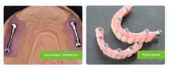
Figure 5. Auricular prosthesis: a) healthy ear; b) plaster model; c) calculation of the symmetrical position of the ears, d) polyester impression; e) beam structure with consoles, f) beam structure; g) preparation of the beam structure.
Figure 6. Auricular prosthesis: a) position of the future ear; b) production of a prosthesis; c) staining; d) beam structure with clamps; e) clamps; f) auricle prosthesis; g) patient after rehabilitation.
conclusions
Extraoral implants provide an effective treatment alternative for patients requiring facial prostheses.
Implants can help overcome many of the difficulties encountered when fixing large facial prostheses. Our data indicate a predicted high percentage of implant survival in the auricle area; pyriform/nasal region and orbital region. The following advantages of installing magnetic implants and facial prostheses can be noted. Optimal stability of the prosthesis guarantees the patient's return to social life. Three magnetic attachments are used in the design, placed in the jaw, glabella, etc. Extraoral implants provide a wider surface for osseointegration. Magnetic attachments provide better surface stability than ball-shaped attachments, and at the same time do not interfere with the patient or make handling of the prosthesis difficult. For auricular prostheses, a beam design is much more convenient due to the load from the surrounding muscles. Facial prostheses using three implants are the most preferred method of replacing missing hard and soft orofacial tissue. The shape of the prosthesis, color and texture should, as far as possible, be indistinguishable from the surrounding natural tissues. Rehabilitation can only be successful when patients are able to appear in public without attracting unwanted attention. The use of implant-retained dentures gives the patient confidence in social life.
Tatiana Dostalova (Charles University, 1st and 2nd Faculty of Medicine, Prague, Czech Republic), Jiri Kozak, Milan Hubacek, Jiri Golakowski, Pavel Kriz, Jakub Strnad and Michaela Seidlova