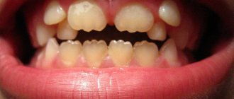
Reason for breakage
The canals in our teeth have a very complex structure; they are curved, very thin and branched in different directions. To clean the canal to the very end, the doctor uses special devices, which are also very thin and flexible. Unfortunately, it is impossible to completely protect yourself from item damage. Even the most experienced professional cannot be 100% insured against such an incident.
The process of removing a foreign object is quite difficult, so not every master can cope with the task. The final decision on the need to remove a part should be made only after an adequate assessment of all the pros and cons.
There are many nuances, for example, if part of the instrument is lost in a living tooth and at the final stage of treatment, no complications should arise in the future. The patient will not encounter an allergic reaction, inflammatory processes or increased sensitivity to various irritants in the future.
The instrument is broken in the tooth canal.
Breakage of a tool in a canal is an extremely unpleasant thing. In the practice of any dentist involved in root canal treatment, such a complication has occurred more than once. Why is this bad? By itself, a metal fragment inside a tooth cannot cause any harm - you won’t be ringing at the airport, the alloy from which the files are made (“canal needles”) is completely inert, and an allergy to it is also unlikely. So what's the problem? The problem is that these fragments clog the root canal and the dentist cannot properly clean the space behind the instrument. This may cause problems in the future. It is possible to remove a fragment from the canal without using a microscope only if it is located at the very surface. And even then, if you are lucky, it should be considered great luck. But if the “bummer” happened deep in the channel, then there is nothing to do there blindly. Rather, you can break new wood.
So, in this case, we see a “spiral” in one of the channels in the picture... It is shown by an arrow. The inflammatory focus around the tooth root is circled with dots.
In order to thoroughly clean the root canals, the task was to remove this fragment. At the same time, it was located quite deep and had a small size. Without an eye equipped with a microscope, the task would be difficult to accomplish. In the photo below, the red arrow shows a fragment deep in the canal. Seeing it, it is much easier for the dentist to remove the fragment and bring it out (green arrow).
Ultimately, the instrument was removed and the canals were completely cleaned and sealed.
Now there is no doubt that the granuloma that grew on the root of the tooth and threatened to remove it will go away in a few months. And the existence of the tooth will no longer be threatened.
Possible complications
It is worth noting that a piece of the instrument itself does not pose any threat to the patient’s health. This is explained by the fact that such items are made from hypoallergenic materials. However, the part may prevent complete cleaning of the dental canal, which will negatively affect the outcome of the entire treatment. As a result, inflammation may develop, severe pain will occur, and in rare cases, tooth extraction may be necessary.
Parts remaining in the canal can negatively affect recovery after treatment. In rare cases, the patient may develop an allergy; sometimes it is accompanied by a strong tooth, swelling and redness of the soft tissues and mucous membrane located next to the diseased tooth.
The final prognosis of endodontic treatment will depend on many factors, for example, on the stage at which the working instrument broke, as well as on the degree of preparation and sufficient treatment of the canal with medications at this stage.
Closing tooth perforations.
Perforation is a hole in a tooth, and an artificial one. It occurs if the doctor worked too zealously and carelessly with the tooth. This most often happens when preparing canals for the installation of various types of pins.
In our case, the lower chewing tooth was restored after treatment with a filling on a pin... and more than one. Apparently, the principle “you can’t spoil porridge with butter” worked. But that was a mess, but for the tooth, the “multiple pins” actually turned out to be murder: one of the pins was installed past the root canal and pierced right through the bottom. Perhaps the tooth was mobile and the doctor wanted to strengthen it a little in this way, but then it was worth choosing a longer pin...
Here in the photo, after removing the old filling, 2 pins are found screwed into the tooth (on the left), and after their removal we see the bottom of the tooth cavity in the holes (on the right).
As it turned out, one of these holes was, as the sailors would say, “below the waterline.” In dental terms, this is root perforation . The tooth channel is in one direction (the channels are shown by green arrows), the hole for the pin is completely in the other direction (the perforation is shown by a red arrow).
A puddle of blood seems to hint that a crime has taken place here. The killer doctor tried to drill a tooth to death and he almost succeeded. Most often, teeth with perforations are sent by dentists for removal. But with good visualization, which is provided by an operating microscope, it is quite possible to carefully treat the wounds inflicted on the tooth and mend the poor thing. Which is what was done.
On an x-ray, the same picture looked like this: red lines show 3 root canals, a black dotted line shows the perforation zone, covered with a special material, and a red dotted line shows the inflammatory process in the bone tissue around the perforation zone.
After closing the root perforation , the main problem of this tooth, which led it straight into the hands of the surgeon, the root canals were re-treated and the root was thus preserved for subsequent prosthetics.
The process of removing debris from the canal
Even if the fragment does not affect the outcome of treatment in any way, a conscientious doctor will definitely inform his patient about the problem. This is necessary so that if the slightest discomfort is detected, a person immediately goes to the clinic, and does not treat the tooth with folk remedies at home. If the instrument breaks at the initial stage of the intervention before the infection is eliminated, the dentist must try to remove the part from the canal.
This will require a special dental microscope; without it, it will be simply impossible to detect such a small detail in a thin canal. At the first stage of the procedure, the doctor must create direct access to the foreign particle; for this he can use a dental bur.
Next, ultrasonic instruments with different tips come into play; it is with their help that the fragment can be removed from the dentin with minimal consequences. After this, the canal is thoroughly cleaned and sealed again, and medications are placed inside if necessary. A temporary filling is first placed on the tooth to ensure that there is no inflammation. After some time, you can put a permanent filling.
previous post
The dangers of teeth whitening at home
next entry
Introduction
A broken instrument in the tooth and one untreated canal is the background to this clinical case, which was encountered in the practice of the German Implantology Center.
A direct participant in the events, the doctor who saved the patient’s tooth, endodontist of the German Implantology Center Melikov Azer Fuadovich, tells about this interesting case, when it was necessary to remove an instrument previously forgotten by one of the doctors from the root of a tooth:
The patient was referred to me by a colleague from another clinic after obturation of 2 root canals of a tooth, to remove a broken instrument during the work. A broken and forgotten instrument in a canal is not good. During the initial CBCT diagnosis, we identified the fact that the broken instrument was located in the root canal.
The fact that an instrument was left
and in this form it was sealed - this is an extremely negative point. But the situation was complicated by the undertreatment of the tooth as a whole, since during the examination a missed 4th root canal was discovered:
The patient was sent to me with a temporary filling, which I immediately removed. Here is a view of the tooth cavity after removing the temporary filling and monitoring the location of the fragment of the broken instrument in the root canal:
It is necessary to evaluate the situation with an open, unfilled canal missed during treatment for the first time. All work of this level is carried out exclusively using a microscope
, about which there will be a short video at the end of the article. In the following image you can see the appearance of the tooth cavity after removing the temporary filling and checking the missed canal before starting work:
Hardware cleaning: help of depophoresis and ultrasonic scaler
Treatment of the tooth canal using a special device is indicated in the presence of severe curvature of the passages and signs of their obliteration. Depophoresis can also be used as a supplement to manual cleaning: the action helps remove pulp residues, which dissolve and disappear under the influence of the healing fluid. The impact is produced by special elongated tubes that have wires connected to the body of the device equipped with a control panel. The most famous models of devices: Original 2, EndoEst, AOK series - 1.0, 2.1 and Endo-lux.
Reference! Depophoresis should not be used if you are allergic to copper.
Additional indications for the use of depophoresis:
- sealed canals;
- wide apical foramen;
- cyst;
- gangrene-like contents in the canal;
- the presence of a broken tool in the passage.
The main acting liquid is hydroxide from a combination of copper and calcium. Before starting the procedure, the orifice is widened as much as possible to increase the likelihood of penetration of the solution to the top of the canal. Together with the influence of the electromagnetic field, effective sterilization of passages is carried out.
The use of a scaler or pneumatic handpiece, which uses ultrasonic influence to remove dentinal canals and pulp debris, helps produce more effective irrigation than using a cannula. At the same time, the specialist is able to provide both mechanical and acoustic effects, which leads to a reduction in the number of auxiliary devices used. This tool can also be used to clean canals from the remains of an old filling, but its functionality in this situation is significantly limited.
Preparatory procedures
In order for the dentist to choose the right treatment tactics, he should obtain maximum information about the condition of the tooth. Additionally, it is necessary to find out if the patient has an allergic reaction to medications, since they are required to be used when cleaning canals.
Collection of information:
- interviewing the patient (nature of pain, sensations);
- conducting an initial examination;
- sending a patient to get an image.
If the patient is a pregnant woman, then it is more rational to choose a study performed using a visiograph. The most informative is a 3D image created by a tomograph. Based on visual data, the dentist determines the length of the canal, allowing you to accurately select the instrument. There is a more accurate way to determine the distance using an apex locator, which displays a picture on the screen that allows you to determine the distance of the inserted instrument from the apex of the root. This method prevents breakage of endodontic devices during cleaning.
Fact! The most difficult cleaning is carried out when the canals are obstructed and are strongly curved.
Price for removing a broken instrument from a dental canal in Partner-Med
| Dental service | Price* for cash in rubles |
| Extraction of 1 foreign body, or 1 pin, or 1 broken instrument from the root using a microscope (Promotion) | 5 500 ₽ |
| Extraction of 1 foreign body, or 1 pin, or 1 broken instrument from the root using a microscope | 8 690 ₽ |
*The indicated cost of the service is valid for DECEMBER 2020 for patients visiting our clinic for the first time. Please check the current cost with individual prices available for you by calling + 7
Just pick up the phone and call us!
8
We will definitely make you an offer that you cannot refuse!
There is an instrument left in the dental canal: what to do?
The main reason for the need to remove a foreign body from the dental canal is the possible breakage of an endodontic instrument during treatment. Part of it can get into the tooth canal when:
- Cleaning of blood vessels or dead nerves.
- A narrow and curved channel, as a result of which the instrument cannot cope with the tension.
- The use of hand tools that can break due to defects in the material and other factors.
Root canal treatment is not always fraught with chipping of the instruments, but we must remember that this is possible. Foreign bodies must be removed.
The main reason for this is corrosion of the metal components of endodontic instruments. It provokes irritation of the walls of the canal, which is why cracks in the root are possible, and subsequently loss of the tooth, causes constant pain, and also leads to swelling of the soft tissues nearby.
Other troubles are also possible. So, if the instrument breaks off and the fragment remains in the cortex, this makes filling the rest of the canal impossible. Inflammatory processes are also a consequence of foreign objects. Under the debris or on the fragment there may be part of the pulp, which, being infected, begins to rot and needs to be removed. In view of this, filling and refilling, installation of prostheses and other manipulations are carried out only after the removal of any foreign objects.
Foreign bodies also include titanium pins and components of stump inlays. This can be used to name all objects that should not be in the canal after the end of treatment. They do not allow for high-quality filling and increase the risk of tooth loss, so if an instrument remains in the dental canal, it is important to remove it in a timely manner.