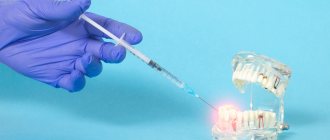
Home » All about modern dentistry » Pain relief in dentistry » Pain relief in the upper jaw For various operations on the upper jaw, dentists prefer to use infiltration anesthesia. This method is most convenient because the thin layer of the compact lamina of the alveolar process of the upper jaw is porous. If you use modern painkillers that have a high diffusion ability, then with this method of their administration the desired anesthetic effect is achieved.
Impregnation of tissues and alveolar processes with an anesthetic is carried out by injection, injecting the solution under the mucous membrane at an angle of 40-45o in the projection of the upper areas of the dental roots along the transitional fold of the vestibule of the oral cavity in the area that is supposed to be operated on. If this area is too large, the needle is moved along the transitional fold and the anesthetic solution is slowly injected. Because rapid administration of the substance can cause pain in the patient.
To anesthetize the palatal tissues, an injection of an anesthetic drug is made into the angle formed by the palatine and alveolar jaw processes, at a distance of 10-15 mm from the edge of the gum.
Regional (conduction) anesthesia is used quite rarely; it includes anesthesia on the tubercle of the upper jaw, in the area of the greater palatine, incisive and infraorbital foramen.
Intraoral anesthesia
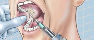
The patient's mouth is in a half-open state, the doctor inserts a mirror into the oral cavity, moving the cheek to the side. This ensures an excellent overview of the arch of the vestibule of the mouth, as well as tension in the mucous transitional fold near the molars. The needle is inserted into the mucosa above the projection of the apexes of the teeth at the level of the 2nd and 3rd molars. If there are no molars, then the needle should be behind the zygomaticalveolar ridge and move up, back and inward. The insertion angle should be 45°. It is necessary to control that the needle always moves along the bone with the beveled surface of its tip. During this movement, the anesthetic should be gradually injected. This method will help prevent injuries to the vessels of the pterygoid plexus. Having deepened the needle by 2-2.5 cm, the anesthetic solution is deposited. Thus, the molars located on the side of the vestibule of the mouth, the mucous membrane, periosteum and the outer posterior bone wall of the maxillary sinus are perfectly anesthetized.
Non-injection methods
For short, small-scale interventions, it is sufficient to superficially numb the soft tissues; this can be achieved using non-injection methods of local anesthesia in dentistry. These include:
- Application method . Applying a special gel or spray containing a certain anesthetic in a small concentration to the desired area of the oral mucosa. This method is often used in pediatric dentistry in the treatment of baby teeth, when treating gingival margins, opening small superficial abscesses, and also to prepare the patient for injection-type local anesthesia (to numb the injection site).
- Physico-chemical method . Injection of the required amount of anesthetic into soft tissues using electrophoresis. It is usually used for trigeminal neuralgia.
- Physical method (rarely used in recent years). Anesthesia of a specific area by applying a freezing reagent (chloroethyl), exposure to a laser beam or electromagnetic waves of a certain frequency.
All other techniques for performing local anesthesia in dentistry belong to the injection group, that is, they require an injection in a certain area of the oral cavity or even outside it.
Extraoral anesthesia
Having made a puncture with a needle in the area of the lower anterior corner of the cheek bone, the needle is then directed upward at an angle of 45° and again towards the tubercle of the upper jaw, bringing it to the bone. And then the anesthetic solution is deposited. The anesthetic effect occurs in approximately the same time as with the intraoral method of anesthesia.
However, during the process of anesthesia, a hematoma appears on the tubercle of the upper jaw, which is a consequence of injury to the veins of the pterygopalatine plexus by the needle.
Computed tomography studies of the path of solution propagation during tuberal anesthesia confirmed that such complications are most likely.
This tomogram, which was taken 5 days after intraoral anesthesia, shows that there are contours of a hematoma on the tubercle of the upper jaw in the pterygopalatine fossa. Approximately 40-60% is allocated to the fact that this cellular formation will fester and turn into phlegmon.
When using the tuberal method of anesthesia, injury to the veins of the pterygopalatine plexus is almost impossible to avoid, and there is also a fairly high risk of complications, in particular if the intraoral method is used. All this threatens the health and even the life of the patient. That is why it is recommended to use this type of anesthesia extremely rarely.
Conductor technology
Conduction local anesthesia in dentistry is used when infiltration anesthesia is ineffective (for example, if volumetric manipulations on the lower jaw are to be performed), when several teeth need to be treated simultaneously, or a long-term intervention is expected. In this case, the anesthetic solution is supplied to the branches of the nerve innervating the area of the upcoming intervention. The drug can be administered endoneurally (directly into the nerve, used rarely and for special indications) or perineurally (next to the nerve, so that the solution gradually permeates the nerve fibers, the most commonly used method of administration). There are also extraoral and intraoral methods of performing the injection - the doctor decides which method to choose in a particular case.
Depending on the site of administration of the anesthetic drug and the branches of the nerves that are to be blocked, the following types of conduction local anesthesia in dentistry are distinguished:
- Infraorbital – anesthesia of the branches of the infraorbital nerve.
- Palatine – blocking impulses from the greater palatine nerve.
- Tuberal - blocking impulses from the superior posterior alveolar nerves.
- Mental, or mental – anesthesia of the mental nerve.
- Mandibular - blocking impulses of the lower alveolar and lingual nerves in the lower jaw. One of the varieties is considered to be torsal anesthesia (blocking the branches of the lingual, inferior alveolar and buccal nerves).
- Incisive – blocking impulses from the nasopalatine nerve passing in the incisive canal.
Typically, the conduction technique requires preliminary preparation of the patient for local anesthesia - applying the anesthetic by application method to numb the injection site, as well as explaining to the patient how best to position his head for this or that type of administration of the anesthetic drug.
Anesthesia of the infraorbital nerve
The purpose of this anesthesia is to block the branches of the inferior orbital nerve, which forms the lesser “crow's foot” in the area of its exit from the bony canal, as well as the middle and superior anterior alveolar branches.
The result of such anesthesia, performed at the lower orbital foramen, is pain relief:
- — Reztsov,
- - Fang,
- — Premolars,
- — The gums adjacent to the premolars in the area of the vestibule of the mouth,
- — Bone tissue of the alveolar process and nasal septum,
- — Shells of the mucous and bone structures of the walls of the maxillary sinus,
- - Skin of the infraorbital region of the lower eyelid and wing of the nose,
- — Mucous membrane and skin of the upper lip.
The projection of the infraorbital foramen, which is oriented towards during an anesthetic injection, is located 5 mm below the edge of the orbit, which corresponds to the axis drawn through the center of the eye pupil when the eye looks forward.
Using a vasoconstrictor
The choice of dilution of the vasoconstrictor depends on the expected duration of the intervention and the presence of concomitant pathology in the child. As a rule, the duration of treatment for a child does not exceed 20-30 minutes. Longer intervention negatively affects the child’s psyche and his attitude towards dental treatment. In particular, fatigue increases, attention decreases, and the need for physical activity arises. All this disrupts cooperation between the doctor and the child, which is sometimes achieved with great difficulty.
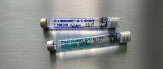
In most cases, when treating caries and its complications, removing temporary teeth, there is no need to provide long-term anesthesia with anesthetics with a high content of epinephrine (1: 100,000) (Rabinovich S. A., Zoryan E. V., 2006).
After dental treatment, it is important to warn parents that the child can injure the tissues of the lips and cheeks without feeling them after local anesthesia. The duration of the “numbness” period is on average 2 hours for anesthetics 1:200000 and 5-6 hours for anesthetics 1:100000. Naturally, a long period of “numbness” of soft tissues is unfavorable and can lead to injury (the child, without feeling pain, bites the “numb” tissue), which in some cases can lead to extensive wound defects and subsequent deformation of the red border of the lips.
The doctor must be aware of the full extent of responsibility when using anesthetics 1:100000, which provide unreasonably long anesthesia. When administering anesthesia to children, especially for the first time, it is imperative to warn parents and talk with the child about the possibility of self-injury of soft tissues that are “numb” from anesthesia.
ANESTHETICS WITH HIGH VASOCONSTRICT DILUTION 1:100000
In pediatric practice, they are used only for a number of surgical interventions for the purpose of hemostasis. The duration of anesthesia is 75 minutes for dental pulp and 360 minutes for soft tissues.
Such outpatient interventions include the removal of an impacted, supernumerary tooth, apperculectomy, cystectomy, plastic surgery of the frenulum and vestibule of the oral cavity, and removal of a tumor. The use of anesthetics with high dilutions of epinephrine for other types of dental interventions is unjustified and disproportionate to the volume of intervention.
ANESTHETICS WITH LOW VASOCONSTRICT DILUTION 1:200000
Indicated for most outpatient interventions in pediatric dentistry. 4% articaine 1:200000 provides anesthesia for soft tissues for 180 minutes and dental pulp for 45 minutes, which satisfies the protocol for most outpatient interventions.
Currently, anesthetics based on 4% articaine with epinephrine 1:400000 have appeared in European countries. They provide anesthesia for dental pulp tissue for 20 minutes and soft tissue for 1 hour. This anesthetic provides the duration of anesthesia required by the doctor and a short period of “numb” tissue, which is so important for pediatric patients. Currently, these anesthetics are not certified in the Russian Federation, but work is underway to introduce them into domestic dentistry.
It is also worth noting that there is no difference in the depth of anesthesia and effectiveness between anesthetics with vasoconstrictors 1:100000, 1:200000 and 1:400000. There is a difference only in the duration of local anesthesia of the dental pulp: 25, 45 and 75 minutes, respectively. Many dentists mistakenly divide anesthetics into “strong” (1:100,000) and “weak” (1:200,000). This statement is misleading.
For short-term interventions in children with concomitant pathologies, the use of anesthetics without a vasoconstrictor is indicated. However, their use does not guarantee complete safety and does not reduce the risk of complications. It is worth noting some of their pharmacological features. Vasoconstrictors are added to the local anesthetic solution not only to increase the duration of pain relief, but also to reduce their toxicity.
The fact is that all anesthetics have a vasodilating effect and undergo fairly rapid absorption into the bloodstream. The addition of a vasoconstrictor slows the absorption of the anesthetic and prolongs its action. When using an anesthetic without vasoconstrictors, this effect does not occur. The anesthetic is forced into the blood, which can lead to a toxic reaction. This complication is possible if the permissible dosage is exceeded, which is different for anesthetics with and without a vasoconstrictor. In summary, it should be noted that anesthetics without a vasoconstrictor do not affect the functioning of the cardiovascular system, are less allergenic since they do not contain preservatives, but due to accelerated absorption they are toxic and safe to use only if the dosage is observed.
ANESTHETICS WITHOUT VASOCONSTRICTER
Provide varying durations of pain relief for dental tissues. In particular, 2% lidocaine provides anesthesia of the dental pulp within 5 minutes, while the rate of onset of anesthesia is also 5 minutes, which is unsatisfactory for the doctor. Therefore, the use of 2% lidocaine without a vasoconstrictor is inappropriate for dental anesthesia.
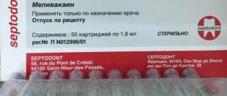
3% mepivacaine, in comparison with other anesthetics, has a less pronounced vasodilator effect, which makes it possible to use it without adding a vasoconstrictor. The anesthetic provides pain relief for 10-20 minutes, while treatment must be carried out from the 5th to 20th minute during therapeutic interventions and from the 10th to 20th during tooth extraction surgery.
Articaine 4% is currently available in the Russian Federation. This anesthetic is short-acting: anesthesia of dental pulp for 6 minutes, soft tissue for 45 minutes. Its widespread use in pediatric dentistry is limited due to its short action, which is not suitable for most interventions.
DOSAGE
In all cases of local anesthesia, it is necessary to calculate the dosage of the administered anesthetic in terms of the child’s body weight. For articaine preparations with a vasoconstrictor, the recommended dosage is 5 mg per 1 kg of weight. Before performing local anesthesia, the child’s weight is checked with the parents. In clinical practice, it is convenient to use a table with weight and the maximum permissible dose of the administered anesthetic (Table No. 1, 2).
Table No. 1
| WEIGHT | MG | ML | CARPOOLS |
| 10 | 44 | 1.5 | 0.8 |
| 15 | 66 | 2.2 | 1.2 |
| 20 | 88 | 2.8 | 1.4 |
| 25 | 110 | 3.6 | 1.7 |
| 30 | 132 | 4.4 | 2.4 |
| 35 | 154 | 5.1 | 2.9 |
| 40 | 176 | 5.9 | 3.2 |
| 45 | 198 | 6.6 | 3.6 |
| 50 | 220 | 7.3 | 4.0 |
| Mepivacaine 3% without vasoconstrictor. Maximum dose 4.4 mg/kg. 3% solution in 1 carpule 1.8 ml (54 mg). | |||
Table No. 2
| WEIGHT | MG | ML | CARPOOLS |
| 10 | 50 | 1.2 | 0.69 |
| 15 | 75 | 1.9 | 1.0 |
| 20 | 100 | 2.5 | 1.4 |
| 25 | 125 | 3.1 | 1.7 |
| 30 | 150 | 3.7 | 2.1 |
| 35 | 175 | 4.4 | 2.4 |
| 40 | 200 | 5.0 | 2.8 |
| 45 | 225 | 5.6 | 3.1 |
| 50 | 250 | 6.2 | 3.4 |
| Articaine 4% with a vasoconstrictor. Maximum dose 5 mg/kg. 3% solution in 1 carpule 1.8 ml (72 mg). | |||
Quite often, at outpatient dental appointments we encounter children suffering from obesity and metabolic syndrome, which is largely due to changes in the nutritional culture of the population. The dosage of the administered anesthetic in these cases has some peculiarities. In particular, if the doctor is going to administer anesthesia to an overweight child, the dosage of the administered anesthetic is calculated without taking into account adipose tissue.
Intraoral access
Using the thumb and forefinger of the left hand, push the upper lip up and out, and with the middle finger hold the projection of the infraorbital foramen. With this type of access, it is located at the intersection of two axes. One of them, horizontal, runs 5-7 mm below the lower orbital margin, and the second, vertical, runs on the corresponding side along the axis of the second upper premolar. The needle must be inserted, retreating 5 mm from the upper edge of the attachment of the transitional fold between the lateral and middle incisors. Then it is pushed upward, forward and outward in the direction of the lower orbital foramen until it stops at the bone. And only there the anesthetic solution is released.
How to improve the quality of pain relief?
The patient, for his part, can also prepare for the upcoming intervention and thereby improve the quality of pain relief. To do this you need to follow these simple rules:
- Postpone a visit to the dentist if you have infectious diseases or (for women) during menstruation.
- Be sure to inform your doctor about allergic reactions to medications.
- The day before visiting the dentist, refrain from drinking alcohol and visiting the sauna.
- The evening before your visit, you may take a small dose of a sedative to relieve tension.
Anesthesia at the greater palatine foramen
This method blocks the innervation of the greater palatine nerve, thus achieving an anesthetic effect on the mucous membrane on the desired side of the hard palate, as well as on the alveolar process from the palate from the third molar to the middle of the short part of the canine. The anesthetized area can reach the lateral incisor, as well as the vestibular surface in the area of the third molar. In some patients the area extends to the second premolar.
The greater palatine foramen is located in the horizontal plate of the palatine bone and its pyramidal process at the base of the alveolar process, 5 mm anterior to the border of the soft and hard palate. Above the hole in the mucous membrane there is a small depression. The projection of the hole onto the mucous membrane of the hard palate is located at the intersection of two mutually perpendicular lines. The one that runs vertically goes through the middle of the line that connects the crest of the alveolar process and the center of the upper jaw, and the horizontal one runs through the middle of the coronal part of the third molar.
Anesthesia technique. The mouth should be opened wide, the syringe needle is directed from the opposite corner of the mouth and inserted 1 cm forward and inward in the direction from the projection of the opening of the palate into the mucous membrane. The needle is advanced until it comes into contact with the bone, and 0.5 ml of solution is injected. After a couple of minutes, the analgesic effect occurs. In the case when the solution is injected directly near the greater foramen of the palate, as well as into the opening of the pterygopalatine canal, the effect captures the posterior nerves of the palate emerging from its lesser foramen. As a result, the soft palate is numbed. Injecting the solution into this area may cause nausea and vomiting. Another side effect that may occur due to excessive injection of a solution under pressure is necrosis of the soft tissues of the hard palate. This can occur in patients with vascular atherosclerosis.
Infiltration technique
Infiltration local anesthesia is most often used in dentistry. In this case, the normal transmission of nerve impulses at the site of injection of the anesthetic is blocked. Experts distinguish two types of infiltration anesthesia:
- Direct – impulse transmission is blocked directly at the site of drug administration.
- Indirect - the anesthetic gradually penetrates into the tissue surrounding the injection site, due to which the “freezing” area increases.
Infiltration anesthesia is successfully used to anesthetize manipulations on the upper jaw; on the lower jaw, the effect of anesthesia in some cases may not be sufficiently expressed. This difference in effectiveness is due to the anatomical features of the alveolar processes of both jaws. If on the top the compact plate of the alveolar process is thin and there are many small holes in it (which facilitates rapid and sufficient diffusion of the drug into the bone tissue), then on the bottom the compact plate is thicker and noticeably denser, and there are much fewer holes in it. Therefore, during volumetric manipulations on the lower jaw, the doctor usually chooses another method to numb the area requiring treatment.
The main advantages of infiltration anesthesia in dentistry can be considered:
- Rapid achievement of analgesic effect.
- Safety for the patient, since low concentrations of anesthetic solutions are sufficient to achieve the required effect. In this case, if necessary, you can increase the amount of anesthetic without any harm to the patient’s body.
- The drug is eliminated from the body quickly.
- In addition to the desired nerve, nearby fibers of neighboring nerves are also anesthetized, so the treatment is painless and completely comfortable.
A variation of the infiltration method is intraligamentary (intraligamentous) anesthesia. The anesthetic solution is injected using a very short needle directly into the periodontium under measured pressure (the area of distribution of the anesthetic depends on the magnitude of the pressure). With this technique, the soft tissues of the oral cavity will not become numb, so it is often used in the treatment of baby teeth in young children - so that the child, who does not feel the numb areas of the soft tissues, does not accidentally injure them.
Anesthesia at the incisive foramen
Such anesthesia is carried out by neutralizing the nasopalatine nerve in order to anesthetize the anterior region of the mucous membrane of the hard palate in the area of the anterior teeth.
The incisive foramen is located between the front incisors 7-8 mm from the edge of the gum at the intersection of the lines that connect the distal edges of the median palatal suture and the necks of the canines.
Anesthesia technique: the patient is in a chair, his head is thrown back, his mouth is open wide. The needle is inserted into the mucous membrane near the incisive opening to a depth of 3-4 mm. The anesthetic solution is released slowly. The process of inserting a needle into the papilla itself is very painful, so thin needles are used for such injections, and additional pain relief is performed. The anesthetic effect is achieved within a few minutes.
General principles of pain-relieving procedures
In order for local anesthesia in dentistry to bring the expected effect and not become a source of complications, it is important for the doctor to follow certain principles of its implementation:
- You should first assess the patient’s condition and find out if there are any allergic reactions to painkillers.
- The correct choice of anesthetic drug and place for its administration.
- The use of exclusively sterile preparations that are compatible with oral tissues.
- The temperature of the solution for administration should be close to normal human body temperature.
- The rate of drug administration should be minimal, and the patient should not experience any unpleasant sensations (burning, itching, pain) during the process.
- Only sharp needles should be used to avoid tissue injury.
- The area of the upcoming injection must be pre-treated with an antiseptic.
- The injection should not be unexpected: the patient must be prepared for local anesthesia.
Complications of infraorbital anesthesia
1. Injury to the vessels located in the area of needle advancement. With this anesthetic injection, you can injure, firstly, the angular artery, the external maxillary artery and the anterior facial vein, art. angularis, art. maxillaris externa et vena facialis anterior (these vessels are crossed by the course of the needle). But we know that the vessels move to the side when the needle approaches them (this creates difficulties with intravenous injections), especially when the anesthetic solution is continuously released. Secondly, the infraorbital artery and accompanying veins can be injured here. Injury to these vessels occurs, according to some authors, in 2-3% of cases. In our practice, vascular injury with this anesthetic injection is a rarer occurrence.
The prevention of vascular injury and other complications that may arise from it will be discussed below.
2. The needle gets into the eye socket. To avoid this complication, you should not advance the needle into the depths of the infraorbital canal to a distance of more than 8-10 mm. If it happens that the end of the needle enters through the infraorbital canal into the orbit, for example, when the needle is advanced deeper into the canal or if its length is insignificant, then transient double vision and some swelling of the soft tissues surrounding the eyes can only occur; there are no more serious consequences. Only with extremely rough and deep advancement of the needle through the infraorbital canal into the orbit can the eyeball be injured.
3. Paresis of the muscular nerves of the eye (most often mi inferiores nervi oculomotorii). This complication occurs when an anesthetic solution gets into the eye socket. It is accompanied by transient double vision. But, as we said, the needle entering the orbit through the upper wall of the infraorbital canal when entering the canal no more than 10 mm is almost completely excluded.
4. If the solution enters the orbit through the lower orbital margin, it is revealed by swelling of the lower eyelid. There are no serious consequences from this.
5. Pallor of the facial skin at the beginning of the injection can also occur with this anesthesia, but here this phenomenon is not surprising, since the injection is performed near the vessels that directly supply the facial skin.
Area of spread of anesthesia. Typically, after infraorbital conduction anesthesia, the area from the middle of the upper central incisor to the middle of the upper second premolar on the labio-buccal side, including the corresponding area of the upper jaw and teeth, is anesthetized. Medial and lateral to this area, anesthesia does not occur due to anastomoses from the anterior superior alveolar nerves of the opposite side (medially) and from the posterior superior alveolar nerves of the same side (laterally).
Sometimes (although very rarely) the teeth are anesthetized only up to the canine (both incisors and the canine). This can be explained by the fact that the solution did not reach the nerve behind the infraorbital canal, where the middle superior alveolar nerves, which play a major role in nerve supply to the upper premolars, are almost always separated, which usually happens with extracanal infraorbital conduction anesthesia, or by the fact that the middle superior alveolar nerves arise outside the infraorbital canal and the groove of the same name (before the infraorbital nerve enters the orbit through the inferior orbital fissure), or, finally, by the fact that the upper premolars in this case are innervated by branches from the posterior superior alveolar nerves. In the mucous membrane on the palatal side, anesthesia does not occur, since this is the area innervated by the nasopalatine and anterior palatine nerves.
It must be added that, in addition to anesthesia of the upper jaw within the mentioned boundaries, with infraorbital conduction anesthesia we also achieve anesthesia of the lower eyelid (except for the lateral part), the side of the nose (except for the back and tip), half of the upper lip and the front part of the cheek. This is explained by the fact that after injection into the infraorbital canal, not only the area of branching of the anterior and middle superior alveolar nerves is anesthetized, but also the area of branching of the infraorbital nerve, protruding through the infraorbital foramen onto the anterior surface of the upper jaw (lesser pes anserine).
The intraoral technique of infraorbital conduction anesthesia is less successful than the extraoral one. This is due not only to the possibility of introducing an oral infection during the intraoral technique of this anesthesia, which is especially dangerous in a heavily infected oral cavity, but also to the fact that the technique of intraoral infraorbital conduction anesthesia is much more complicated than extraoral one. With an intraoral infraorbital anesthetic injection, it is necessary to deviate from the most important rule of conduction anesthesia on the jaws, which requires that the needle advanced into the depths of the perimaxillary soft tissues be kept close to the bone at all times. It should be emphasized that the need to insert a needle into the very infraorbital canal makes the intraoral technique of this anesthesia even more complex and reduces the likelihood of success.
The extraoral technique of infraorbital conduction anesthesia is easier and, therefore, more accessible.
Infraorbital conduction anesthesia, as indicated at the beginning, plays an important role in anesthesia of the upper jaw. When carried out intracanal, it completely anesthetizes almost two-thirds of the upper jaw. It can and should take pride of place among the methods of conduction anesthesia of the dental system. It can and should play almost the same role for anesthesia of the upper jaw as mandibular conduction anesthesia plays for conduction anesthesia of the lower jaw.
In case of failure, when we were unable to get into the infraorbital foramen and, consequently, into the infraorbital canal (this happened during the first time we developed this anesthesia), we released the anesthetic solution outside the infraorbital canal. This was sometimes relatively sufficient to anesthetize minor interventions (routine tooth extraction, etc.), and in cases where a significant and very painful intervention was to be performed, we used central conduction anesthesia at the round hole (about anesthesia at the round hole and the ways of its implementation, see . Further). Failures, i.e. cases of failure to enter the infraorbital canal, even with the extraoral technique of infraorbital conduction anesthesia are relatively common.
Wanting to reduce the percentage of such failures and bring it to a minimum even among inexperienced doctors, we set ourselves the task of modifying the technique of extraoral infraorbital conduction anesthesia. Having studied a large number of skulls and corpses for this purpose, we were convinced that the infraorbital foramen does not always open exactly obliquely downward and inward. In a significant percentage of cases, this hole is directed either only inward, or, more often, almost completely downward.
Considering the existence of these options for the direction of the infraorbital foramen, we recommend the following technique for performing extraoral infraorbital conduction anesthesia.
From the usual injection site, the needle is first directed obliquely, from below and from the inside, up and out. If it does not hit the infraorbital foramen, this way they switch to the direction from bottom to top, for which they slightly extend the needle and again, after moving the soft tissue at the injection site, slightly move it outward in the indicated direction (from bottom to top). In case of failure, take the direction from the inside to the outside, for which they again slightly extend the needle, lift the tissue at the injection site slightly upward, then direct the needle from the inside to the outside and enter the infraorbital foramen.
Our research, based on the study of over a hundred skulls for this purpose, has established the following regarding the options for the direction of the infraorbital foramen. In approximately 60% of cases, the orifice is directed obliquely downward and inward, in 25% - mainly downward and only slightly inward, and in 15% - mainly inward.
In the technique of extraoral infraorbital anesthesia, the depth of the canine fossa should also be taken into account. With a very deep canine fossa, getting the end of the needle stuck in it can prevent successful entry into the mouth of the infraorbital foramen. To avoid this, the injection begins closer to the projection site of the infraorbital foramen and, thus, has the opportunity to reach the area of the bone located above the canine fossa. With such an injection site, you can almost always easily get into the infraorbital foramen, even with a very deep canine fossa.
INJECTION INSTRUMENTATION
For local anesthesia in children, carpule syringes of various designs are used. Preference should be given to injectors intended for carrying out an aspiration test (Rabinovich S. A., Vasilyev Yu. L., Sokhov S. T., 2013; Tarasenko S. V., Kuzin A. V., Belyaeva E. A., Kurtyshov A. A., 2013). Local injection anesthesia in children carries the risk of intravascular injection of local anesthetic. This fact is explained by the high degree of vascularization of the tissues of the maxillofacial region of children. Thus, the frequency of intravascular administration of an anesthetic during mandibular anesthesia in adults is 10-15%, and in children - 20-25%.
Syringes with a plunger in the form of an anchor and a corkscrew have the best technical characteristics. The choice of injection needle depends on the method of anesthesia. For conductive methods, needles with a diameter of at least 0.4 mm (27G) should be used. When conducting conduction anesthesia, 0.3 mm (30G) needles bend excessively in the tissues (deflection), which leads to the deposition of the anesthetic away from the intended end point of pain relief (Rabinovich S. A., Vasiliev Yu. L., 2011).
Needles 0.3 mm (30G) are advisable to use for infiltration anesthesia and periodontal methods of pain relief.
Do not forget that when performing local anesthesia, the needle may break off. This complication, as a rule, occurs when the child makes sudden movements: withdrawing the head, abruptly closing the mouth. In most cases, these severe complications occur when using 30G needles when performing mandibular anesthesia in children.
There is an opinion that the thinner the needle, the less painful the patient perceives the stage of puncturing the mucous membrane and advancing the needle into the tissues. This opinion can be classified as a misconception. There are studies confirming that the diameter of the needle does not affect the reduction in the degree of pain of the anesthesia (Malamed SF, 2002).