When considering in detail the issue of biomechanics of movements of the lower jaw, one involuntarily thinks about the perfection and harmony of everything that is created by the hands of nature.
In the human body, as well as in other biological natures, everything is aimed at the implementation of the fundamental principle - expediency.
From the molecular structure of any substance to the complex biological structure, everything is tied to the implementation of a single plan and the answer to the question - for what and in the name of what?
Without such a strict organization, the biological functioning of any organism is impossible.
Purpose, structure and functioning of the dental system
Understanding the complex process, which is called the biomechanics of the masticatory apparatus of the dentofacial system, contributes to the timely detection of pathology in the development of muscles, articular structures, closure of teeth and the condition of the periodontium (tooth - Greek odontos, Latin dente - hence the formation: odontology - the science that deals with the description of odontos , periodontitis is a disease of periodontal tissues).
It is on a healthy periodontium - the complex of tissues surrounding the tooth, which is a single component of the temporomandibular joints - that its normal functioning depends. It follows from this that the biomechanical functions of the periodontium are determined by the anatomical and physiological features of its structure and are closely related to the work of other elements.
The laws of biomechanics of the dental system are successfully applied in orthopedics at the stages of design and creation of various prostheses, as well as some auxiliary devices.
Devices that reproduce the movements of the lower jaw include:
- Occluder . A device for assisting the patient, allowing to simulate and make the correct adjustment of the orthopedic structure.
- Facial bow . This device allows you to make an impression as accurately as possible for further correction of the bite.
- Articulator . They come in different types: universal, medium (simplified). This device is used for the manufacture and fitting of removable and fixed dentures and bridges, crowns and caps.
Photo:
Occluder
Facebow
Articulator
It is worth noting that the articulator is an extremely important device that helps to correctly make an exclusive fit of various prostheses. After all, it is articulation, in dentistry perceived as a multi-vector movement of the lower jaw (lat. mandibula) relative to the upper one, which occurs during compression and stretching of the masticatory muscles, which decisively determines intelligible and articulate pronunciation.
If some pathology associated with low frequency develops, then speech, chewing food, laughter, and swallowing are immediately impaired.
The table of movements of the lower jaw in a concise form sets out the basic provisions and determining factors of the dominant theories of articulation, the authors of which are Hanau, Gysi, Monson. Despite some discrepancies in the interpretation of the processes, their authority is indisputable, and their role in the development of orthopedics is beyond doubt.
| Articulatory theories of dentition construction | Basic provisions | Determining factors |
| Gysi's theory | The inclination of the articular path determines the displacement vector of the mandibula, which is influenced by the size and shape of the articular tubercle | Accurate determination of the articular path. Recording the incisal path. Determination of the sagittal compensation curve. Determination of the transversal compensation curve of the line. |
| Monson's theory | Complex vector movements of the LF are determined not by the articular paths, but by the surfaces of the dental cusps, which give direction to the movements | |
| Hanau's theory | The theory is similar to Gysi's theory, which analyzes the entire system of articulation. In particular, she highlights the differences between the position of the prosthesis in the articulator and in the mouth due to a decrease in the elasticity of muscle tissue | Inclination of the articular path Depth of the compensation curve Inclination of the reference plane Inclination of the upper incisors Cusp height |
| Balancing theory | Takes into account:
| |
| Spherical theory | Provides:
|
In addition, correct and healthy breathing and aesthetic emotions (expression) are impossible if the muscles that push the lower jaw forward are subject to obstruction (spasm, remission).
Complete chewing of food occurs only if the teeth of the upper and lower jaws come into correct contact - occlusion. Therefore, it is the closure of the dentition that is the defining characteristic of chewing movements.
All connecting elements of the LF move as a result of the synchronous interdependent action of the temporomandibular joint (TMJ), muscular chewing tissues and teeth. Their actions are organized, coordinated and controlled by the central nervous system.
Displacements of a spontaneous and reflex nature are entirely subordinate to the neuromuscular system and can be reproduced consistently.
The initial voluntary movements include the process of biting food and directing it into the mouth. And the following chewing and swallowing are reflexive-unconscious actions.
Due to the tasks that are defined for the jaw, its complex structure is determined.
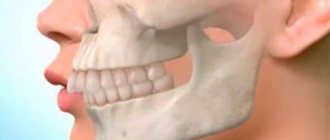
First of all, it is the only movable bone of the facial skull, which vaguely resembles a horseshoe.
This structure is due not only to its defining purpose as a responsible component of the chewing process, but also to its development, which originates from the first gill arch.
Mandible structure:
- Body.
- The edge of the body where the cells for teeth (alveoli) are located is the alveolar process.
- Chin hole. It serves as a communicator for nerves and blood vessels.
- Corner.
- Head.
- Mandibular canal and foramen.
- Branches.
- Articular and coronoid processes.
The bone formations would remain constantly in a static position if it were not for the muscle tissue connecting them.
The muscles that move the lower jaw are called mastication.
Moreover, each muscle structure, or rather their groups, produces certain movements:
- The medial pterygoid, masseter and temporal muscles raise the jaw.
- The digastric, mylohyoid, and geniohyoid are involved in the process of descent.
- Lateral movements are possible thanks to the lateral pterygoid muscles.
Directions of movement of the lower jaw
During the active phase, the biomechanics of the masticatory apparatus ensures the operation of the LF in three vector directions or planes of movement, simultaneously producing rotational and sliding displacements of its heads:
- vertical;
- sagittal;
- transversal.
Translational movements of the heads of the lower jaw forward and downward
Vertical movement
It is possible with the active work of bilateral muscle tissues going from the MF to the hyoid bone. This movement is characteristic of opening and closing the mouth.
The weight of the bone itself in this case acts as an auxiliary factor.
Three phases characterize this process, i.e. the opening of the mouth itself:
- insignificant;
- significant;
- maximum.
The maximum displacement in the vertical plane can reach 5 centimeters.
The reverse movement is carried out thanks to the same muscle group, but with their contraction.
Raising and lowering occurs at the bottom of the joint between the head of the bony structure and the cartilage disc.
To identify anomalies in the structure of the jaws of the dentition in the vertical vector of movement, as well as to calculate the linear and angular values of the skull and TMJ, in 1884, at the congress of anthropologists in Frankfurt, the term “Frankfurt horizontal” was adopted and consolidated.
Sagittal movement
The sagittal axis of displacement is expressed by the forward-backward movement vector. It is realized as a result of the work of the lateral pterygoid muscle tissues in the upper part of the joint, between the articular surface of the temporal bone and the cartilaginous disc.
At first glance, forward bone movement is a simple biomechanical process. In fact, it consists of quite complex components, which are divided into two phases:
- First . The cartilaginous disc, together with the head, moves along the articular surface of the tubercles.
- Second . At this stage, the sliding displacement of the head is simultaneously combined with its articulated movement around its own axis. The vector of this axis itself passes directly through the head of the main bone structure.
This traffic is synchronized on both the left and right. The structure of the LF allows the head to be pushed down and forward along the articular tubercle to a distance of up to one centimeter.
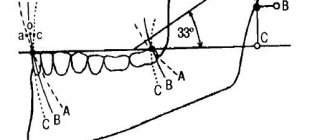
The distance covered by the articular head when moving forward is called the sagittal articular path.
It is worth recalling that this movement or path is not purely linear, but passes at a certain angle, which is formed at the intersection of vectors lying in the occlusal plane and the sagittal line - in the plane of the sagittal articular path.
A logical question arises: what is the angle of the sagittal articular path equal to in this case?
Alfred Gysi, an authoritative university professor from Zurich, already in the last century - in 1908, measured and substantiated the relationship between the inclination angles of the incisal and articular paths.
According to his statement, which no one disputes, the sagittal path angle is 33°.
The traffic that the lower incisors make when moving the bone structure is called the sagittal incisal path by the same scientist.
When the line of this path intersects with the occlusal plane, the angle of the sagittal incisal path is formed. And it falls within the range of 40 to 50 degrees.
By the way, A. Gisi made a significant contribution to the development of gnathology, a science that studies the coordinated work of the dentofacial apparatus. These and other discoveries allowed the eminent scientist, already in 1912, to create a non-adjustable articulator, which became the prototype of current orthopedic devices.
Transversal movement
Lateral displacements occur in the horizontal or transversal plane and are carried out by contraction (compression) of the lateral pterygoid muscles.
Here you need to correctly understand vector directions. Simply put, horizontal displacement is carried out to the left and right relative to the horizon, but in the frontal plane, when looking at the person’s face (front).
If the joint moves to the right side, then the left lateralis muscle is working and vice versa.
In this case, the jaw head on the displacement side rotates around the vertical axis. It slides simultaneously with the disc along the articular surface of the tubercle - down and slightly inward. Simply put, the head makes a lateral articular path, which is also at an angle to the sagittal plane.
The angle of the transversal articular path in dentistry is called Bennett's angle and is equal to 17°.
The position of the teeth will change if the bass moves to the left or right. These displacements have an angular projection, which is called the transversal incisal path or Gothic angle. With lateral displacements, it determines the span of the incisors, which falls within the range of 100 to 110°.
Lateral movements of the lower jaw (Gothic angle - 110° and Bennett angle - 17°)
Knowledge and understanding of the functioning of the apparatus for moving the lower jaw forward and backward, as well as other vector components, allows us to correctly take into account general factors that are extremely necessary when creating high-quality orthopedic structures.
It is these factors that decisively influence articulation:
- Sagittal occlusal curve.
- The height of the cusps of the chewing teeth.
- Angle of inclination of the sagittal articular path.
- Angle of inclination of the sagittal incisal path.
- Transverse occlusal curve.
Also, without knowledge and consideration of the Bonneville-Hanau articulation laws, which determine the linear arrangement and close synchronous relationship of all components of the LF, it will not be possible to correctly manufacture and install artificial teeth in dentures on edentulous jaws.
The lines connecting the incisal point with the articular heads and the heads themselves form a Bonville triangle
What else is remarkable about the Spee curve?
Also, according to Spee, a researcher and practitioner, the occlusal curve is completely connected with the articular path, since it, together with the curve of the row of teeth, is formed by one radius, and accordingly, the teeth and the articular head slide along one circle forward and the steeper the inclination of the articular path, the more uniform the occlusal curve is concave. Such assumptions made by the scientist met with many objections. Other scientists argue that the occlusal curve cannot be called a segment of a circle. In its continuation, the sagittal curve of Spee often passes below or above the articular path, and its center is not in the orbit.