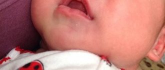
Reasons for the formation of bumps behind the ears
There are a number of health problems that can cause lumps and lumps to form behind the ears. It is most likely that this problem may occur with the following diseases:
- mastoiditis;
- otitis media;
- infection;
- abscess;
- lymphadenopathy;
- acne;
- fat cyst.
If you find any suspicious growths, for example, a ball behind the ear, you should immediately consult a doctor. Our clinic specialists are ready to conduct an examination, determine the causes of the disease and prescribe the necessary treatment.
Ear cysts
Earlobe cysts - sebaceous cysts
Some patients report that they find a bulbous appearance on their earlobe or behind their ear (a pimple inside the earlobe). The doctor can determine whether this lump is a lymph node of inflammatory etiology (various scalp infections, pharyngitis, otitis media, etc.), lipomas localized behind the ear, parotid tumor, etc.
But the most common diagnosis is atheroma or epidermal cyst in the ear area.
The patient, using his fingers, feels in the area of the lobe a round, small and hard lump that appears in the ear area or behind the ear. Although the word "epidermal cyst" may seem a little scary, lobular cysts are not considered dangerous.
An epidermal cyst is actually a type of sebaceous blister and is a sac formed in the subcutaneous layer (subcutaneous cyst) by the sebaceous glands. It can be a small, medium or large epidermal cyst.
Causes of cyst formation in the earlobe
It is not entirely clear what exactly causes ear cysts. One explanation is that cysts are caused by overproduction of sebum by the sebaceous glands of the skin. These glands, located under the skin, produce a certain substance called sebum, which lubricates the dermis. The sebaceous glands continue to produce sebum, which eventually accumulates locally, forming a so-called sebaceous blister.
Other possible causes of earlobe cysts include skin trauma, infection, or inflammation of the hair follicles.
Treatment of earlobe cyst
Often cysts behind the ear are asymptomatic, but if there is damage to the skin, infection can occur. In this case, in addition to the usual compaction, the person feels significant discomfort, pain, and the affected area becomes hyperemic and swollen. The solution to the problem in this case is radical - the cyst is removed to prevent the occurrence of any complications.
In a contaminated sebaceous bladder, removal of such a cyst is not always safe, so first the surgeon makes a single hole for drainage, while the cavity is treated with antibacterial agents. The mentioned alarming symptoms become an external reason for carrying out such manipulations.
Please note that a sebaceous bladder cannot degenerate into a malignant tumor. At a late stage (after approximately 15-20 days), in the absence of complications, standard surgical excision is performed.
Contraindications for sebaceous cyst removal:
- cyst obstruction with abscess formation;
- critical increase in cyst size;
- aesthetic reasons (in the absence of complications).
Sebaceous cysts on the ear - removal features
The surgical procedure is simple and is usually performed in a clinic or hospital.
Duration of surgical removal of a cyst in the ear: about 10-20 minutes.
Removal of the cyst should be carried out radically with an intact, intact capsule, so that the sebaceous bubble does not return again in the future. Removal of a cyst inside or behind the ear is carried out under local anesthesia or general anesthesia. An incision is made in the center of the cyst, the cyst is discovered, after which it is excised.
After the cyst is removed, the area is cleaned and disinfected. For a better aesthetic result, a cosmetic suture is made. Antibiotics are prescribed to prevent possible infections. The sutures are removed after 7-10 days.
Otitis media
Otitis media is another type of ear infection that can be of either viral or bacterial origin. This disease is characterized by the appearance of a lump behind the ear, which is quite painful and can cause swelling. This disease leads to a tumor noticeable even to the naked eye.
Treatment of such pathologies involves the use of potent antibiotics, which can not only alleviate the symptoms, but also fight the infection. Correct treatment can only be prescribed by an experienced doctor who will conduct a full examination to confirm the diagnosis.
Treatment and prevention of atheroma
Treatment of atheroma is predominantly surgical, with removal of the cavity along with its contents. To do this, a small skin incision is made on the body, through which access to the atheroma is provided. After removing the contents, the cavity is washed, and if necessary, a turunda is inserted for several days to facilitate the release of blood, pus and fat.
The operation is performed not in a hospital setting, but on an outpatient basis. Local anesthesia is used for pain relief. For small atheromas, sutures are not applied, since the skin incision heals on its own after 5-6 days. For large atheromas, cosmetic sutures are applied, which are regularly processed after 1-2 days. The operation itself is painless for the patient, but the danger is that without eliminating the causes that caused the appearance of atheroma, there is a high probability of relapses (almost 50% of all cases). That is why it is so important not only to eliminate the cause of atheroma, but also to take measures to prevent the disease. These include personal hygiene measures and early consultation with a doctor. It is not recommended to treat atheroma on your own.
Lymphadenopathy
Lymphadenopathy is a secondary infection of the throat or ear that begins its development from the lymph nodes. These organ-like structures are small structures located throughout the human body, including the pelvis, armpits, neck and ears.
With the development of infectious diseases, the lymph nodes will become inflamed, which is the immune system’s reaction to the pathogens. The bumps located behind the ears will gradually increase in size. Therefore, if you suspect lymphadenopathy, you should immediately contact qualified specialists.
Benign tumors of the ear
Benign tumors of the middle ear include hemangioma and various neurogenic neoplasms. Hemangiomas of the middle ear are manifested by the following symptoms:
- Decreased hearing;
- Ear congestion;
- Feeling of noise.
Often the first symptom of the disease is a slowly occurring paralysis of the facial muscles on the side where the hemangioma is located. For middle ear hemangioma, otolaryngologists usually perform abdominal surgery or widely remove the mastoid process.
Chemodectoma of the middle ear develops from glomus bodies, which are normally located at the bottom of the tympanic cavity, on the dome of the bulb of the internal jugular vein and in the temporal bone. They differ in structure from glomus bodies, which are located in other areas. Depending on the histological structure and the ratio of cell accumulations, there are 3 types of glomus tumors: adenoid-like, alveolar and angioma-like. According to the clinical course, limited and widespread forms of chemodectoma are distinguished.
Diagnosis of chemodectoma is carried out using radiography of the jugular fossa, pyramid of the temporal bone, attic-antral region, mastoid process. X-ray examination includes radiography of the temporal bone in three main projections and tomography in direct and lateral projections.
Chemodect treatment is surgical. Small tumors that do not destroy the eardrum are removed or exposed to ultra-low temperatures. Tumors that have spread to the external auditory canal, mastoid process, or antrum are also subject to surgical treatment. Otolaryngologists perform operations of varying scope - from tympanotomy to extended radical surgical interventions on the ear. Sometimes cryotherapy is used. For tumors that destroy the pyramid and spread into the cranial cavity, external gamma irradiation is performed, which often causes growth arrest or a decrease in chemodectoma.
Acne
Acne occurs as a result of clogged hair follicles and occurs mainly in teenagers. Once oil and dead skin cells accumulate in the pores, pimples or nodules can form. In some cases, neoplasms can be quite impressive in size, hard in structure, and quite painful.
In our clinic, you can make an appointment with an experienced doctor who will conduct an examination, tell you what to do if there is a lump behind your ear, and, if necessary, prescribe additional examinations.
Osteoma in the ear
Osteoma in the ear (exostosis, osteophyte) develops mainly from the compact layer of the posterior wall of the bony part of the external auditory canal. Much less often, neoplasms are found on the lower and upper walls of this section. Endophytic osteomas penetrate into the mastoid process. Osteoma is a benign tumor that grows rather slowly.
Osteoma has the appearance of a round formation, which is covered with a skin layer, very dense when palpated with a Vojacek probe. It is treated surgically. The operation is performed after the tumor has grown to medium size. In this case, removing the tumor is technically as convenient as possible. If the tumor is small, there is a risk of not completely removing the pathological tissue. If the osteoma is large, it is possible to capture a significant part of the healthy bone tissue during surgery. This will cause a large bone defect.
General information
Oncology of the auricle is a less common type of pathology, however, we should not forget the threat it can pose. In 85-90% of cases, tumors are localized on the auricle and in approximately 10-15% the tumor is localized in the middle ear.
Among the most common benign tumors of the outer ear are fibromas and papillomas , and more rare ones are chondromas , lipomas and angiomas . Malignant formations are represented by sarcomas (oncology is more often diagnosed in children) and directly cancerous tumors ( carcinomas , melanoblastomas ), which are more susceptible to adults and elderly people.
Diagnostics
To find out the reason for the appearance of a lump on the jaw, a comprehensive examination is prescribed. First, the doctor examines and collects the patient’s complaints. If a patient has lymphadenitis, at the initial stage of the disease the following symptoms appear:
- slight enlargement of lymph nodes, their mobility;
- pain of the nodes on palpation, turning the neck (pain can radiate to the ear or other parts of the body nearby);
- general weakness;
- low-grade temperature ;
- decreased sleep quality;
With inflammation of the lymph nodes, accompanied by purulent processes, the patient has complaints of:
- increased pain when moving the jaw;
- redness of the skin in the area where pus accumulates;
- severe swelling of the lymph nodes, their pain even without pressure;
- temperature rise to 38 ºC or more.
In the last stages of lymphadenitis, the patient’s well-being deteriorates significantly: inflammatory processes spread to the neck and shoulders, fever appears, and bluish skin is observed at the site of inflammation.
Since, in addition to inflammation of the lymph nodes, there are other reasons for the appearance of lumps on the jaw, it is impossible to make a correct diagnosis based on the clinical manifestation of the pathology. Therefore, laboratory and instrumental diagnostic methods are used.
The patient is prescribed:
- blood test (general, advanced);
- bacteriological examination of mucus from the tonsils and upper respiratory tract;
- radiography of the jaw (in different projections);
- ultrasound examination of pathological formation;
- CT or MRI of the head;
- biopsy (if necessary).
Regardless of the mechanism of occurrence of a lump on the jaw, it is important to begin taking therapeutic measures in a timely manner. The earlier treatment is started, the lower the risk of complications.