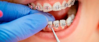
After wearing braces and poor oral hygiene, a complication may appear in the form of white spots on the tooth enamel. Such complications lead among the consequences of orthodontic treatment. Hygiene during this type of treatment requires great attention. One third of orthodontists admitted to removing braces from their patients early due to poor oral hygiene.
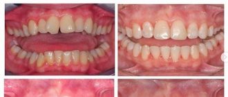
stains on teeth after removing braces
It has been found that white spots appear after orthodontic treatment in twenty percent of patients. In this regard, many dentists strictly monitor the oral hygiene of their patients during the treatment process and prescribe regular professional teeth cleanings. And immediately after removing the braces, it is recommended to take a course of enamel remineralization.
Clear marks from braces are visible, and there are white spots around them
Reasons for changes in enamel color
Lighter areas of enamel often remain in the places where the locks are attached. Various factors lead to the appearance of such marks.
- Improper oral hygiene. If you brush your teeth irregularly, plaque accumulates on them, which leads to darkening of the enamel.
- Smoking and eating dye products - coffee, strong black tea, grapes, etc. Dyes penetrate the enamel and cause it to turn yellow. At the same time, the areas of the teeth on which the braces are attached remain white.
- Insufficient hydration. Due to the tight fixation of the lock, the enamel is no longer wetted by saliva, loses its shine and becomes matte.
- Caries. One of its manifestations is white spots.
The doctor will determine the exact reason for the change in enamel shade based on the results of the examination.
Myth No. 1. Correcting an overbite is very painful.
This is greatly exaggerated. Most patients who wear braces note that they felt some discomfort for the first 2-3 days after installation/activation. The level of this discomfort is individual and depends on the person’s pain sensitivity threshold. In the vast majority of patients, the intensity of pain is minimal. Some sensitivity when eating at first is normal, choose something softer to reduce the stress.
In the vast majority of patients, the intensity of pain when wearing braces is minimal.
Ways to solve the problem
Have white spots appeared on your teeth after removing braces? Various dental techniques will solve the problem.
- Professional oral hygiene. Removing tartar and bacterial plaque is the first procedure that is indicated after completion of orthodontic treatment. Allows you to eliminate all deposits that cannot be removed with a toothbrush.
- Remineralization of teeth. Includes grinding of damaged areas, fluoridation, treatment with special solutions to saturate dental tissues with minerals, and taking medications to eliminate calcium and fluoride deficiency.
- Enamel whitening. It is carried out in the clinic using gels that are activated by special lamps. Indicated no earlier than a month after removal of the orthodontic apparatus. The technique allows you to significantly lighten the enamel - up to 10-12 tones.
- Installation of veneers or lumineers. It is carried out if no other methods of restoring the color of enamel have given the desired result. Veneers and lumineers are thin ceramic veneers. They are fixed on the outer surface of the teeth, creating a beautiful smile without defects.
Preventing stains
If you approach orthodontic treatment correctly and strictly follow the dentist’s recommendations, you can avoid the formation of white spots on your teeth.
- Follow the recommended diet. Fruits and vegetables with high acidity, which destroy teeth, should be excluded from the diet. Do not eat cold or hot foods that contribute to the formation of cracks. Hard and sticky foods should also be excluded.
- Brush your teeth regularly. Traditionally, dentists recommend brushing your teeth twice a day, but with braces, the number of procedures increases. After each meal, it is necessary to use a brush with paste, brushes, or a single-tuft brush. It is important to use dental floss and fluoride-based mouth rinses.
- Professional oral care should be performed every 6 months. Plaque forms within a few hours. After a day it becomes hard, and a regular brush can no longer remove it. Even if professional hygiene is neglected several times, a stone will form around the bracket, which will start the process of caries development.
After getting braces, you should learn more about oral care recommendations and ask about ways to help avoid white spots in the future.
Ceramic crowns and veneers for prosthetics of all teeth
It was decided to restore the shape of the teeth and strengthen them using ceramic crowns. Ceramic crowns will also be installed on the implants. When installing crowns on implants, individual ceramic abutments will be used, that is, a ceramic base will first be screwed into the implant, replacing the tooth prepared for the crown, and then a thin ceramic crown will be installed on the abutment.
Before the ceramic crowns and veneers were made, the patient wore temporary crowns and veneers to protect the prepared teeth and provide a normal appearance.
Ceramic crowns are made from pressed e.max ceramic. e.max ceramics have excellent performance in both aesthetics and durability. Ceramic crowns look natural, do not change the taste in the mouth, and do not cause allergic reactions.
After the ceramic crowns and veneers were made in Dial-Dent’s own dental laboratory, they were fixed on the teeth.
Enamel loss after removal of ceramic brackets: quantitative in vitro analysis
Orthodontics, as a specific and independent branch of dentistry, has undergone a significant number of changes in the last century, accompanying its progress as an independent specialty. One of the most significant innovations was the use of enamel etchants and bonding systems to secure brackets to the surface of teeth. Before this discovery, the only reliable way to secure braces was to alloy each individual tooth in the arch with a metal wire. Although the introduction of the method of direct bonding of braces was an important step in the course of complex progress, the new approach required solving new related problems and practical issues. On the one hand, due to the strong adhesion of the braces, their tight connection with the tooth structure is ensured, which prevents the loss of structures during operation, but on the other hand, too high a bonding force provokes the risk of damage to the surface of the teeth during the debonding stage. This problem is unsolved and still remains a fairly pressing clinical issue in orthodontics.
Particular attention is drawn to the issue of disruption of the enamel structure during debonding of ceramic braces, which have become popular since 1980, primarily due to their aesthetic characteristics, which metal braces simply could not boast of. However, some of the first generation ceramic brackets caused significant enamel damage during debonding, and in vitro studies have shown that 63.3% of the enamel structure at the bracket interface is damaged during debonding. Hence the conclusion: enamel damage during debonding of orthodontic units is significant. However, it should be noted that since their introduction, ceramic braces have been improved all the time, and manufacturers of new analogues assure that their current generation is safe for debonding, while the risk of damage to the enamel is minimized. However, there is not yet enough quantitative data to support these advertising slogans.
Modern technology allows quantitative measurement of small changes in the surface of the teeth that may occur during the debonding phase of braces. In the case of metal braces, after their removal, adhesive residues were found on the surface of the tooth. Removal of the adhesive provoked loss of enamel to a depth of 20-50 microns. The purpose of this study was to examine two ceramic bracket systems and to quantify changes in the enamel surface after the debonding process. To visualize the surface of the teeth before bonding braces, after their removal, and also after complete cleaning of the contacting surface, a three-dimensional (3D) optical scanner was used, which helped to objectify quantitative changes in the enamel structure.
Materials and methods
Forty premolars extracted for various orthodontic reasons in the maxillofacial surgery clinic were selected for the study (the procedure was performed with the prior consent of the Expert Council 13-02375-xm). The teeth were previously examined for the presence of signs of enamel damage, caries, tartar or restorations, and if any could be found, the samples were excluded from the study. Then the selected samples were washed with water and soaked in a 10% formaldehyde acetate solution until immediate use. In the study, tooth surfaces were etched with a 38% phosphoric acid solution (Reliance Orthodontic Products, Itasca, Ill.) for 30 seconds, rinsed, and dried until the enamel began to appear snow-white. This shade of enamel after etching and drying was confirmed using an optical scanner. Initial dental scans were obtained using a 3D optical scanner (COMET xS, Steinbichler Vision Systems, Neubeuern, Germany). Its accuracy is 5 microns, and its spatial resolution (the distance between the points under study) is 60 microns. Transbond XT adhesive (3M Unitek, Monrovia, Calif) was applied to the facial surface of the teeth and then air dried. After this procedure, two different bracket systems were bonded to the surface of the teeth using Transbond XT orthodontic bond (3M Unitek). To simulate clinical conditions, the brackets were secured with moderate pressure using a hand instrument until the brackets were completely immobilized. All excess bond was removed to ensure a completely clean enamel surface adjacent to the bracket. The adhesive was cured for 45 seconds with a halogen lamp (VIP Junior, BISCO Dental Products, Schaumburg, Ill). The teeth were divided into two experimental groups depending on the type of braces used. Group 1 (19 teeth) contained teeth with fixed metal-reinforced polycrystalline ceramic brackets (Clarity, 3M Unitek), which are made of white opaque material. Group 2 included 21 specimens that were bonded with pure monocrystalline ceramic brackets (Inspire-ICE, Ormco, Orange, Calif). After fixing the braces, the teeth were placed in a dark, air-conditioned chamber with room temperature inside for a period of 7 days until the adhesive completely polymerized.
Polycrystalline ceramic brackets were removed using Weingart forceps (Ortho-Pli, Philadelphia, Penn), and monocrystalline ceramic brackets were removed using a special plastic debonding instrument (Ormco), following the manufacturer's protocols. After removal of the braces, the number of detached fragments of the structures was recorded, as well as the number of fragments of the braces that remained fixed to the surface of the teeth after the debonding procedure. After this, a second scan of the surfaces of all teeth was carried out to evaluate the changes immediately after debonding. Adhesive residue on teeth was also assessed visually using a modified Adhesive Remnant Index (ARI). This indicator is often used in research because it helps to evaluate those localizations in which the bonding procedure was ineffective. All remaining adhesive was removed using a high-speed handpiece and a carbide bur (H48LQ, KOMET of America, Schaumburg, Ill). After this, we scanned for the third and final time to record the changes after complete cleaning.
All scans were saved in standard STL format and exported to Cumulus software (Regents of the University of Minnesota, Minneapolis, Minn). To determine changes in the enamel surface at the site of fixation of the bracket, scans of teeth after debonding and scans after complete cleaning were superimposed on one another and on the base scan. The intact, intact tooth surfaces of the base scan were used as reference reference areas for precise image alignment (Figure 1A) during superimposition. The alignment algorithm built into the Cumulus software itself helped minimize the root mean square difference when matching unaltered surfaces (undamaged after bracket debonding) when superimposing the base and subsequent scans. So at least 90% of the relevant points of the reference surfaces fit within the scanner's dimensional accuracy of 5 µm. Once the scans were compared, changes in the enamel surface were visualized using a linear color scale. In this way, it was possible to identify areas whose surface area had increased (due to adhesive residues) and surfaces whose area had decreased (due to loss of enamel). Changes in the volume of affected enamel and the depth of the lesion itself were also measured along the contours of the areas where braces were fixed. These procedures were carried out using Cumulus algorithms. Areas where both loss of tissue volume (due to loss of enamel) and its increase (due to adhesive residues) were recorded were analyzed separately in order to avoid the effect of leveling objective indicators of changes due to the superposition of unrelated causes of changes physical parameters of the surface (photo 1B). Volume and depth measurements of lesions using two different bracket systems were compared using a t test, and ARI scores were compared using the Mann-Whitney test (significance level was 0.05).
Figure 1. (A) Post-debonding scans and post-clearance scans were compared to the initially acquired base image using the occlusal, proximal, and lingual surfaces as reference areas for superimposition (highlighted in yellow). (B) The volume and depth of the enamel lesion (area 1) as well as the area with adhesive residues (area 2) were analyzed independently to avoid the leveling of objective parameters due to the different nature of changes in the enamel surface (for color interpretation, the reader should consult the web -program version).
results
Metal-reinforced polycrystalline ceramic brackets were bonded to 19 teeth, while monocrystalline ceramic brackets were bonded to 21. However, two samples of polycrystalline ceramic brackets and two monocrystalline ceramic brackets were excluded from the study due to incomplete scanning of the fixation area, resulting in a sample size of the former group was reduced to 17 samples, and the second - to 19.
Debonding of braces causes the loss of enamel on the surface of the tooth, or the formation of a small amount of adhesive residue at the bonding site. Five teeth in the group with fixed polycrystalline ceramic brackets and one in the group with monocrystalline ceramic brackets showed enamel loss after bracket removal and before the cleaning procedure. The depth of enamel damage after debonding (average depth) was 21 µm for polycrystalline ceramic brackets and 33 µm for monocrystalline ceramic brackets. The volume of damaged enamel in the first group was 144 µm3 and 36 µm3 in the second group, respectively.
As a result of the 3D scan, it was discovered that in the first group there were adhesive residues on 16 teeth (that is, on all teeth except one). The average thickness of the residual bond layer in the group of polycrystalline braces was 188 µm, while in the second group it did not exceed 120 µm. The difference in the average thickness of the residual bonding layer between the two bracket systems was statistically significant (t-test, p = 0.0164), but the difference in the volumetric indicators of the adhesive residue was no longer statistically significant (t-test, p = 0.2381).
Using the ARI classification, it was found that the amount of adhesive residue after the debonding procedure was significantly higher in the group of monocrystalline ceramic brackets compared to polycrystalline ceramic brackets (Mann-Whitney test; P = 0.016): 2 teeth met criterion 2 - more than half of the adhesive layer remained on tooth, and in 17 cases the situation was assessed by parameter 3 - the entire adhesive layer remained on the tooth. When using polycrystalline braces, in 2 cases the amount of residual adhesive was assessed by criterion 0, in 6 – by criterion 1, in 1 – by criterion 2, and in 8 – by criterion 8 (according to the ARI scale). The remaining adhesive was removed using a carbide bur. Only on two teeth from the group of polycrystalline braces and on one from the group of monocrystalline braces were bond residues found even after the cleaning stage. The average thickness of their layer in the first group was 16 µm, and in the second – 15 µm. However, the cleaning process also inadvertently removed a small amount of enamel, resulting in an average enamel loss (i.e. lesion depth) of 28 µm for polycrystalline ceramic brackets and 18 µm for monocrystalline ceramic brackets. Enamel loss rates after the cleaning phase were significantly different between the two bracket models, both in terms of volume (t-test, p = 0.0245) and mean lesion depth (t-test, p = 0.0191).
The fracture characteristics of brackets during the debonding procedure differed significantly between polycrystalline ceramic units and their monocrystalline counterparts (Mann-Whitney test, p < 0.0001). Of the monocrystalline ceramic bracket group, 95% (18/19) were removed in one single piece without any fractures. One monocrystalline ceramic unit (5%) broke into two parts. 35% (6/17) of polycrystalline brackets broke into four or more pieces during debonding, and 65% (11/17) broke into exactly two pieces. The ratio of bracket fragments remaining fixed after the debonding procedure to the tooth surface in the first and second groups was 24% and 5%, respectively.
Discussion
Typically, enamel loss due to braces is only assessed after the bonded area has been completely cleaned. However, debonding is a two-step procedure: removing brackets and cleaning off any remaining adhesive. And the manipulations carried out at each of these stages can affect in their own way the final amount of enamel damage. Therefore, we assessed the loss of enamel after each stage of debonding.
Using the power of 3D scanning, we were able to quantify and compare the parameters of enamel loss immediately after bracket removal and after a full cleaning procedure. For both bracket systems, minor damage was reported after the debonding phase, which was slightly worsened after complete cleaning of the bonded area. The loss of enamel after debonding was recorded on 5 brackets from the polycrystalline group, and 1 from the monocrystalline group. After complete clearance, their number increased to 15 and 16 cases, respectively. When using metal-reinforced ceramic units, the depth and volume of enamel damage after debonding were 21±8 µm and 114±183 µm3, and after complete cleaning they increased to 28±14 and 420±287 µm3. Similar indicators when using single-crystal structures after debonding were 33 µm and 36 µm3, and after cleaning – 18±6, and 238±136 µm3, respectively. Similar results, according to the literature, were obtained after removing metal braces and completely cleaning the places where they were fixed. In this study, both the volume (area times depth) and the mean depth of enamel lesions were determined using two ceramic bracket systems. It should be noted that the depth of the lesion is a more clinically important indicator than the amount of enamel tissue reduction. Since the thickness of the enamel varies in the range from 1500 to 2000 microns, and when carrying out preventive procedures using a brush or rubber cup, tissue loss is 7-14 microns, a loss of enamel to a depth of about 20-30 microns is considered acceptable, and comparable to that after the procedure professional hygiene. Other studies have also reported minimal enamel loss during debonding, but most were based on qualitative analysis using techniques such as scanning electron microscopy. Such an approach cannot provide a differentiated assessment of enamel loss, first after the debonding stage, and then after complete cleaning of the area of fixation. It should be noted that the surface of the enamel that is lost during bracket debonding contains a large amount of fluoride, so doctors should understand that although the amount of enamel loss is small, they should not underestimate the fact of damage to the enamel structure.
In addition, by performing a 3D analysis of enamel loss separately during the debonding phase and after full cleaning, the difference between the use of two different bracket systems can be quantified. Rates of enamel loss after cleaning were significantly higher when using metal-reinforced polycrystalline brackets than when using monocrystalline analogues. This fact can be justified by the thicker residual adhesive layer after the use of braces from the first group of the study (photo 2). Its thickness was 188±113 µm after debonding and 16±5 µm after complete cleaning. In the group of monocrystalline braces, the thickness of the residual bond layer varied within 120±37 µm after debonding and 15 µm after cleaning. If some bracket fragments remain attached to the tooth surface during the debonding process, removing them can be quite a difficult task for the dentist. The procedure for removing such fragments is uncomfortable for both the patient and the doctor; in addition, as a result of such complete cleaning, the risk of damage to the enamel only increases. The latter conclusion is supported by the quantitative results of our study, which demonstrate higher rates of enamel loss when using a polycrystalline ceramic bracket system, when debonded, the number of residual fragments was significantly higher than when using monocrystalline units.
Photo 2. (A) Scan after debonding: the bracket fragment remained fixed on the tooth (black color corresponds to the color gamut indicator at a damage depth of 250 µm) with the adjacent area of the residual adhesive layer (light blue, blue, violet). (B) Scan after complete cleaning of the tooth: removal of a fragment of the bracket provoked additional loss of enamel (green, yellow) (the reader should familiarize themselves with the web version of the program to interpret the color scheme).
Fractures and chips of brackets in most cases occurred when using polycrystalline ceramic units (65% - split into two fragments, 18% - into 4, 18% - more than 4). When using monocrystalline braces, in 95% of cases they were removed as a single fragment. All polycrystalline brackets fractured into two or more fragments, while among monocrystalline brackets, only one bracket (5%) fragmented into two parts during debonding. The findings contradict another study, which found that polycrystalline ceramic brackets were less likely to break than their monocrystalline counterparts. However, it remains a well-known and documented fact that the increase in debonding force is directly proportional to the incidence of enamel cracks and bracket fractures. Debonding of monocrystalline ceramic brackets requires less force to break the relationship with the tooth structure, which is confirmed by lower rates of enamel loss and fewer fractures in the study of the second group of samples. According to the manufacturer, fragmentation of the unit into two parts is the preferred characteristic of polycrystalline ceramic brackets. The design of the bracket provides a vertical slot for debonding using a tool. An area of stress concentration under the action of force is formed at the base of the bracket in such a way that the fracture occurs along the vertical axis along the slot. During the study, it was found that the majority of polycrystalline ceramic brackets (65%) actually fragment into two parts. However, 6 of 17 polycrystalline ceramic units (35%) fractured into four or more pieces, and in the case of 4 brackets (24%), the fragments remained attached to the tooth surface after debonding.
Monocrystalline ceramic braces, on the other hand, are uniquely designed to be removed from the tooth surface in one piece using special plastic pliers. This characteristic of the bracket is ensured by the presence of zirconium microspheres (40 microns in diameter) embedded in the base of the unit. As a result, only in one case a fragment of a monocrystalline bracket (5%) remained on the tooth after debonding. This result is consistent with a previous study in which approximately 20% of used polycrystalline ceramic brackets fractured into four or more fragments, while monocrystalline counterparts remained intact when removed. 3M Unitek offers specially designed forceps for debonding polycrystalline ceramic brackets, but states that Weingart or Hove forceps can also be used with similar success. However, it is obvious that the use of such instruments provokes the fragmentation of brackets into a significant number of parts, since this is the only way to explain the results obtained in our study.
It is possible to challenge the opinion that the brackets are perfectly separated from the adhesive, and almost the entire layer of bond remains on the surface of the tooth. The ARI index, which characterizes the amount of remaining adhesive on the tooth surface, had quite different indicators when using two different bracket systems. When using monocrystalline ceramic brackets, 18 of 19 samples (95%) separated during debonding in such a way that almost the entire adhesive layer remained on the tooth surface with well-defined boundaries. This made the cleaning procedure easier since the bond was visually visible (photo 3). When using polycrystalline ceramic brackets, the ARI values were quite different in different experimental cases. About half of the brackets in this group (47%) separated similarly to monocrystalline ones, leaving almost the entire adhesive layer on the surface of the tooth, while the other half of the samples (47%) retained almost half of the bond layer on the surface of the bracket itself. These results are consistent with another study that tested brackets with a similar debonding protocol and found that nearly half of the samples retained 50% or less of the adhesive on the bonding surface of the bracket itself.
Photo 3. (A) Scan after the debonding procedure of a monocrystalline ceramic bracket: clear outlines of adhesive residues (blue, purple, black). (B) Scan after complete cleaning: no signs of enamel loss detected (gray) (the reader should consult the web version of the program to interpret the color scheme).
Reducing the amount of enamel damage during the debonding procedure of ceramic braces, as well as after complete cleaning of the tooth surface from adhesive residues, is an important and clinically acute issue. Although we found significant differences in the performance of the two different bracket systems, the overall conclusion is that both provide a successful debonding phase, after which there is fairly little enamel loss. When evaluating the results of this study, it is important to remember that data obtained in a clinical setting and in an in vitro experiment may differ to some extent. The forces required to remove brackets may vary depending on the influence of external conditions such as temperature, humidity and other oral parameters, which can reduce the strength of the bond and, therefore, affect the depth of damage to the enamel during the debonding procedure. In addition, the extracted teeth used in our study may have had undetected surface or subsurface cracks caused by forceps during extraction. In this case, the enamel surface was initially damaged even before the braces were fixed. However, the results of this in vitro study are clinically relevant. 3D scanning technology has made it possible to quantitatively analyze very small and visually imperceptible changes in enamel surfaces, the clinical assessment of which is almost impossible. This study found that cleaning the enamel surface after the debonding procedure may cause further damage to the enamel, so the latter must be done with extreme caution. Given the progress of current digital technologies, a future clinical study will be conducted using an intraoral scanner, which will allow us to verify and expand the results of our study in real clinical conditions, and also form an objective picture of what happens to the tooth surface after debonding of orthodontic brackets.
conclusions
- Quantitative assessment of enamel loss during debonding of ceramic brackets can be performed using an accurate 3D scanner.
- Both polycrystalline and monocrystalline ceramic bracket systems can be successfully removed from the tooth surface with virtually minimal damage to the enamel. When using polycrystalline ceramic brackets, it was found that enamel loss rates were slightly higher after the complete tooth surface cleaning step, which could be explained by the fact that a thicker layer of residual adhesive remained on the enamel surface, and the number of bracket fragments remaining bonded to the tooth was also lower. significantly higher than that of monocrystalline analogues.
- The final enamel loss after surface cleaning with a carbide bur was 20-30 µm when using either of the two ceramic bracket systems studied.
Authors: Sam N. Sulimana, Terry M. Trojanb, Daranee Tantbirojnc, Antheunis Versluis
Myth No. 5. An allergic reaction to braces is possible.
A very small percentage of patients are allergic to nickel, which is part of the alloy from which braces are made. This is not a problem. If you have any concerns, you can always conduct a preliminary allergy test and, if an allergy is detected, choose braces from a different material. If you are allergic to latex components necessary for treatment, they can be replaced with latex-free ones.
A very small percentage of patients are allergic to nickel, which is part of the alloy from which braces are made.
Dial-Dent specialists who took part in the treatment:
- Orthopedic dentist S.V. Zukor – drawing up a comprehensive treatment plan, coordination of treatment, dental prosthetics with ceramic crowns.
- Orthodontist M.P. Sleptsova – treatment with braces: elimination of crowding of teeth, correction of bite, preparation for prosthetics on implants.
- Implant surgeon V.P. Alaverdov – installation of Astra Tech dental implants, sinus lifting, bone grafting, removal of wisdom teeth.
- Dentist-endodontist T.I. Matienko – treatment of tooth canals with a microscope, replacement of old fillings, treatment of caries.
- Dental neurologist E.V. Saxonova – consultation on the issue of night grinding of teeth and severe clenching of teeth (bruxism).
- Dental hygienist T. Kondratieva – hygienic preparation for treatment, hygienic support during treatment.
- Dental technician D.V. Wolf – production of ceramic dental crowns and veneers.
- Dental assistants M. Erkimbekova, S. Shchelkova, L. Kharlamova, A. Antoshkina.
The cost of treatment depends on the specific situation, so treatment at Dial-Dent begins with an examination, diagnosis and drawing up an estimate for treatment, where all the necessary procedures and their prices are prescribed. The administrator will not be able to tell you the price of treatment over the phone, since the price in the price list is only the price of individual treatment procedures. Without examination and diagnostics, it is impossible to say what exactly and in what volume will be required. Consultation with a dentist at Dial-Dent is free, you may only need to pay for the images (sight image - 390 rubles, panoramic image - 1300 rubles, computed tomography of the teeth - 2900 rubles). Only after signing the estimate, when the patient understands what will be done and how much it costs, treatment begins.
See other examples of treatment from Dial-Dent specialists here.
Make an appointment for a consultation by phone +7-499-110-18-04 or through the form on the website. You can ask questions about dental prosthetics to the chief doctor of the clinic, Sergei Vladimirovich Tsukor, at