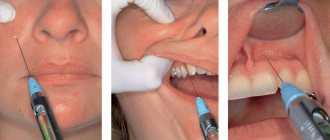
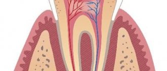
To ensure painless dental treatment, local anesthesia is usually used in dentistry - it is used when performing almost all dental procedures to provide the patient with the most comfortable and painless conditions while in the dental chair. Local anesthesia in dentistry allows you to temporarily block in a certain area the transmission of nerve impulses that signal the brain about the impact on a tooth, so a person does not feel pain during treatment procedures. In this case, the effect of the anesthetic extends only to the peripheral nervous system, without affecting the brain: the patient remains fully conscious.
There are several different types of this kind of anesthesia; the doctor chooses a certain type depending on the volume of the upcoming intervention, the localization of the pathological process and the general condition of the patient’s body. You also need to know that in some cases the patient is given some preparation for local anesthesia - it is necessary for him to endure this procedure as comfortably as possible.
Non-injection methods
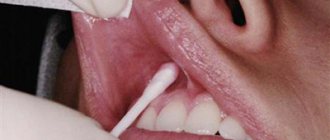
For short, small-scale interventions, it is sufficient to superficially numb the soft tissues; this can be achieved using non-injection methods of local anesthesia in dentistry. These include:
- Application method . Applying a special gel or spray containing a certain anesthetic in a small concentration to the desired area of the oral mucosa. This method is often used in pediatric dentistry in the treatment of baby teeth, when treating gingival margins, opening small superficial abscesses, and also to prepare the patient for injection-type local anesthesia (to numb the injection site).
- Physico-chemical method . Injection of the required amount of anesthetic into soft tissues using electrophoresis. It is usually used for trigeminal neuralgia.
- Physical method (rarely used in recent years). Anesthesia of a specific area by applying a freezing reagent (chloroethyl), exposure to a laser beam or electromagnetic waves of a certain frequency.
All other techniques for performing local anesthesia in dentistry belong to the injection group, that is, they require an injection in a certain area of the oral cavity or even outside it.
Anesthetics without a vasoconstrictor
Provide varying durations of pain relief for dental tissues. In particular, 2% lidocaine provides anesthesia of the dental pulp within 5 minutes, while the rate of onset of anesthesia is also 5 minutes, which is unsatisfactory for the doctor. Therefore, the use of 2% lidocaine without a vasoconstrictor is inappropriate for dental anesthesia.
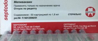
3% mepivacaine, in comparison with other anesthetics, has a less pronounced vasodilator effect, which makes it possible to use it without adding a vasoconstrictor. The anesthetic provides pain relief for 10-20 minutes, while treatment must be carried out from the 5th to 20th minute during therapeutic interventions and from the 10th to 20th during tooth extraction surgery.
Articaine 4% is currently available in the Russian Federation. This anesthetic is short-acting: anesthesia of dental pulp for 6 minutes, soft tissue for 45 minutes. Its widespread use in pediatric dentistry is limited due to its short action, which is not suitable for most interventions.
Infiltration technique
Infiltration local anesthesia is most often used in dentistry. In this case, the normal transmission of nerve impulses at the site of injection of the anesthetic is blocked. Experts distinguish two types of infiltration anesthesia:
- Direct – impulse transmission is blocked directly at the site of drug administration.
- Indirect - the anesthetic gradually penetrates into the tissue surrounding the injection site, due to which the “freezing” area increases.
Infiltration anesthesia is successfully used to anesthetize manipulations on the upper jaw; on the lower jaw, the effect of anesthesia in some cases may not be sufficiently expressed. This difference in effectiveness is due to the anatomical features of the alveolar processes of both jaws. If on the top the compact plate of the alveolar process is thin and there are many small holes in it (which facilitates rapid and sufficient diffusion of the drug into the bone tissue), then on the bottom the compact plate is thicker and noticeably denser, and there are much fewer holes in it. Therefore, during volumetric manipulations on the lower jaw, the doctor usually chooses another method to numb the area requiring treatment.
The main advantages of infiltration anesthesia in dentistry can be considered:
- Rapid achievement of analgesic effect.
- Safety for the patient, since low concentrations of anesthetic solutions are sufficient to achieve the required effect. In this case, if necessary, you can increase the amount of anesthetic without any harm to the patient’s body.
- The drug is eliminated from the body quickly.
- In addition to the desired nerve, nearby fibers of neighboring nerves are also anesthetized, so the treatment is painless and completely comfortable.
A variation of the infiltration method is intraligamentary (intraligamentous) anesthesia. The anesthetic solution is injected using a very short needle directly into the periodontium under measured pressure (the area of distribution of the anesthetic depends on the magnitude of the pressure). With this technique, the soft tissues of the oral cavity will not become numb, so it is often used in the treatment of baby teeth in young children - so that the child, who does not feel the numb areas of the soft tissues, does not accidentally injure them.
Area of influence
Tuberal anesthesia affects the area from the gums of the first molar to the first molar. The needle enters the space between the optic nerve and the fat pad. In some cases, it is possible to reduce or increase the working space.
The anesthetic is injected near the alveolar ending, located behind the fixed jaw bone. The action extends to adjacent mucous tissues, the posterior part of the alveolar process and directly to the molars.
Conductor technology
Conduction local anesthesia in dentistry is used when infiltration anesthesia is ineffective (for example, if volumetric manipulations on the lower jaw are to be performed), when several teeth need to be treated simultaneously, or a long-term intervention is expected. In this case, the anesthetic solution is supplied to the branches of the nerve innervating the area of the upcoming intervention. The drug can be administered endoneurally (directly into the nerve, used rarely and for special indications) or perineurally (next to the nerve, so that the solution gradually permeates the nerve fibers, the most commonly used method of administration). There are also extraoral and intraoral methods of performing the injection - the doctor decides which method to choose in a particular case.
Depending on the site of administration of the anesthetic drug and the branches of the nerves that are to be blocked, the following types of conduction local anesthesia in dentistry are distinguished:
- Infraorbital – anesthesia of the branches of the infraorbital nerve.
- Palatine – blocking impulses from the greater palatine nerve.
- Tuberal - blocking impulses from the superior posterior alveolar nerves.
- Mental, or mental – anesthesia of the mental nerve.
- Mandibular - blocking impulses of the lower alveolar and lingual nerves in the lower jaw. One of the varieties is considered to be torsal anesthesia (blocking the branches of the lingual, inferior alveolar and buccal nerves).
- Incisive – blocking impulses from the nasopalatine nerve passing in the incisive canal.
Typically, the conduction technique requires preliminary preparation of the patient for local anesthesia - applying the anesthetic by application method to numb the injection site, as well as explaining to the patient how best to position his head for this or that type of administration of the anesthetic drug.
Tuberal anesthesia according to the method of P.M. Egorova (blockade of the posterior superior alveolar nerves)
Extraoral method The basis of the method of blocking the posterior superior alveolar nerves according to P.M. Egorov is to determine the individual anatomical landmarks of the injection site, the direction of insertion and the depth of immersion of the needle. To perform the method P.M. Egorov recommends using the extraoral route of needle insertion. With extraoral methods of local anesthesia, the surgical field is more accessible for review and selection of the injection site; there are all conditions for reliable antiseptic treatment of the injection site.
General principles of pain-relieving procedures
In order for local anesthesia in dentistry to bring the expected effect and not become a source of complications, it is important for the doctor to follow certain principles of its implementation:
- You should first assess the patient’s condition and find out if there are any allergic reactions to painkillers.
- The correct choice of anesthetic drug and place for its administration.
- The use of exclusively sterile preparations that are compatible with oral tissues.
- The temperature of the solution for administration should be close to normal human body temperature.
- The rate of drug administration should be minimal, and the patient should not experience any unpleasant sensations (burning, itching, pain) during the process.
- Only sharp needles should be used to avoid tissue injury.
- The area of the upcoming injection must be pre-treated with an antiseptic.
- The injection should not be unexpected: the patient must be prepared for local anesthesia.
Anesthesia at the incisive foramen
Such anesthesia is carried out by neutralizing the nasopalatine nerve in order to anesthetize the anterior region of the mucous membrane of the hard palate in the area of the anterior teeth.
The incisive foramen is located between the front incisors 7-8 mm from the edge of the gum at the intersection of the lines that connect the distal edges of the median palatal suture and the necks of the canines.
Anesthesia technique: the patient is in a chair, his head is thrown back, his mouth is open wide. The needle is inserted into the mucous membrane near the incisive opening to a depth of 3-4 mm. The anesthetic solution is released slowly. The process of inserting a needle into the papilla itself is very painful, so thin needles are used for such injections, and additional pain relief is performed. The anesthetic effect is achieved within a few minutes.
How to improve the quality of pain relief?
The patient, for his part, can also prepare for the upcoming intervention and thereby improve the quality of pain relief. To do this you need to follow these simple rules:
- Postpone a visit to the dentist if you have infectious diseases or (for women) during menstruation.
- Be sure to inform your doctor about allergic reactions to medications.
- The day before visiting the dentist, refrain from drinking alcohol and visiting the sauna.
- The evening before your visit, you may take a small dose of a sedative to relieve tension.
Intraoral access
Using the thumb and forefinger of the left hand, push the upper lip up and out, and with the middle finger hold the projection of the infraorbital foramen. With this type of access, it is located at the intersection of two axes. One of them, horizontal, runs 5-7 mm below the lower orbital margin, and the second, vertical, runs on the corresponding side along the axis of the second upper premolar. The needle must be inserted, retreating 5 mm from the upper edge of the attachment of the transitional fold between the lateral and middle incisors. Then it is pushed upward, forward and outward in the direction of the lower orbital foramen until it stops at the bone. And only there the anesthetic solution is released.
Dosage
In all cases of local anesthesia, it is necessary to calculate the dosage of the administered anesthetic in terms of the child’s body weight. For articaine preparations with a vasoconstrictor, the recommended dosage is 5 mg per 1 kg of weight. Before performing local anesthesia, the child’s weight is checked with the parents. In clinical practice, it is convenient to use a table with weight and the maximum permissible dose of the administered anesthetic (Table No. 1, 2).
Table No. 1
| WEIGHT | MG | ML | CARPOOLS |
| 10 | 44 | 1.5 | 0.8 |
| 15 | 66 | 2.2 | 1.2 |
| 20 | 88 | 2.8 | 1.4 |
| 25 | 110 | 3.6 | 1.7 |
| 30 | 132 | 4.4 | 2.4 |
| 35 | 154 | 5.1 | 2.9 |
| 40 | 176 | 5.9 | 3.2 |
| 45 | 198 | 6.6 | 3.6 |
| 50 | 220 | 7.3 | 4.0 |
| Mepivacaine 3% without vasoconstrictor. Maximum dose 4.4 mg/kg. 3% solution in 1 carpule 1.8 ml (54 mg). | |||
Table No. 2
| WEIGHT | MG | ML | CARPOOLS |
| 10 | 50 | 1.2 | 0.69 |
| 15 | 75 | 1.9 | 1.0 |
| 20 | 100 | 2.5 | 1.4 |
| 25 | 125 | 3.1 | 1.7 |
| 30 | 150 | 3.7 | 2.1 |
| 35 | 175 | 4.4 | 2.4 |
| 40 | 200 | 5.0 | 2.8 |
| 45 | 225 | 5.6 | 3.1 |
| 50 | 250 | 6.2 | 3.4 |
| Articaine 4% with a vasoconstrictor. Maximum dose 5 mg/kg. 3% solution in 1 carpule 1.8 ml (72 mg). | |||
Quite often, at outpatient dental appointments we encounter children suffering from obesity and metabolic syndrome, which is largely due to changes in the nutritional culture of the population. The dosage of the administered anesthetic in these cases has some peculiarities. In particular, if the doctor is going to administer anesthesia to an overweight child, the dosage of the administered anesthetic is calculated without taking into account adipose tissue.
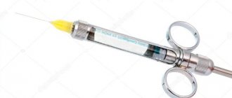
Injection equipment
For local anesthesia in children, carpule syringes of various designs are used. Preference should be given to injectors intended for carrying out an aspiration test (Rabinovich S. A., Vasilyev Yu. L., Sokhov S. T., 2013; Tarasenko S. V., Kuzin A. V., Belyaeva E. A., Kurtyshov A. A., 2013). Local injection anesthesia in children carries the risk of intravascular injection of local anesthetic. This fact is explained by the high degree of vascularization of the tissues of the maxillofacial region of children. Thus, the frequency of intravascular administration of an anesthetic during mandibular anesthesia in adults is 10-15%, and in children - 20-25%. Syringes with a plunger in the form of an anchor and a corkscrew have the best technical characteristics. The choice of injection needle depends on the method of anesthesia. For conductive methods, needles with a diameter of at least 0.4 mm (27G) should be used. When conducting conduction anesthesia, 0.3 mm (30G) needles bend excessively in the tissues (deflection), which leads to the deposition of the anesthetic away from the intended end point of pain relief (Rabinovich S. A., Vasiliev Yu. L., 2011).
Needles 0.3 mm (30G) are advisable to use for infiltration anesthesia and periodontal methods of pain relief.
Do not forget that when performing local anesthesia, the needle may break off. This complication, as a rule, occurs when the child makes sudden movements: withdrawing the head, abruptly closing the mouth. In most cases, these severe complications occur when using 30G needles when performing mandibular anesthesia in children.
There is an opinion that the thinner the needle, the less painful the patient perceives the stage of puncturing the mucous membrane and advancing the needle into the tissues. This opinion can be classified as a misconception. There are studies confirming that the diameter of the needle does not affect the reduction in the degree of pain of the anesthesia (Malamed SF, 2002).