In case of problems with the facial muscles, you will need to be examined by a neurologist; if any studies have been previously performed, be sure to take their results for consultation, incl. the pictures themselves. If studies have not been performed, they will be recommended and performed based on the results of the examination.
- Facial hyperkinesis.
- Facial hemispasm.
- Facial paraspasm.
- Blepharospasm.
- Examination for facial hyperkinesis.
- How to stop facial hyperkinesis. Administration of Botulinum toxin A.
- Treatment of facial muscle spasms.
Facial hyperkinesis
Facial hemispasm and paraspasm, blepharospasm are diseases associated with uncontrolled contraction of facial muscles. They are very similar to neuropathy, neuritis, and facial nerve paresis.
Facial muscle control is structured as follows:
- Nerve cells of the cortex and subcortical nuclei of the brain send nerve impulses to the brain stem, to the control centers of the facial nerve;
- These centers distribute impulses along the nerve fibers and send them along the nerve, like an electrical cable, directly to the facial muscles;
- Each tiny nerve fiber causes its own microscopic section of muscle to contract.
Causes of facial hyperkinesis:
- An error in the functioning of the subcortical nuclei of the brain, as a result of which excess uncontrolled nerve impulses are sent to the facial muscles, which lead to spasm;
- Excessive stimulation of the facial nerve centers in the brain stem;
- Irritation of the facial nerve itself after it leaves the brain.
In the first and second cases, this is the result of damage to the nucleus or center due to neuroinfection, multiple sclerosis, blood supply deficiency or tumor. In the third case, the disease occurs even with slight compression of the root of the facial nerve in the area of its exit to the base of the brain by an altered vessel (entanglement of the nerve by an artery), a tumor, or compression in the thickness of the parotid salivary gland in some of its diseases.
Even doctors often confuse facial hyperkinesis with neuropathy, neuritis of the facial nerve. We will definitely understand the causes of the disease and provide the necessary treatment.
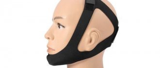
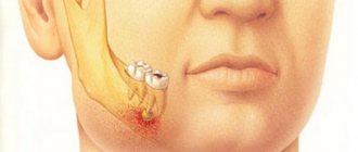
The mechanism of trismus occurrence
The chewing muscles set our jaw apparatus in motion. A sudden muscle contraction, accompanied by a strong closure of the jaws, limitation of the movement of the lower jaw, temporary loss of the ability to speak and eat, is called trismus. Strong clenching of teeth often causes breathing problems.
Excessive muscle tension leads to hardening. The disease can become a factor in a significant decrease in the quality of life and deterioration of the psycho-emotional background. A person’s appearance changes, the digestive tract organs suffer, so it is important to see a doctor as soon as possible. Without timely help, your general health may deteriorate, and it is also necessary to find out the causes of spasms.
Ask a Question
Examination for facial hyperkinesis
The success of treatment directly depends on an accurate diagnosis of the location and cause of damage to the pathways and brain centers of the facial nerve. Therefore, we carefully examine each patient. Your doctor will determine the cause of excessive stimulation of the facial muscles, and based on the data obtained, select the most effective treatment.
MRI and X-ray computed tomography of the brain and facial skull. On tomograms, especially if they are performed with preliminary contrast, the brain centers of the facial nerve and its area of exit (root) to the base of the brain, blood vessels, and skull bones are visible. They help to see the cause of compression of the facial nerve root and evaluate the structure of the salivary glands. Circulatory disorders, cysts and tumors are easily recognized.
Blood tests to check for infections and biochemical changes that damage the brain and facial nerve. The suspicion of the presence of a neuroinfection and its activity can be easily verified using a blood test.
Electromyography, Blink reflex - electrophysiological techniques based on measuring the electrical potentials of the facial muscles. They help assess the function of impulse transmission along the facial nerve, the degree of its impairment, judge the effectiveness of treatment, the presence of complications, and help in choosing the correct treatment tactics.
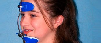
Treatment methods
Patients with hypertension should receive help as soon as possible, since trismus can accompany a hypertensive crisis. It is also important to call an ambulance if you suspect rabies and tetanus. In the absence of severe concomitant pathologies, you can consult a dentist.
Knowledge of how to relax the jaw muscles on your own is necessary for self-help directly during spasm - but in the future, examination and consultation with a specialist is necessary. A warm compress can help with spastic jaw closure. Its use is not recommended if there are foci of inflammation in the oral cavity, for example, with pulpitis, periodontitis, periostitis, or suspected abscess. A light massage of the muscles can also help to relax, but remember that the movements should be light, and in no case should you put any force on the muscles.
The approach to the treatment of trismus includes a detailed analysis of the condition and a search for the causes of the disease. To eliminate the causes, it may be necessary to remove the inflamed tooth or surgical treatment of purulent inflammation. In case of a fracture, the doctor will immobilize the jaw.
Physiotherapeutic methods are widely used as a supplement. Laser, ultrasound therapy, electrophoresis with the use of painkillers can be used.
Antibacterial therapy is used for the inflammatory nature of spasms, for example, inflammation of the trigeminal nerve.
Treatment of spasm of the masticatory muscles often includes the use of sedative medications if the pathology was caused by diseases of a neurological nature. However, a neurologist prescribes such drugs.
Also, if muscle tone disorders are suspected of being infectious, vaccination is mandatory. Infection with the rabies virus requires immediate attention.
How to stop facial hyperkinesis. Administration of Botulinum toxin A
Botulinum toxin A (Botox, Dysport) is a means of reducing the excitability of the muscles involved in hyperkinesis. The drug is one of the most convenient options for the symptomatic treatment of spasms of the muscles of the face and eyes, and other hyperkinesis. We inject Botulinum Toxin A using a syringe with a very thin needle directly into the affected muscle, after which the muscle stops engaging in tics or other hyperkinesis. The effect develops within 3-5 days and lasts up to 4-8 months. During this time, a course of treatment of the causes of the disease and rehabilitation procedures (massage, gymnastics, physiotherapy, psychotherapy, cosmetic procedures) are carried out.
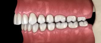
Types of trismus and diagnostic methods
There are two main types of spasm of the masticatory muscles:
- Unilateral. Most often it is associated with an inflammatory process or injury to the mandibular joint and adjacent tissues. The result of unilateral pathology is a displacement of the lower jaw to the side when opening the mouth, as well as facial asymmetry.
- Bilateral. The cause of the disease is neuralgia and common infectious diseases. With this type of trismus, the jaws close together with a slight movement of the lower jaw back. There is an inability to open the mouth, difficulty speaking and eating.
To diagnose trismus, the doctor will find out information about previous diseases, operations, injuries, collect anamnesis, and listen to complaints. External examination is supplemented with radiography and other methods of clarifying the diagnosis.
Treatment of facial muscle spasms
Depending on the detected causes of the disease, your doctor will choose the appropriate combination from a wide range of treatment methods:
- Prescription of drug therapy aimed at reducing irritation of the facial nerve root, restoring blood supply, eliminating infection if it is detected;
- Injection of Botulinum toxin A into muscles subject to involuntary contractions;
- Acupuncture, massage, gymnastics;
- Special neuropsychological gymnastics, which allows you to normalize the relationships between different parts of the brain;
- Carrying out neurosurgical intervention to eliminate contact between the modified vessel and the root of the facial nerve (microvascular decompression) or remove the tumor.
Causes of trismus
The problem of spasm of the lower jaw can be associated not only with damage to nerve endings and reflex contraction, but also with other factors:
- infectious and inflammatory diseases of the oral cavity;
- previous injuries;
- unsuccessfully performed anesthesia of the teeth of the lower jaw;
- arthrosis of the mandibular joints;
- pathologies of ENT organs;
- irritation, inflammation of the trigeminal nerve;
- purulent processes, etc.
In addition, trismus can be a consequence of pseudobulbar palsy, meningitis, epilepsy, calcium deficiency, etc. Symptoms may first appear after injury, temperature changes, or sudden hypothermia. Sometimes the jaw cramps after opening the mouth wide, for example, after removing a wisdom tooth.
Why do muscles cramp?
The reasons can be divided into two groups. The first includes those that do not have any underlying diseases. So, if the calf or gluteal muscles cramp in a dream, there is no physiological pathology in this. The seizure in this case is a parasomnia, that is, a phenomenon associated with sleep. Spasms that occur during increased physical activity also do not indicate an abnormality.
In other cases, such discomfort may be caused by:
- neurological disorders;
- compaction of muscle fibers against the background of physical inactivity;
- metabolic disorders;
- micronutrient deficiency;
- dehydration;
- poisoning (including cramps from a hangover).
There is also a disease called hypoparathyroidism. It is characterized by a convulsive syndrome that immediately affects muscle groups. This condition is caused by hypofunction of the parathyroid glands.
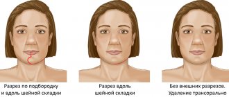
The main reasons why the oval of the face “floats”
Age-related changes
Skin aging is the most common cause of changes in facial contours. Most often this is expressed after 35 years. The production of collagen and elastin, which are responsible for the firmness and elasticity of the skin, begins to decrease.
Weight fluctuations
If extra pounds appear, the skin, accordingly, stretches. In case of sudden weight loss, it begins to sag because it does not have time to adapt to the new volumes.
Edema
The main causes of edema can be a sedentary lifestyle, poor diet, and bad habits such as alcohol and smoking. All this retains fluid in the body and swelling develops. Since water metabolism is disrupted, the contours of the face become rounded, and the skin becomes looser.
Improper skin care
If your skin is not cleansed and moisturized enough, it will begin to show signs of aging very early. Moreover, it is important to remember that the choice of skincare products should be based on a specific skin type. This will help achieve maximum effect from cosmetics and prevent your skin from aging early.
Incorrect posture
Constant slouching can lead to more than just spinal curvature. This also leads to the fact that the oval of the face becomes more drooping, the cheeks begin to sag, and the corners of the lips begin to droop down.
Exposure to direct sunlight
Ultraviolet radiation has a very adverse effect on the skin. Due to prolonged exposure to the sun, the skin ceases to produce enough collagen and this leads to rapid aging.
Hypertonicity of the masticatory muscles and its correction with BTA for aesthetic problems of the lower half of the face
Soykher M.I., Orlova O.R., Mingazova L.R., Soykher M.G.
The aesthetic appearance of the lower half of the face reflects the morphology of the dental system and the function of the masticatory muscles. In aesthetic medicine, the problems of correction of the lower third of the face remain relevant, despite the accumulated many years of experience. The functional state of the masticatory muscle is associated with the configuration of the lower third of the face and possible asymmetry, which hides a combination of dental and neurological problems that require a detailed examination and adequate treatment of the patient. Some diseases, such as bruxism and oromandibular dystonia, are accompanied by hypertrophy of the masticatory muscles, which occurs as a result of their forced contraction, and the massive lower third of the face becomes an aesthetic manifestation here.
Keywords:
Facial disproportion; hypertrophy of the masticatory muscles; bruxism; oromandibular dystonia; myofascial pain syndrome; botulinum toxin type A
Soykher Marina Ivanovna, candidate of medical sciences, dentist, head. Doctor of the Center for Interdisciplinary Dentistry E-mail
Orlova Olga Ratmirovna, Doctor of Medical Sciences, Professor of the Department of Nervous Diseases of the Faculty of Faculty of Physics, First Moscow State Medical University named after. I. M. Sechenova, President of the MoESBT E-mail
Mingazova Leniza Rifkatovna, candidate of medical sciences, neurologist, employee of the department of nervous diseases of the State Educational Institution of Higher Professional Education MMA named after. I. M. Sechenova E-mail
Soykher Mikhail Grigorievich, candidate of medical sciences, dentist, leading specialist of the Center for Interdisciplinary Dentistry E-mail
INTRODUCTION
“Everything in a person should be beautiful.” Today, more and more people are striving to implement this principle in their own lives <2>. Almost every person pays attention to his appearance, and attaches special importance to how his face looks <3, 4>. Dissatisfaction in this case can become quite a serious problem and affect both the psychosomatic state (causing depression, uncertainty, neuroses...), and professional status, family and personal relationships.
In aesthetic medicine, the problems of correction of the lower third of the face remain relevant, despite the accumulated many years of experience. According to a survey of patients, the main complaint they present is facial disproportion, in particular the “square face” problem. What is meant by the expression “square” (or “trapezoidal”) face?
In domestic and foreign medicine, for a brief answer to this question, they use such characteristics as protruding angles of the lower jaw, hypertrophy of the masticatory muscles themselves, prominent contours of the lower zone of the face, angular contours of the face (prominent mandibular angle, hypertrophy of the masseter, lower facial contour). The contours and shape of the lower half of the face are determined by the relative position of the upper and lower jaws (occlusal relationship), the size and shape of the lower jaw, as well as the condition of the masticatory muscles.
The lower jaw is suspended in space to the fixed bones of the skull with the help of muscles and ligaments. The only support for it is the chewing teeth. It is the teeth that fix the position of the jaw in three mutually perpendicular planes. When the position of the teeth and, accordingly, the dentition changes or their loss, the position of the jaw in space also changes. In most cases, there is a decrease in the lower third of the face, distalization of the bite, with characteristic facial manifestations. There are disturbances in the coordination of the masticatory muscles and temporomandibular joints. The dentition, the geometry of which is normally designed to compensate for the complex biomechanics of the chewing function of the cranial-maxillary system, simultaneously serves as support for the soft tissues of the face, which must be taken into account during the aesthetic rehabilitation of patients.
The four chewing muscles on each side are interconnected genetically (they originate from one branchial arch - the mandibular), morphologically (they are all attached to the lower jaw, which they move during their contractions) and functionally (they perform chewing movements of the lower jaw, which determines their location) .
M. masseter is a chewing muscle that starts from the lower edge of the zygomatic bone and zygomatic arch and is attached to the tuberositas masseterica and to the outer side of the ramus of the lower jaw. It has the shape of an irregular rectangle and consists of a superficial part and a deep part. The strongest muscle in the human body in terms of force generated - on molars it develops a force of up to 72 kg.
M. temporalis is the temporal muscle, with its wide origin it occupies the entire space of the temporal fossa of the skull, reaching at the top to the linea temporalis. The muscle bundles converge in a fan-shaped manner and form a strong tendon, which fits under the zygomatic arch and is attached to the processus coronoideus of the lower jaw.
M. pterygoideus lateralis - lateral pterygoid muscle, starts from the lower surface of the greater wing of the sphenoid bone and from the pterygoid process and is attached to the neck of the condylar process of the mandible, as well as to the capsule and to the discus articularis of the temporomandibular joint.
M. pterygoideus medidlis - medial pterygoid muscle, originates in the fossa pterygoidea of the pterygoid process and is attached to the medial surface of the angle of the mandible, symmetrically m. masseter, to the tuberosity of the same name..
M. masseter, m. temporalis and m. pterygoideus medialis, with the mouth open, pull the lower jaw towards the upper, in other words, they close the mouth.
With the simultaneous contraction of both muscles of the pterygoidei laterales, the lower jaw moves forward. The reverse movement is produced by the most posterior fibers of m. temporalis, running almost horizontally from back to front. If m. pterygoideus lateralis contracts only on one side, then the lower jaw moves to the side, in the direction opposite to the contracting muscle. M. temporalis gives a certain position to the lower jaw during speech, thereby ensuring the articulation of the latter.
The masticatory muscle, in addition to chewing movements, takes part, together with the facial muscles, in the articulation of speech sounds, facial expressions, yawning, and swallowing. We can say that this muscle is in a state of “chronic fitness”. Excessive prolonged activity of the masticatory muscles leads to their hypertrophy, which is characterized by an increase in strength and muscle mass (Fig. 5).
The functional state of the masticatory muscle is associated with the configuration of the lower third of the face and possible asymmetry, which hides a combination of dental and neurological problems that require a detailed examination and adequate treatment of the patient. Some diseases, for example, bruxism and oromandibular dystonia, are accompanied by hypertrophy of the masticatory muscles, which occurs as a result of their forced contraction, and their aesthetic manifestation is a massive lower third of the face <6–10>.
The purpose of the study is to study the relationship between the condition of the masticatory muscles and the aesthetic appearance of the lower half of the face; to evaluate the effectiveness of using the botulinum toxin type A drug "Lantox" in order to reduce hypertonicity and correct hypertrophy of the masticatory muscles under the control of surface electromyography.
MATERIALS AND METHODS
40 patients were examined. The average age is 35 years. In order to understand whether problems of an aesthetic nature are caused by pathological processes in the dental system, we conducted a dental and neurological study, which included:
analysis of anamnestic data; clinical study of the masticatory muscles, muscles of the neck and upper shoulder girdle, and the area of the temporomandibular joint (TMJ);
- occlusiogram, analysis of the static and dynamic organization of occlusion;
- orthopantomogram;
- TMJ tomogram;
- teleradiography (TRG) of the lateral surface of the head with markers;
- photoanalysis (portrait and intraoral photographs);
- axiography;
- functional analysis of jaw models in an articulator;
- diagnostics of parafunctions using brookscheckers;
- electromyography (EMG) of the masticatory and neck muscles.
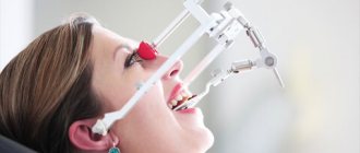
When examining the patient, attention was paid to the following clinical signs: head position, range of active movements in the cervical spine; facial expression, state of the facial muscles when speaking, swallowing, signs of blepharospasm, oromandibular dystonia, facial asymmetry; corneal reflex and reflex from the nasal mucosa, the state of the muscle ridges at rest and when clenching the teeth; volume of active movements of the lower jaw - the distance between the incisors (in cm) when opening the mouth, the trajectory of movement of the lower jaw; mandibular reflex; volume of active movements of facial muscles, brow and orbicular reflexes; Chvostek's sign; sensitivity on the face, oral mucosa and tongue.
Myograph "Synapsis" dental
Electroneuromyograph for maxillofacial studies and monitoring the effectiveness of therapeutic measures.
More details
The study of the state of the musculoskeletal system included: identification of biomechanical static disorders - scoliosis, asymmetry of the shoulders, shoulder blades and other deformities; identification of the “short leg”.
During palpation examination, we used a 3-point scale for assessing muscle tension and soreness:
0 points - no tension and no pain;
1 point - slight muscle tension, no pain on palpation;
2 points - moderate muscle tension and pain on palpation;
3 points - severe muscle tension and sharp pain on palpation, the presence of painful muscle tightness and/or trigger points.
EMG indicators were recorded using an electromyograph “Synapsis” (NMF “Neurotech”, Taganrog), supplemented with special software. Injections of botulinum toxin into the masticatory muscles were carried out under the control of electromyography; for this purpose we used the Mist device (NMF Neurotech, Taganrog).
During the clinical study, 2 groups of participants were identified:
Group 1 - 30 patients who suffered from bruxism: 25 women, 5 men;
Group 2 - 10 patients with signs of focal muscular dystonia (the leading syndrome of oromandibular dystonia): 8 women, 2 (two) men.
"MIST" professional
Myographic control of injections, biofeedback training, anesthesia regimen.
More details
Clinical features
1st group. In dental practice, bruxism is defined as parafunctional activity of the masticatory muscles. The following parafunctions of the masticatory muscles have been described (arranged in descending order of frequency): clenching the teeth, moving the lower jaw forward or to any side, inserting the tongue between the teeth, biting the tongue and lips, grinding the teeth, rhythmic movements of the tongue and submandibular muscles <12–13> . Only 10 patients from group 1 noted grinding and grinding of teeth at night. The rest had the habit of clenching their teeth tightly during the day in response to even minor emotional stress. A hereditary factor (bruxism in close relatives) was determined in all patients in this group. The history also included episodic pain in the face of muscular origin (myofascial pain syndrome of the face), frequent headaches caused by tension in the pericranial muscles. All patients complained of fatigue of the masticatory muscles in the morning. When studying the aesthetic appearance of the face, a massive lower third was determined due to hypertrophy of the masticatory muscles, which caused considerable concern, especially in women.
Dental status in patients of group 1: violation of the integrity of the dentition (absence of one or more teeth), pathological abrasion of teeth, the presence of wedge-shaped defects in the cervical area.
Clinical examination of the masticatory muscles revealed signs of hypertrophy; the muscles are dense, tense, with the presence of painful muscle compactions (myofascial trigger points). When palpating the masticatory muscle itself, in 20 patients the pain radiated to the upper and lower jaws, upper and lower molars, ear, frontal region, TMJ, and neck. In 22 patients, palpation in the area of the tubercle of the upper jaw was sharply painful.
Tenderness or discomfort in the medial pterygoid and digastric muscles was also noted. All patients had muscle tension in the floor of the mouth and limited mobility of the hyoid bone; in 10 patients there was tension and slight hypertrophy of the sternocleidomastoid muscle. Palpation of the muscles on the opposite side was painless or moderately painful.
In 40% of patients, pain occurred on palpation of the lateral pole of the joint head during rotation on both sides, and the temporomandibular ligament on both sides. Discomfort when palpating the lateral pole of the joint head in static conditions on both sides.
In 25% of patients, there was a restriction in mouth opening due to pain to 1.5±2.2 cm between the incisors (normally from 4.6 to 5.6 cm). Further lowering of the lower jaw due to the appearance of sharp pain became almost impossible. There was also a restriction in the movement of the lower jaw forward and to the side.
Only 20% of patients experienced a clicking or crunching sound when opening their mouths. Pain sensitivity of the facial skin and oral mucosa was not changed.
When conducting surface electromyography of the masticatory muscles and neck muscles, the following results were obtained: asymmetry in the work of the temporal, masticatory muscles, high rates of the total biopotential of the muscles under study.
When analyzing a chewing test, a violation of the symmetry of chewing, frequency, amplitude, phase and total biopotential of chewing is noted.
2nd group. Oromandibular dystonia (OMD) is hyperkinesis involving the muscles of the perioral region and masticatory muscles. In patients of group 2, the following clinical forms of OMD were determined: spasm of the muscles that close the mouth and compress the jaw (dystonic trismus) - in 6 people; constant trismus with lateral jerking movements of the lower jaw, bruxism and hypertrophy of the masticatory muscles - in 4 people. Subjectively, all patients of the 2nd group complained of unpleasant sensations, which were described as “periodic movement of the lower jaw”, “the jaw moves to the side”, “it is impossible to find a comfortable position of the jaw”, “forcible clenching of the teeth”, “teeth chattering against each other” friend."
Clinical examination of the masticatory muscles revealed signs of hypertrophy; muscles are dense, tense, with the presence of painful muscle thickening (myofascial trigger points). As a rule, the masticatory muscle itself undergoes pronounced changes, while the temporal and pterygoid muscles undergo smaller changes. Three patients had asymmetric dystonia. Visually, this phenomenon was manifested by asymmetry of the lower half of the face (the volume of the hypertrophied masseter muscle on one side is more pronounced than on the other).
Also, in all patients of group 2, subcompensated signs of dystonic phenomena in other areas were determined: blepharospasm, mild and moderate forms of cervical dystonia, dystonic tremor of the head and upper extremities, writer's cramp.
Dental status: in patients of the 2nd group, there was a violation of the integrity of the dentition (the absence of one or more teeth), pathological abrasion of teeth, and the presence of wedge-shaped defects in the cervical region. When conducting surface electromyography of the masticatory muscles and neck muscles, the following results were obtained: asymmetry in the work of the temporal, masticatory, and neck muscles, torsional twisting of the lower jaw, an increase in the functional activity of the neck muscles and the total biopotential of the muscles under study. When analyzing the chewing sample, a violation of the symmetry of chewing, frequency, amplitude, phase and total biopotential of chewing was noted.
For therapeutic and aesthetic purposes, all patients received injections of botulinum toxin type A (BTA) “Lantox” (Lanzhou Institute, China). It was injected into the masseter proper, temporalis, medial and lateral pterygoid muscles. Large doses were injected into the masticatory muscle itself from the outside (5–10 units at one point). For a more uniform distribution of the drug throughout the muscle, it is injected into several points (from 4 to 8), 1-2 injections are carried out from the oral cavity. In patients with an asymmetric form of dystonia, large doses of the drug were also administered on the side of the larger muscle. The average total dose of BTA (Lantox) was 100 units per procedure.
RESULTS
Analysis of clinical data showed that in patients of group 10 suffering from bruxism, on days 2–10 after injections, the feeling of fatigue in the masticatory muscles in the morning disappeared and headaches stopped. By days 14–21, a decrease in the massiveness of the lower half of the face became noticeable. Against this background, the necessary dental procedures were successfully carried out.
"Hummingbird" dental
Express assessment of the level of bruxism, study of facial muscle tone, wireless technologies.
More details
Due to the weakening of the masticatory muscles, the phenomena of bruxism and the phenomenon of clenched jaws ceased for some time. During this time, patients were advised to fix their attention on the facial muscles, consciously relax the lower jaw, open the dentition and try to form a new motor stereotype of the masticatory muscles (based on the principle of biofeedback). For this purpose, disconnecting splints were used on the lower jaw.
In patients of group 2 (OMD), the signs of hyperkinesis were also leveled out already on days 10–12 after injections due to weakening of the activity of the masticatory muscles, which significantly improved the quality of life of patients. Unpleasant subjective sensations disappeared, and the opportunity for dental treatment appeared. In patients with asymmetric dystonia, the symmetry of the lower half of the face was restored.
The study demonstrated the positive effect of BTA (Lantox) on the functional and morphological state of the masticatory muscles, and a good clinical effect alleviated the condition of patients and made it possible not to use any medications.
CONCLUSIONS
The aesthetic appearance of the lower half of the face reflects the morphology of the dental system and the function of the masticatory muscles. In aesthetic practice, it is necessary to conduct a detailed clinical analysis of the condition of the masticatory muscles, especially in patients with a massive lower half of the face. Patients with increased tooth wear and failing dentures require a detailed neurological examination to exclude bruxism and oromandibular dystonia. Injections of botulinum toxin into the masticatory muscles are the method of choice for this group of patients. The drug botulinum toxin type A (Lantox) at a dose of 100 units per procedure is effective and safe for the treatment of hyperactivity of the masticatory muscles in neurological, dental and aesthetic practice.
LITERATURE
1. Budylina S.M., Degtyareva V.P. Physiology of the maxillofacial region. - M: Medicine, 2001. - P. 87–156.
2. Goldstein R. Aesthetic dentistry. Volume 1. - M.: Stbook. - 2005. - P. 10–14.
3. Kalyuzhny D.V. Physiological mechanisms of regulation of pain sensitivity. - M.: Medicine, 1984. - P. 102–114.
4.Karlov V.A. Neurology of the face. - M.: Medicine, 1991. - 284 p.
5. Kupriyanov V., Stovichek G. Human face. - M.: Medicine, 1988. - 272 p.
6. Mingazova L.R. Pathogenesis and treatment of myofascial pain syndrome of the face // Clinical materials. conference of young scientists FPPO MMA named after. I.M. Sechenov “Current issues of clinical medicine.” - M., 2002. — pp. 54–58.
7. Mingazova L.R. Clinical and physiological analysis and treatment of myofascial pain syndrome of the face. Abstract of thesis. dis…. Ph.D. honey. Sci. - M., 2005. - 25 p.
8.Orlova O.R. Focal dystonia: clinical picture, pathogenesis, treatment using botulinum toxin // Diss.... Dr. med. Sci. - M., 2000. - P. 13–29.
9.Orlova O.R., Mingazova L.R., Vein A.M. Facial pain of a muscular nature: clinical and physiological characteristics and treatment with botulinum toxin type A (dysport) // Abstracts of the Russian scientific and practical conference “Clinical and theoretical aspects of acute and chronic pain”. - Nizhny Novgorod, 2003. - pp. 113–115.
10.Orlova O.R. Focal dystonia: clinical picture, pathogenesis, treatment using botulinum toxin. Dissertation for the degree of Doctor of Medical Sciences. - Moscow, 2000. - pp. 13–29.
11.Orlova O.R., Yakhno N.N. The use of Botox (botulism toxin type A) in clinical practice. - M., 2001. - P. 143–147, 161–163.
12. Orlova O.R., Mingazova L.R., Vein A.M. Myofascial pain syndrome of the face: new aspects of the clinic, pathogenesis and treatment // New in dentistry. - 2003. - No. 1. — pp. 25–29.
13.Petrov E.A. Electrophysiological characteristics of pain syndrome of temporomandibular joint dysfunction // Ross. dental magazine. - 2002. - No. 6. - P. 34–35.
14.Petrosov Yu.A., Skorikova L.A. Prevention of TMJ dysfunctions by eliminating parafunctions of the masticatory muscles // Abstracts of the V All-Russian. Congress of Dentistry: Prevention of dental diseases. - Novosibirsk - M., 1988. - P. 156–157.
15.Al-Ahmad HT, Al-Qudah MA. The treatment of masseter hypertrophy with botulinum toxin type. — A. Saudi Med. J. - 2006. - 27. R. 397–400.
16.Gurney CE Chronic bilateral benign hypertrophy of the masseter muscle //Am. Surg. - 1947. - No. 73. - P. 137.
17.Moore AP, Wood GD The medical management of masseteric hypertrophy with botulinum toxin type A // Br. J. Oral Maxillofac Surg. - 1994. - No. 32. - P. 26–28.
18. Sannomiya E., Goncalves M., Masseter muscle hypertrophy // Bras. Dent. J. - 2009. - No. 17 (4). — R. 347–350.
19. Slavicek R. The masticatory organ: Function and Dysfunction.
— Kloster neuburg: Gamma Med. — wiss. Fortbildungs - GmbH, 2006. - R. 59–90. toxin type A // Saudi Med. J. - 2006. - No. 27. - P. 397–400. come back
Causes of seizures in different parts of the body
Muscle spasm in a specific area of the body is caused by specific factors. Most often it occurs in the legs. The culprits may be overexertion (including due to intense training), varicose veins and hypothermia.
It can cramp not only the calf muscle, but also the femoral and even gluteal muscles. Sometimes the unpleasant sensation will spread throughout the entire leg.
Abdominal cramps are also common because most abdominal organs are composed of highly contractile smooth muscle cells. Most often, the organs of the digestive system spasm. This is how the well-known abdominal pain occurs. In women, contractions of the uterus (during menstruation, pregnancy or gynecological diseases) cannot be ruled out.
Muscle spasms in the back indicate diseases of the musculoskeletal system. This could be osteochondrosis, intervertebral hernia, degenerative changes in the spine.
Who usually experiences muscle cramps?
This trouble is familiar to everyone: men and women, children and the elderly, athletes and office workers. Only some people get to know her for natural reasons and rarely encounter her, while for others she becomes a frequent companion.
The risk group includes:
- children under 3 years of age who have experienced a rise in temperature above 38 degrees;
- elderly people suffering from vascular diseases and muscle atrophy;
- men engaged in heavy physical labor;
- athletes (football players, swimmers, runners);
- people who abuse alcohol and often experience hangovers;
Predisposes to muscle spasms and pregnancy. In mild cases, they are caused by a lack of vitamins due to changes in the body; in severe cases, they are caused by eclampsia.
What to do if a muscle is severely cramped
It is not always possible to easily endure such an attack by limiting yourself to rubbing the affected area. The pain can be unbearable. It can be removed with a sharp impulse.
For cramps due to a hangover, the patient should be placed in a horizontal position and the legs should be raised to reduce blood flow to the extremities. At the same time, it is important to make sure that we are not talking about an epileptic attack (they are not uncommon in alcohol intoxication).
In older people, involuntary muscle contractions are often a warning sign of stroke. Convulsions may give way to paralysis. Timely treatment is important here, so it is better to call an ambulance immediately.
How to correct a swollen oval face
What can you do at home?
First, prevention is the best cure. The better the quality of your skin, the longer your facial contour will be clear and even. And you can improve the quality and density of your skin yourself with daily and proper care. Retinol is recommended as an anti-aging component - it affects both the dermis, triggering collagen formation processes, and the epidermis, stimulating cellular renewal processes. Currently, retinol is a very popular anti-aging component and for good reason - its effect is noticeable and pronounced, but it is important to remember about the peculiarities of its use, because it irritates the skin and if used incorrectly, complications can occur (it will be especially difficult to deal with vascular problems). Retinol is always applied at night; its use should always be accompanied by the use of SPF during the day.
Another important ingredient in skin tightening is vitamin C. Not only does it have vascular strengthening and brightening effects, but collagen formation processes also cannot take place without it! Even when taking collagen internally, you must make sure that you consume enough vitamin C, because otherwise all your efforts are in vain! By tightening the skin, vitamin C has a lifting effect and significantly improves the quality of the skin.
What can be done in a cosmetologist's office
Massages and microcurrents
They give a good effect and are also able to relieve the lymphatic system, preventing the formation of edema. It is important to follow the course and regularly.
Peelings
Stimulates cellular renewal processes. Cosmetologists often encounter patients’ fear that peelings thin the skin, but when done correctly, they, on the contrary, thicken and renew the tissue.
Mesotherapy and biorevitalization
An excellent way to influence the dermis - they stimulate collagen formation processes, saturate the skin with amino acids - the building material of proteins, and replenish the deficiency of hyaluronic acid. It is also important to follow the course of these procedures.
Fillers
A popular procedure, during which it is especially important to pay attention to the qualifications and working methods of the specialist. When tissues sag downwards, as a rule, the corrective method of choice will be not to work in the lower third of the face (not filling the nasolabial folds), but, first of all, to fill the deficit in the areas in which it has formed, that is, in the temporal and zygomatic areas. In any case, everything is individual and when installing fillers, it is extremely important to take into account the patient’s personal data, so look at the work of the cosmetologist you are contacting, read reviews, and ask what actions will be taken in case of failure. And, of course, for this procedure you can only consult a doctor!
Hardware techniques
Currently, the most popular type of oval restoration devices are ultrasonic face lift devices (non-surgical SMAS lifting). The principle is based on the fact that ultrasonic waves penetrate to a depth of 5 mm, causing targeted thermal contraction and tissue tightening, which leads to tightening of not only the skin, but also subcutaneous fat. As mentioned above, we are all individual and only a doctor can determine whether this technique is right for you after an individual consultation.
SMAS-lift
A surgical facelift can give an amazing result; in case of pronounced tissue prolapse, surgical intervention can help eliminate the problem, however, it is important to remember that here, too, a lot depends on the qualifications of the surgeon, so choose a specialist carefully. And let me remind you that the operation will not affect the quality of the skin in any way, so you should not wait for pronounced age-related changes to undergo surgery in the hope that it will solve all the problems at once.