Diagnostic methods
To accurately determine the nature of dental damage, doctors use many methods. The main types of diagnostics include:
| Inspection of the tooth surface with staining to identify carious spots. |
| Probing. Allows you to determine the depth of the carious cavity, assess the condition of the pulp chamber, the density of the affected tissue, and the degree of pain. |
| Electroodontometry. A test to determine the degree of excitability of the pulp. Normally, in an inflamed or necrotic state, it reacts differently to irritation. |
| Percussion (tapping). In case of caries, the procedure is painful for the patient; a healthy person will not pay attention to it. |
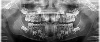
| X-ray. Using interproximal radiography, it is possible to identify hidden caries and determine the depth and extent of tooth damage. The method is used only in combination with other diagnostic methods. |
These methods are applied only to carious teeth; healthy teeth are not subject to unnecessary diagnostics. Only a doctor with special education can determine what the research should be like. The patient can assist by providing the results of previously performed diagnostics. Using them, dentists determine how long ago the problems arose, how quickly dental caries and its complications develop.
Complications of caries
The destroyer of tooth enamel and dentin, caries, can lead to tooth loss and also cause severe inflammation of the pulp, peri-apical tissues and jaw.
Cariogenic bacteria streptococci, which form hollow cavities in untreated teeth, tend to infect the connective tissues that make up most of our organs. And this is fraught with the development of diseases such as endocarditis, chronic tonsillitis, lymphadenitis and abscess.
How quickly advanced forms of caries develop
Typically, from the moment a carious spot appears on the enamel to the formation of deep caries, it takes from 1 to 4 years; the infection affects root canals 2 times faster.
But there are cases when complete tooth destruction and the formation of complications occur in just 3-4 months; this is acute caries. The reason for this is individual resistance, that is, the susceptibility of teeth to caries.
Signs that caries has reached the deep layers of dentin:
- the appearance of short-term pain;
- increased sensitivity to cold and hot;
- bad breath.
And not always, when the form is neglected, the tongue rests on a deep relief cavity in the tooth - sometimes it is just a small hole in the enamel, behind which lies an almost completely destroyed tooth.
Caries classifications
In dentistry today, thanks to the efforts of scientists and specialists from all over the world, there are several classifications of caries. The basis for diagnosis is the international classification of diseases and related health problems, tenth revision of the World Health Organization (ICD-10). In it, caries is highlighted in a separate section, where it is divided into classes, each of which has a corresponding code:
K02.0 Enamel caries. Stage of “white (chalk) spot” (initial caries). This class is characterized by: a change in enamel color (dullness) due to demineralization, and then a change in texture (roughness). In this case, there is no carious cavity, pathological changes do not extend beyond the enamel-dentin border. There is no toothache.
K02.I Dentin caries. This class implies destructive changes not only in enamel, but also in dentin, while the pulp is covered with a larger or smaller layer of preserved dentin. In many cases, at this stage, an uncomfortable reaction of the teeth to hot, cold, sour, and sweet foods may appear.
K02.2 Cement caries. In addition to the signs characteristic of the above classes, it is distinguished by damage to the exposed surface of the tooth root in the cervical region. This stage of caries in most cases is accompanied by sudden pain due to temperature, chemical, mechanical irritation, which disappears after a short period of time. Patients pay attention to the obviously changed aesthetics of the dentition, they may experience pain when eating, and if the chewing teeth are damaged, they may experience inconvenience associated with the chewing function.
K02.3 Suspended caries. It is characterized by the presence of a dark pigmented spot within the enamel as a result of local demineralization of the enamel.
K02.4 Odontoclasia (caries of the roots of primary teeth): childhood melanodentia and melanodontoclasia.
K02.5 Caries with pulp exposure (added: January 2013).
K02.8 Other caries.
K02.9 Unspecified caries. 2
Also, there is a modified classification of carious lesions according to their location - the Black classification, which includes six classes of carious formations:
Class 1 – caries of natural convexities and depressions of the chewing, palatal and buccal surfaces of the teeth.
Class 2 – caries of the contact surfaces of molars and premolars.
Class 3 – caries of the contact surface of incisors and canines.
Class 4 – intense caries of incisors and canines with a violation of the integrity of their cutting edge.
Class 5 – caries of the vestibular surface of all groups of teeth.
Class 6 – caries affecting the cutting edges of the incisors and canines and the cusps of the molars.
In addition, there are classifications of caries according to the depth of the lesion - initial, superficial, medium, deep; according to the presence of complications - uncomplicated (typically occurring superficial, medium or deep caries) or complicated (a carious process that affects the deeper structures of the tooth and the tissues surrounding its root).
At the same time, the Vinogradova classification involves dividing the disease into degrees of activity. Thus, caries can be compensated (sluggish), subcompensated (moderately progressive) and decompensated (progressive or, as it is also called, transient).
The clinical classification of caries distinguishes the disease according to the nature of its course - acute, chronic, acute and recurrent; according to the intensity of the process - single, multiple (generalized) and systemic; according to the localization of the process - fissure, aproximal/interdental caries (or caries of contacting surfaces), cervical, circular and hidden. Thus, caries is a complex and diverse polymorphic pathological process that can stabilize and worsen over a long period, acquire varying degrees of activity with different stages of compensation. This explains the difficulty in its classification. The variety of different classifications suggests that today there is no single system that would 100% satisfy the needs of all doctors and reduce this diversity to a common denominator. However, in global clinical practice this is seen as an obvious advantage, as it allows dentists to approach the issue of diagnosis and treatment more broadly.
In our clinical practice, we work according to international standards and approved clinical protocols. When making a diagnosis, the international classification of diseases and related health problems, the tenth revision of the World Health Organization (ICD-10), is used, which allows for clear and complete documentation of all aspects of the disease. For each specific patient and each dental unit, taking into account the degree of development of the pathology, appropriate treatment methods are selected. This approach to diagnosis and treatment is the most complete and effective.
Causes and types of complications
The culprits in the development of caries and its complications are streptococcus bacteria – Streptococcus sanguis, mutans, viridans. These pathogenic microorganisms oxidize and destroy tooth enamel, and then penetrate deep into the hard tissue through the dentin canals.
In the absence of proper treatment, the infection affects the coronal part of the soft tissue, the pulp, and the first stage of complicated caries occurs - acute pulpitis.
Pulpitis
When the infection reaches the pulp, an inflammatory process begins in the neurovascular bundle of the tooth in the form of:
- acute focal;
- diffuse (general) pulpitis,
a characteristic symptom of which is severe, throbbing pain in the tooth and increased sensitivity to cold.
If you do not go to the dentist with pulpitis in the first 2-3 days, the acute stage of the complication becomes chronic and develops:
- fibrous;
- hypertrophic;
- concretory;
- or gangrenous chronic pulpitis.
With chronic pulpitis, the inflamed tissue of the neurovascular bundle degenerates and is gradually destroyed, dentin formation in the tooth stops, and it becomes dead.
Over time, decay processes reach the peri-apical tissues and ligamentous apparatus - periodontium.
Periodontitis
Between the root of the tooth and the alveolus there are periodontal tissues, they act as a shock absorber during chewing and hold the tooth in the socket.
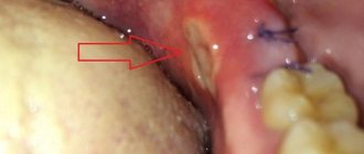
With periodontal inflammation, periodontitis, the integrity of the tooth ligaments is disrupted, the jawbone becomes infected and damaged.
Symptoms of acute periodontitis:
- acute pain when biting or touching a tooth with the tongue;
- pain is transmitted along the trigeminal nerve - to the temple and jaw;
- general malaise, deterioration of health;
- increase in body temperature to 37-38 degrees.
The acute phase with severe pain lasts from 2 to 14 days, after which the disease takes on a chronic form.
With chronic fibrous or granulating periodontitis, a fistula, that is, a through hole, can form in the gum. Patients also complain of a feeling of the diseased tooth moving out of its row and attacks of aching pain.
Granuloma
An advanced form of chronic periodontitis, granulomatous, leads to the formation of a capsule with exudate (pus) at the apex of the tooth - granulomas.
The gums of the diseased tooth become inflamed, patients complain of a feeling of bulging bone and persistent, severe pain.
A granuloma that is not eliminated in time can cause the formation of gumboil or a cyst at the root of the tooth.
Multiple caries
Another common type of complication is multiple (acute, blooming) caries, in which the infection simultaneously affects the coronal part of eight or more teeth.
In record time, caries destroys the enamel-dentin junction, penetrates the pulp and leads to tooth loss. The infection affects the cement on the roots of adjacent teeth, and the patient feels as if the entire dental arch hurts. Most often, multiple caries is detected in people with diabetes and preschool children.
Cost of caries treatment
Treatment of caries is a very individual process, and its cost is determined depending on a particular combination of certain clinical measures. These measures include the diagnostic measures carried out, the selection of a treatment method depending on the established stage of the disease, the methods, technologies, materials, equipment and instruments used in treatment, the qualifications of the attending physician, and consultations with specialists if necessary.
You should not consider caries a “mild” disease and ignore its signs. After all, this is a rather serious pathology that, in the absence of treatment, can leave a person literally “without teeth.” But, fortunately, modern dentistry has studied both caries itself and methods of treating it quite well, and with timely treatment from the patient, this process can be made reversible. Only one thing is required from patients - attention to the health of their teeth, since not a single pathology in the dental system goes away on its own; today there is not a single case of a person self-healing from caries or other diseases. Therefore, visit the dental office regularly to detect and eliminate caries in a timely manner, and then nothing will ruin your healthy and beautiful smile.
According to antiplagiat.ru, the uniqueness of the text as of October 16, 2018 is 99.8%.
Key words, tags: pulp diseases, periodontitis, tooth extraction, oral hygiene, OPTG image, dental microscope, pulpitis, periodontitis.
1 Clinical recommendations (Treatment protocols) for the diagnosis of dental caries. Approved by Resolution No. 15 of the COUNCIL OF THE ASSOCIATION OF PUBLIC ASSOCIATIONS “DENTAL ASSOCIATION OF RUSSIA” DATED SEPTEMBER 30, 2014. 2 https://mkb-10.com 3 Clinical recommendations for the diagnosis of dental caries.
Complicated caries in children
In children and adolescents, complications, like the caries that precede them, develop at lightning speed. This is due to insufficient mineralization of enamel in childhood and the tendency of young patients to infectious diseases caused by streptococci - sore throat, bronchitis, measles and scarlet fever.
Advanced forms of caries in children lead to:
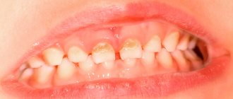
- the development of its circular shape, when, starting from the neck of the tooth, decay spreads around its crown, and over time the tooth breaks off;
- spread of infection along the surface of the crown (planar caries). In this case, the tooth completely darkens, the child complains of aching pain;
- acute and chronic pulpitis;
- infection of the rudiments of permanent teeth, resulting in multiple caries after their eruption.
Contraindications
Caries is a disease that has no absolute contraindications to treatment. But if certain factors are present, treatment may be delayed or interrupted until they are eliminated. These factors include: the presence of intolerance to drugs and materials that are planned for use in treatment; concomitant diseases aggravating treatment; inadequate psycho-emotional state of the patient before treatment; acute lesions of the oral mucosa and red border of the lips; acute inflammatory diseases of organs and tissues of the mouth; a life-threatening acute condition/disease or exacerbation of a chronic disease (including myocardial infarction, acute cerebrovascular accident), which developed less than 6 months before seeking this dental care; periodontal tissue diseases in the acute stage; unsatisfactory hygienic condition of the oral cavity.
Types of disease
Acute caries has two forms:
- medium spicy;
- deep spicy.
The difference between medium and deep acute forms lies in the size of the carious cavity. With moderate acute caries, there is no need to remove the nerve, and the tooth can be treated and restored. In case of deep acute caries, depulpation is usually required, and in case of severe tooth decay it is necessary to remove it.
Since in the acute form of the pathology the destruction of tooth tissue occurs very quickly, and children are most susceptible to the disease, in pediatric dentistry the following grouping is accepted:
- Compensated form (I group);
- Subcompensated (group II);
- Decompensated (group III).
Groups have been created to carry out clinical observation.
Pediatric dentist T. F. Vinogradova identified several dispensary groups:
- practically healthy teeth;
- compensated form;
- subcompensated form;
- decompensated form.
Children with a compensated form are examined once a year, with an undercompensated form - 2 times, with a decompensated form - 3 times. Planned sanitation reduces the risk of complications in the development of caries, the number of fillings and extracted teeth decreases. The need for treatment is also reduced by almost half, and the number of annual scheduled examinations is reduced.
Monitoring risk groups allows you to keep records based on a number of criteria:
- general prevalence of caries;
- anamnesis of life;
- health status;
- severity of the disease.
Planned sanitation and timely diagnosis of caries in adults and children make it possible not only to cure it in the initial stages, but also to prevent the development of blooming caries.
Prevention
To prevent caries, you should follow a number of simple rules:
- visit the dentist once every six months;
- Brush your teeth twice a day;
- use dental floss and mouthwash;
- eat less sweets;
- Perform ultrasonic cleaning periodically;
- stop smoking;
- limit your consumption of coffee and tea;
- Change your toothbrush every 2-3 months.
To further cleanse the oral cavity, you can use irrigators - devices that clean the teeth and interdental spaces using a pulsating stream of water. However, such devices should not become an alternative to a toothbrush.
If you notice symptoms of tooth decay or other dental disease, consult your doctor immediately.
4th stage of caries development
The last stage of caries development is called deep. It occurs as a result of progression of the previous stage or due to the chipping of a previously installed filling. Excessive consumption of sweets and poor hygiene can speed up the process of tissue destruction.
Deep caries is considered an advanced form of the disease. At this stage, the carious cavity extends to all hard tissues and almost reaches the pulp. In addition, the patient's crown is destroyed. The tooth may break into several pieces. This stage is often complicated by pulpitis or periodontitis. My mouth constantly smells bad. Painful sensations are observed not only during eating and palpation, but also at rest. If a pathological process develops under the filling, it becomes loose and falls out.
Tip: only healthy teeth can be beautiful
And for this you don’t need much at all, just brush your teeth correctly and regularly. The hygienist will tell you about this in detail after the “Professional Oral Hygiene” procedure. Our clinic specialists recommend doing it once every 6 months.
Home oral hygiene includes three-minute brushing of teeth 2 times a day, followed by additional hygiene products such as irrigators, dental floss, rinses, etc. Don't forget about cleaning your tongue. But what you definitely need to forget about is HORIZONTAL movements; train yourself to only do sweeping movements.
Adjust your diet, do not abuse carbohydrates, and after eating sweet or sour, it is better to drink a glass of water or rinse your mouth.
Come for regular preventive examinations for good results.
What happens if you ignore a toothache or the appearance of a small spot on a tooth?
The main cause of caries is the activity of acid-forming bacteria.
With initial caries, minerals and trace elements are “washed out” in the enamel, the so-called demineralization of the enamel. This provokes the formation of small spots and is the first signal about the beginning of the carious process. At the very beginning, as a rule, there is no pain. This is where the danger lies. Caries develops, and we continue to live without knowing it.
A dentist can detect carious cavities:
- at preventive examinations;
- when undergoing professional oral hygiene.
Caries has several stages of development.


