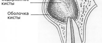
Patients with chronic periodontitis often encounter a situation where a cyst forms at the apex of the root - a cavity in the bone tissue filled with pus. Most often, a cyst develops due to infection in the root canals; sometimes the cause of a cyst is trauma, inflammatory processes in the periodontium (gums) and adjacent areas. It can also occur due to undetected caries, pulpitis or periodontitis, as well as due to poorly filled root canals.
Such a neoplasm may not bother a person for a long time. As a rule, a cyst makes itself felt during periods of weakened immunity, against the background of a cold. In such cases, the infectious process in the cyst cavity worsens, which leads to abundant pus formation. The patient feels acute pain, swelling and swelling of the gums appears, and a fever may appear. The main thing is to seek dental care on time. In these cases, resection of the apex of the tooth root and dental surgery to remove the cyst are used. However, you can often get by with therapeutic treatment.
Treatment of dental cyst: methods
Most often, treatment of dental cysts is carried out jointly by dentists - a therapist and a surgeon.
Therapeutic treatment of a dental cyst is aimed at sanitation, or cleaning of the root canals - it is necessary to remove the source of infection and the cause of the cyst. First, the dentist-therapist processes the root canals and fills them with antiseptic paste for a period of 1 week to 1 month, as in the treatment of periodontitis. Then he fills the root canals with gutta-percha and sealer. Sometimes, especially in young patients, therapeutic treatment of the cyst is quite sufficient.
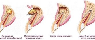
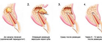
Surgical treatment is an operation to remove a cyst from bone tissue, sometimes this operation is accompanied by resection of the apex of the tooth root. The technique of apex resection is generally simple, but the operation is a complex surgical procedure and, like any operation, it must be taken seriously.
If resection is not done in time, the infection will spread upward through the root canals and can spread to the entire jaw. The consequence is that you may have to schedule the removal of all inflamed teeth. Therefore, you should not joke with such inflammations!
Diagnostics: basic methods
It is possible to notice a cyst during a routine examination only at the stage of obvious symptoms (swelling of the gums, tubercle, etc.).
Only an X-ray examination can definitively diagnose a cyst and confirm a specialist’s suspicions. As a rule, the picture is taken in the area where the cyst is located.
In some cases, a panoramic photograph of the jaw or an orthopantomogram is additionally required. OPTG is necessary for the maxillofacial surgeon in case of surgical intervention and if the X-ray image does not allow the full picture of the disease to be seen. Such cases often occur when a dental cyst is caused by infections of the nasopharynx and maxillary sinuses.
Help from other specialists
Cysts caused by gum disease require further consultation and treatment with a periodontist. For a cyst caused by ENT diseases, consultation with an otolaryngologist is necessary.
How is tooth root apex resection and cyst removal performed?
Typically, the entire procedure takes about an hour. In this case, the position of the tooth is important: surgery on distant teeth requires more skill and time.
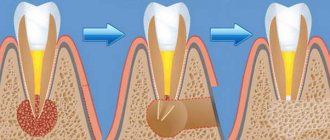
During the operation, which is performed under local anesthesia, an incision is made in the gum near the diseased tooth to peel it away to expose the bone tissue. A small hole is then made in the bone using a special bur. Through it, the doctor should see the apex of the tooth root and the cyst attached to it.
Using a drill, the tip of the root is cut off and then removed from the wound along with the cyst attached to it. After the cyst is removed, synthetic bone is placed into the empty space to restore jaw bone tissue. At the end of the procedure, the mucous membrane of the gums at the incision site is sutured, sometimes a drainage is inserted between the sutures to drain the ichor from the operation site for several days.
When performing a tooth resection, only the inflamed and infected part of the root is removed, healthy tissue is not affected.
Symptoms of anterior tooth cyst
This disease is especially dangerous due to its practically asymptomatic course at the initial stage - few people will worry if they notice a slight change in the color of the enamel or a slight displacement of the tooth. But a front tooth cyst is much more likely to be noticed than other types, since the frontal units have to “work” more often, because they are located only on one side of the jaw. However, other types of formations can go unnoticed for years, for example, if the patient is used to chewing on only one side. A cyst on the front upper or lower tooth is noticed much earlier, because pain is immediately felt.
Dangerous symptoms!
When the size of the granule with pus begins to exceed 10 mm, pronounced symptoms of a front tooth cyst arise:
- constant nagging pain with a feeling of fullness
- high fever and painful condition, which sometimes continue after the procedure for removing the cyst of the front tooth has already been completed
- swelling of the gingival tissue at the location of the granule, in severe pathologies it spreads to the face
- protrusion of a tubercle in the affected area
- enlarged lymph nodes - occurs if bacteria from the granule with pus begin to spread through the blood
Cystectomy / root apex resection: indications, contraindications and causes
- The appearance of any neoplasm, for example, fibroma or other, larger than 1 cm.
- Incomplete or poor-quality root canal filling in the treatment of pulpitis, for example;
- Stump tab or pin in the dental canal;
- Damage to the root walls during tooth filling;
- Periodontitis, granulomas, cysts, fibromas;
- Perforation of the tooth root, fracture of the root or its apex;
- Curvature of the root canals or the apex of the tooth root;
- Presence of a foreign body;
- Inflammation near the site where the denture is installed;
- A fragment of a dental instrument in the canal;
Contraindications include periodontal disease, periodontitis, and the presence of a tumor at the location of the tooth.
Why do cysts form on teeth?
Dental cysts are a polyetiological disease. Slow inflammatory reactions around the apical zone of the root can cause the development of radicular tumors. A follicular dental cyst is formed due to a disruption in the development of the dental germ of a permanent tooth from the dental sac.
The reason for the disruption of the development of the tooth germ is chronic periodontitis of temporary teeth, due to which the division of epithelial cells occurs around the crown of the unerupted segment. The pathogenesis of a retromolar cyst involves the formation of a bone pocket. Due to inflammation, it closes to form a cavity.
Provoking factors leading to the formation of a cyst under a tooth:
- complicated course of caries, pulpitis, periodontitis;
- incorrectly installed orthopedic design;
- maxillofacial injuries;
- tooth root fracture;
- complicated infections of ENT organs;
- pathology of teething, etc.
Possible complications after cyst removal
Removing a cyst is a necessary procedure; the longer the treatment of a cyst is delayed, the more likely complications are to develop. There is no need to be afraid of removing a cyst; according to statistics, the percentage of possible adverse consequences is immeasurably less than complications from an unoperated cyst (abscess, phlegmon, sepsis).
After cystectomy, the service life of the tooth is not reduced. Both after therapeutic and surgical treatment of cysts, dynamic monitoring of the condition of the bone tissue is required.
To avoid complications, it is important to maintain oral hygiene, follow your dentist's recommendations, as well as eat a healthy diet and avoid alcohol, smoking, carbonated drinks and solid foods in the first days after surgery.
What happens if you don't treat a dental cyst?
The formation does not manifest itself clinically for a long time. The progression of the disease leads to discomfort in the pathological area (especially when chewing food). Over time, a tubercle forms on the surface of the gum near the root area, which gradually increases. The mucous membrane changes color and swelling appears.
When the process suppurates, a person experiences symptoms of inflammation and intoxication: hyperthermia, headache, toothache, chills, tachycardia. A fistula may form in the middle of the protrusion, from which the contents of the cavity are released.
If you do not start treating cystic formation in a timely manner, serious complications may develop:
- phlegmon;
- lymphadenitis of nearby lymph nodes;
- inflammation of the maxillary sinuses;
- periostitis (spread of infection to the periosteum);
- osteomyelitis;
- abscess;
- deformation of the dentition;
- blood poisoning.
What do patients say about apex resection? Reviews are usually only positive!
“Resection of the top of the tooth took about an hour, the operation was performed under pain relief. It didn’t hurt very much near the tooth for about a month. A few months later I had to take an x-ray, the doctor showed that all the tissues had been restored."
“The cyst grew, they told me to remove it, it was a little unpleasant, but quite tolerable - the doctor quickly and accurately carried out the procedure. Everything was restored soon, now it’s completely unnoticeable that there was anything there.”
At Ritsa Dentistry, cyst removal operations are performed by experienced surgeons with extensive experience in performing such procedures. Therefore, you can rely on our professionalism and entrust the treatment of the cyst to our doctors.
What is the difference between a cyst and a granuloma?
There are other diseases in the oral cavity that are symptomatically similar to a cyst. Specifically, a granuloma is an outwardly swollen round projection in the root zone of a tooth. The doctor must correctly diagnose the disease, since the sequence of exposure, removal techniques, and pharmacology have significant differences. Granuloma is eliminated with the help of medications, as well as accompanying mouth rinsing with phytocomponents. The cyst has deeper roots and does not go away without surgery.
What is the “insidiousness” of pathology?
The tumor develops slowly. The cavity gradually increases under the pressure of the produced pus. In the early stages it is difficult to diagnose, since the symptoms are not clearly expressed and occur periodically (with exacerbation). Signs of pathology:
- pain when biting;
- discomfort when palpating the gums;
- swelling of the gums and face;
- increase in body temperature.
If the tumor grows, bone deformation is possible. The bone tissue of the upper jaw is porous, so resorption occurs quickly, with pronounced deformations. Large cysts of the molars and premolars of the upper jaw often occupy the entire volume of the maxillary sinus.
Formation on the root of the tooth complicates subsequent implantation:
- During removal, there is a risk of damage to the tumor membrane. Then its contents spill out onto the surrounding tissues, and the infection spreads.
- For large cystic tumors, osteoplasty is required before implantation to restore bone deficiency and ensure reliable fixation of the artificial root.
Advantages of the classical method
The classic protocol involves 2 stages, separated by time:
Implantation of an artificial rod
The implantologist installs a titanium root into the healed and restored socket and closes it with a plug screw. The implant is not subjected to load until complete engraftment.
Prosthetics
After 4-6 months, the doctor places the gum former for 2 weeks. The orthodontist then installs the crown or denture.
The classic two-stage regimen is preferable for patients with cysts because it allows time for:
- complete drainage of the cystic tumor cavity;
- eliminate infection and inflammation;
- bone tissue growth.
After complete treatment, the risks of complications in the early and late postoperative periods are minimized.
Don't let your fears of doctors get the better of your common sense.
Patients, due to their disapproval of dental procedures, are ready to make not entirely correct, but quick, express decisions. Please take the time to carefully study the guarantees section in the contract before deciding to carry out such procedures.
Levin Dmitry Valerievich
Chief physician, Ph.D.
Clinical cases that served as the prototype for this experimental treatment were demonstrated at several surgical conferences. But with a detailed analysis of information and a request for facts of “ideal results” of long-term observation of patients, external information independently received and processed by us cannot be considered reliably confirmed by us.
Undoubtedly, this technique is an interesting new technology that requires additional monitoring for several more years.
In cases of surgery with a lifetime guarantee, these scientific experiments are unacceptable when providing medical care on the principles of high-quality paid medicine.
The method of installing an implant immediately after removing a tooth with a cyst at the Dr. Levin Center for Private Dentistry is not used until successful and confirmed results of such treatment are obtained.
Types of dental cysts
A molar cyst is distinguished by several characteristics:
- depending on the reasons for the appearance;
- according to location.
Distributing according to the reasons that provoke the appearance of a dental cyst, the following varieties are distinguished:
- A keratocyst appears on the lower jaw. Older people are susceptible to the disease. It can turn into a malignant form - it grows deeply into the body and destroys the bone.
- Odontogenic or radicular dental cyst is the most common type. After periapical inflammation and necrosis of the pulp, a granuloma of the dental root is formed in the upper part. The size of the purulent focus ranges from 3 mm to 3 cm. The process does not affect bone tissue and does not provoke tooth displacement.
- A cyst that forms at the site of teething is a retention cyst. It appears during the period of replacement of milk teeth with molars. A purulent fistula with bloody contents looks like a dark blue abscess. Education is formed if the process of eruption is slow. The purulent contents of the abscess should be opened and cleaned out.
- A calcifying odontogenic tumor occurs in the area of the support of the lower jaw. Experts do not have a consensus on the reasons for its formation - they have not yet been fully studied.
- A residual formation with pus occurs after tooth extraction as a result of careless work by the doctor. If a piece of root or a fragment of a tooth remains in the wound, a granuloma forms.
- A follicular cyst forms in place of soft tissues that resist the eruption of a new tooth. A cyst grows around a tooth hidden by the gum, enlarges and can spread to other teeth. The result of the development of a cyst can be tilting, tooth displacement or root resorption.
- A lateral periodontal cyst looks like a small formation, most often found on the lateral part of the root.
Depending on the location of the purulent tumor, there are:
- A cyst is formed on a tooth root. Three location options: basal, inter-root or peri-root zones. When a purulent capsule forms, the disease manifests itself well.
- The purulent focus is located in the maxillary sinus. Making a diagnosis is extremely complicated - you cannot do without an image of a certain area. In the advanced phase, the cyst provokes the appearance of sinusitis.
- At the initial stage, a cyst on the gum is asymptomatic. At the moment of filling with purulent contents, the bubble begins to grow rapidly. In treatment, medications are used, and they are also fought by puncturing the capsule, treating with saline solution and cleaning until complete healing.
- The formation of a cyst under the tooth crown occurs after unsuccessful disinfection during its installation after treatment. As a result of a loose fit, food debris gets under the crown, rots and creates a favorable environment for cyst growth. To treat it, you need to remove the crown.
- A cyst on the front teeth has practically no room for development and localization. It comes out at the initial stage of development.
- A wisdom tooth cyst forms at the site of a long-erupting “figure eight.” As a treatment for this category of teeth, a proven effective method is recommended - removal of the cyst and wisdom tooth together.
The mechanism of formation and growth of a dental cyst
The cystic cavity, located in the jaw, has external resistance from the surrounding jaw bone. But the epithelial cells of the lining begin to produce fluid, which gradually fills the cavity, creating excess pressure.
This pressure acts on the surrounding bone tissue, causing its gradual peripheral resorption, giving the cyst the opportunity to grow even larger, increase fluid secretion, and therefore put even more pressure on the walls. This is why cysts can grow to very large sizes, sometimes asymptomatically, if not accompanied by periodic inflammation. This may be due to the patient’s good immune status, low pathogenicity of the microflora in the lesion and other factors.