From this article you will learn:
- what are gum diseases,
- how do they differ from each other,
- treatment and symptoms of gum disease in adults.
The article was written by a dentist with more than 19 years of experience.
Gum disease is a whole group of diseases, among which the most common are “gingivitis” and “periodontitis”, and much less frequently are diseases such as periodontal disease. The latter term is known to everyone, but in most cases patients use it incorrectly, using it to refer to inflammation of the gums. Although real “periodontal disease” occurs without inflammation at all and is only a degenerative gum disease.
In professional language, gum disease is usually called “periodontal disease,” and dentists who specialize in the treatment of this pathology are proudly called periodontists. Therefore, if you have one of the gum diseases, we recommend that you contact periodontists for advice and treatment, and not regular dental therapists. The same applies to regular removal of dental plaque.
What is periodontium (periodontal disease) –
This is a complex of tissues surrounding the tooth, which includes - 1) the gums around the tooth, which is part of the mucous membrane of the oral cavity, 2) periodontium - the ligamentous apparatus due to which the tooth is attached to the bone, 3) the bone tissue around the tooth. In accordance with the fact that various gum diseases are accompanied by inflammation and/or destruction of the constituent parts of the periodontium, these diseases are called the specific term “periodontal diseases”.
In addition to gingivitis, periodontitis and periodontal disease, periodontal diseases include 2 more groups of diseases. Firstly, quite rare “idiopathic periodontal diseases” (these include desmontosis, histocytosis-X and Papillon-Lefevre syndrome). Secondly, these are benign tumor-like formations, called “periodontomas”. These include, for example, diseases such as gingival fibromatosis, epulis and periodontal cysts.
Why is the tooth root exposed?
The reasons for the development of gum recession can be different, and in some cases there is a combination of several factors at the same time.
- aggressive brushing with a toothbrush - there is such a thing as brush gum recession. They occur when there is excessive pressure on the toothbrush when brushing, or when using a brush of unsuitable stiffness (hard). If you have increased tooth sensitivity when brushing, if your toothbrush becomes deformed over time (the villi move in different directions), you should consult a dentist about proper self-hygiene.
An example of brush gum recession on the upper jaw.
- incorrect position of the tooth in the dentition. Sometimes even a small bite problem can lead to gum pathology. This occurs because the tooth bears excess load when chewing or when teeth are closed, which over time leads to atrophy of the tissues of the supporting apparatus of the tooth, including the gums. In addition, excessive protrusion of the tooth root from the dentition also leads to gum atrophy.
Multiple gum recessions caused by malocclusion.
- caries - in cases of gingival caries, gum loss often occurs due to the presence of an infectious focus in the immediate vicinity of the gingival margin.
Gum recession caused by tooth decay in the immediate vicinity of the gum margin.
- Tartar can also cause root exposure.
- anatomical features - for example, a thin biotype of gingival tissue in combination with other factors can lead to recession. Shallow vestibule of the oral cavity (in which the muscles of the lips attach very close to the gingival margin and gradually “pull back” it, exposing the roots of the teeth). In addition, improper attachment of the lip and tongue frenulum can contribute to gum recession.
The frenulum of the lower lip, a thin tissue biotype, caused the exposure of the lower incisor root
- bad or professional habits - for example, chewing a pen/pencil, biting threads (for a seamstress), pinching metal objects with teeth, etc.
- lip or tongue piercing - constantly touching the gums and regularly injuring it, metal jewelry can lead to exposure of the roots of the teeth.
- periodontitis - with inflammatory diseases of the gums, the roots are often exposed due to the resorption of the tissues surrounding the tooth, including gum tissue. Unlike “normal” gum recession, periodontitis is treated differently and the prognosis for such recessions is somewhat worse (as a rule, it is impossible to restore the volume of lost gum by completely covering the exposed root).
Gum disease: symptoms and treatment
Gum diseases in adults and children occur in the same way. Below we will analyze each disease - with a description of its characteristic symptoms and photographs of the patients’ teeth and gums. The information will allow you to easily distinguish one disease from another, and understand how much professional treatment you will need in different situations.
The author of the article has more than 10 years of experience as a periodontist, and therefore our recommendations really work (state-issued documents on advanced training in the Periodontology program can be viewed in the editorial section).
How to treat gum recession?
To cover the exposed root, a small operation is performed in the gum area.
There are many methods of gum plastic surgery, but they are fundamentally divided into 2 types:
- gum plastic surgery with local tissues (when the existing gum is “stretched” or moved to the root in a certain way so that the exposed area is closed, and fixed with small sutures, which are removed after healing).
- gum plastic surgery using a “patch” (the patch can be a special collagen-based material or the patient’s own tissue from the hard palate or the area of the far upper tooth - gum transplantation).
The choice of method is at the discretion of the doctor, according to each specific situation.
Important for patients with cardiovascular pathology -
Many patients with gum inflammation try to self-treat, which involves regular use of antiseptics. Antiseptic solutions are not able to completely “disinfect” dental deposits (consisting of pathogenic bacteria) - to their entire depth, because antiseptics affect only the most superficial layer of microbial plaque or tartar. Therefore, firstly, the pathogenic effect of bacteria will continue even against the background of antiseptic rinses. But this is not the most dangerous thing.
Scientific research has shown that antiseptics interfere with the ability of oral bacteria to convert nitrates (found in foods) into nitrites. This bacterial activity is called “nitrate-reducing” and is one of the mechanisms that regulates our blood pressure. The fact is that nitrites formed by bacteria are then absorbed into the blood, and they help lower blood pressure. Therefore, if you regularly use antiseptic products (even toothpastes), this will reduce the production of nitrites by oral bacteria and may cause an increase in blood pressure.
This may not be noticeable in young healthy people, but will be significant in patients with chronic cardiovascular pathology. All these data are confirmed by scientific research - “The increase in plasma nitrite after a dietary nitrate load is markedly attenuated by an antibacterial mouthwash. Nitric Oxide" (2008), authors – Mirco Govoni, Emmelie Å. Jansson, Eddie Weitzberg, Jon O. Lundberg. If you wish, you can read this study using the link above using a browser translator. We hope that our article was useful to you!
Sources:
1. Dental education of the author of the article, 2. Based on personal experience as a periodontist, 3. American Academy of Periodontology (USA), 4. “Therapeutic dentistry. Textbook" (Borovsky E.V.). 5. “Non-surgical periodontal treatment” (Roncati M.).
Is gum grafting painful?
No . Operations to eliminate gum recession are performed under local anesthesia; the surgical intervention is 100% painless. In the postoperative period, especially with gum plastic surgery using local tissues, patients often do not experience pain at all. If pain occurs, it is usually only on the day of surgery, a couple of hours after the intervention, when the effect of the local anesthetic wears off. During this period, the pain is perfectly eliminated with painkillers prescribed by the doctor. During the entire rehabilitation period, on average, 1-2 painkiller tablets are required.
In cases of gum plastic surgery using a “patch,” unpleasant sensations may be added due to the presence of a donor zone, which, according to patients, resembles the feeling “as if you grabbed hot tea,” but this does not always occur. The presence/absence of pain is determined by the area from which the “patch” is taken. If anatomical conditions allow, doctors can remove the graft from the donor area absolutely painlessly.
Gum diseases: prevention
Prevention of gum pathology consists of following simple rules:
- Correct and regular brushing of teeth.
- Balanced diet.
- Quitting smoking and drinking alcoholic beverages.
- Use of dental gels and rinses.
It is also necessary to visit the dentist regularly to diagnose diseases at an early stage. Unfortunately, restoration of destroyed tissue is impossible, so it is important to prevent the deterioration of oral health.
What are the features and complications after gum recession surgery?
With a mild course of the rehabilitation period, patients do not note any peculiarities . But sometimes it is possible:
- the appearance of white plaque on the gums (this is normal, one of the healing options after gum surgery).
- swelling of the soft tissues of the face (from minor swelling to significant) depending on the extent of the surgical intervention (number of teeth involved), the size of the recession, the method of its closure, anatomical features and the patient’s body.
- hematomas on the facial skin (rarely encountered with gum surgery).
- increase in body temperature.
- pain (relieved by painkillers).
Gum disease: treatment
It is unfortunate, but the most adequate patients who follow absolutely all the recommendations of a periodontist are those who have self-medicated for a long time with various rinses, ointments, and also used different toothpastes “for the gums.” When such patients come with loose teeth and suppuration from periodontal pockets, they no longer need to be convinced: 1) of the ineffectiveness of such self-medication, 2) of the need to regularly spend money on removing dental plaque, 3) of the need to brush their teeth after every meal, including flossing rather than twice a day.
Yes, such patients have a difficult situation in the oral cavity, but they are easy to work with. And when, after just a few days of adequate therapy, they see the first positive results, and you further recommend splinting their mobile teeth with fiberglass or curettage of periodontal pockets - they sign up without any questions and do it. But there are other patients for whom “not everything is so bad,” and who almost always listen with distrust to your recommendations - remove dental plaque, do curettage, splinting, prosthetics. They always think that they are being deceived and cheated out of money, and finally they always ask “how much does it cost” and then disappear for 1-2-3 years.
When they come again, they are also ready to do anything to avoid having their teeth removed and wearing removable dentures, but in many cases this is all that remains to be done. Therefore, when reading this article, you have 2 choices. The first option is that you can take only the anti-inflammatory therapy regimen for yourself, and then treat yourself at home. The second option is to also undergo a course of home anti-inflammatory therapy, but before that, visit a periodontist at least to remove the causative factor of gum inflammation, i.e. supra- and subgingival dental plaque.
Important: remember that gingivitis is reversible, and its proper treatment leads to a complete cessation of inflammation in the gums, and if after treatment, oral hygiene is good and regular, then you will never have gingivitis again. The development of periodontitis begins with the destruction of the dentogingival attachment (i.e., the attachment of the gingival margin to the necks of the teeth). As soon as the fibers of this attachment are destroyed, a path is opened for pathogenic bacteria under the gum, and there they do what they do best - they begin to destroy bone tissue, as well as the periodontal attachment (due to which the roots of the teeth are attached to the bone).
It is no longer possible to restore the destroyed dentogingival attachment and periodontal attachment. The resulting periodontitis can only be stabilized at the stage at which you consulted a doctor. Below we will talk about the general principles of treating gum disease, and you will also find useful links where some treatment issues are discussed in much more detail (than in this review article).
How to treat gums with gingivitis -
In general, you need to start with a consultation with a periodontist, and if periodontal pockets are found during the examination, the doctor will refer the patient to a panoramic photograph of the teeth - with a preliminary diagnosis of “chronic generalized periodontitis.”
If a diagnosis of “catarrhal gingivitis” is made, then this is a reason for joy, because in this case, treatment will consist only of removing dental plaque, a short course of anti-inflammatory therapy, as well as training in oral hygiene. Removal of microbial plaque and tartar is most often carried out using ultrasonic cleaning. This is a completely painless procedure, provided there is no increased sensitivity of the necks of the teeth (otherwise, anesthesia may be required). Usually, with gingivitis, only 1-2 visits to the periodontist are enough, who will remove dental plaque, polish your teeth, and also prescribe antiseptic rinses and gel applications to the gums. Well, he will definitely teach you how to use dental floss and brush your teeth correctly.
Video of dental plaque removal with ultrasound –
Anti-inflammatory therapy –
Anti-inflammatory therapy is prescribed by the dentist immediately after ultrasonic teeth cleaning, and its duration will be 7-8 days for the treatment of catarrhal gingivitis, and 10 days for periodontitis. In most cases (with the exception of patients with purulent discharge from periodontal pockets), there is no need to carry out antiseptic rinses and treat the gums with gel at a dentist’s appointment; you can do this just fine at home.
The procedures are carried out 2 times a day - in the morning and in the evening (after breakfast/dinner and subsequent oral hygiene). Please note that the sequence should be “breakfast → brushing teeth”, and not vice versa. First, you should perform an antiseptic mouth rinse with chlorhexidine solution. Depending on the severity of gingivitis, the effective concentration of chlorhexidine should be from 0.12 to 0.25%, but if we are talking about periodontitis, then rinsing your mouth with only 0.2-0.25% chlorhexidine.
Why exactly these concentrations - you can read at the link above, and in the same article you will find information on specific mouthwashes that contain chlorhexidine in this concentration. To rinse, you need to put 10-12 ml of solution in your mouth (this is about 1 sip) and, without spitting, rinse your mouth for 1 minute. After spitting the solution, it is important not to rinse your mouth with water! And then you will need to apply an anti-inflammatory gel to the gums.
Gel application regimen - I have treated thousands of patients with gingivitis and periodontitis, and in my opinion, the most effective anti-inflammatory gel is the drug Cholisal. If its price is high for you, then an alternative may be Parodontocid gel (however, it will work effectively only if used with antiseptic rinses, where the concentration of chlorhexidine will be at least 0.2%).
Before applying this gel to the gums, it is advisable to dry the wet mucous membrane of the gums using a dry gauze pad, because in this case the gel will stick better. Apply the gel only in front of a mirror, with your teeth bared properly (so that you can clearly see the gingival margin around the necks of the teeth). The gel should be applied only to the marginal part of the gum around the teeth, i.e. on the gingival margin - using a clean finger. To do this, you will squeeze portions of the gel onto the tip of your index finger and transfer the gel from it to the gum.
It is optimal to first treat the gums around the lower teeth, and then around the upper ones. On the gingival margin from the front surface of the teeth, the gel is best applied in 2 stages. First, rub small portions of the gel with light massaging movements, after which the second stage is to apply the gel without massage and leave it on the gum. On the palate/tongue side, it is also advisable to apply the gel, but here it is enough to only rub in small portions of the gel.
Important: we draw your attention to the fact that saliva will always be released during the application of the gel, but it does not need to be accumulated in the mouth or specially spit (swallow it as usual). In addition, after treating the gums with gel, it is undesirable to drink for 30-40 minutes, and it is also undesirable to eat or not rinse your mouth for 2-3 hours. The second treatment should be carried out in the evening according to a similar scheme (dinner → oral hygiene → antiseptic rinse → application of gel to the gums). And so on for 7-8 or 10 days, depending on the type of gum inflammation.
Useful links -
→ The best mouthwashes, → Rating of the best gels for gums.
Oral hygiene training –
Remember that the effect of treatment will not last long if you continue to not floss and brush your teeth after every meal. You can see how to brush your teeth correctly in the following video, and you will find comprehensive recommendations on oral hygiene in the article, the link to which is under the video.
→ Detailed recommendations for oral hygiene
How to treat gums with periodontitis -
You need to start with a consultation with a periodontist and analysis of a panoramic X-ray. Basic therapy will continue to consist of removing dental plaque and anti-inflammatory therapy, which may additionally include systemic antibiotic therapy. Periodontitis differs from gingivitis by the presence of not only supragingival, but also subgingival dental deposits, which are localized in periodontal pockets.
The problem is that not only ordinary dentists, but even many periodontists remove subgingival deposits quite poorly. The fact is that searching for subgingival dental plaque and removing it requires a large amount of doctor’s time (much more than for removing supragingival dental plaque), and time in modern dentistry is money. Therefore, with moderate/severe periodontitis, even 3-4 visits to the dentist may be required for high-quality removal of deposits. Removal of plaque is usually carried out using ultrasound (ultrasonic scaler), but we do not recommend using the Vector system apparatus for primary removal of dental plaque.
Depending on the degree of destruction of bone tissue and the depth of periodontal pockets, splinting of mobile teeth, depulpation of teeth, removal of highly mobile teeth, curettage of gum pockets, prosthetics of missing teeth, etc. may be required. Thus, further treatment for each patient will be individual, depending on the specific situation in the oral cavity. Read more about special additional treatment methods in the article:
→ Treatment of moderate and severe forms of periodontitis
How to treat gums with periodontal disease -
Treatment of this form of the disease is fundamentally different from the treatment of gingivitis or periodontitis. because The cause of periodontal disease is tissue degeneration and sclerosis, and not microbial factors and inflammation. The main treatment will be aimed at stimulating blood circulation in the gums (primarily physiotherapeutic methods are used for this), as well as at normalizing occlusal contacts between the upper and lower teeth.
Effective methods of physiotherapy for periodontal disease -
- vacuum massage of gums (including creation of vacuum hematomas),
- electrophoresis of drugs,
- the use of diadynamic currents,
- use of helium-neon laser.
All of the above methods are carried out only in a physiotherapy office; your dentist can give you a referral for the procedure. In addition to physiotherapeutic methods, regular massage of the gums, which can be done with a finger, can have an effect. An additional effect can be provided by regular courses of using Asepta gel containing beekeeping products, which will improve blood circulation in the gums.
What are the recommendations and restrictions after gum recession surgery?
- Do not disturb the wound surface (with tongue, food, foreign objects, etc.) this is the main condition for good healing.
- Do not bite (food eaten should be soft or finely chopped).
- Do not brush your teeth in the surgical area with a toothbrush (hygiene in this area should be maintained only as permitted by the attending physician).
- Do not eat or drink hot foods.
- Avoid physical activity.
- Follow the recommendations of your doctor and take prescribed medications.
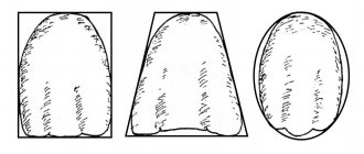
Gum diseases in adults
Gingivitis
Inflammation of the mucous membrane is localized around the diseased tooth; the pathological process can be acute or chronic.
Types of Gingivitis:
- catarrhal,
- atrophic,
- hypertrophic,
- ulcerative-necrotic.
If you start treatment on time, you can defeat the disease very quickly and without consequences in the form of tooth loss. If the situation is neglected, the disease can develop into a more dangerous form - periodontitis.
Periodontitis: what is it?
Periodontitis is a gum disease that destroys the supporting structure of the affected tooth, as well as the ligament between the bone and the root of the tooth. As a result of the inflammatory process, the gum partially peels off from the tooth, forming a gum pocket in which food debris accumulates, which is a breeding ground for pathogenic microorganisms. It is not possible to remove these accumulations with a toothbrush.
Without proper treatment, bleeding and swelling of the gums quickly develops, and pus can form in periodontal pockets. Teeth become loose and eventually fall out. The acute phase is characterized by general malaise, weakness and severe pain in the affected area.
Periodontal disease: photo
Periodontal disease, like periodontitis, affects the supporting part of the tooth, but inflammation does not occur. There is a disruption in the blood supply to soft tissues, their dystrophy develops, the necks of the teeth are exposed, and the gums are slowly destroyed. Periodontal disease does not remain local - over time it affects both jaws, so it is very difficult to fight this disease. For periodontal disease, physiotherapy, local treatment and splinting of loose teeth are effective.
What happens if gum recession is not treated?
- root caries (the root, unlike the crown of the tooth, is not covered with enamel - a dense “pearl” shell, and therefore is more vulnerable). Normally, the root is covered by the gum and is not exposed to the aggressive environment of the oral cavity (acids, humidity, microorganisms, enzymes). With gum recession, the exposed root is susceptible to destruction.
- abrasion of root tissues (again, due to the lack of enamel, tooth root tissues are not resistant to abrasion).
- increased sensitivity of teeth.
- the transition of a recession to a class that is not subject to complete closure.
- more plaque (the surface of the root is rougher than the surface of the tooth crown, which means that plaque is retained more when the roots are exposed).
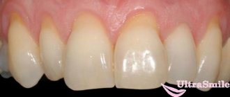
Large gum recession, caries of the exposed tooth root, an abundance of soft plaque on the root surface.
Gingivitis –
Gingivitis is an inflammation of the gingival margin, including the interdental gingival papillae (i.e., that part of the gum that is directly adjacent to the necks of the teeth). The main symptoms of this disease are periodic bleeding and soreness of the gums when brushing teeth, redness or cyanosis of the gingival margin, swelling of the gingival papillae, and often there is bad breath. Gingivitis with a predominance of such symptoms is called “catarrhal” by dentists.
Gum disease: photo of teeth with gingivitis
The cause of the development of catarrhal gingivitis is exclusively insufficient oral hygiene. As a result, soft microbial plaque accumulates at the necks of teeth, and hard dental deposits can also form (we are talking about tartar). In this case, the development of inflammation is due to the fact that pathogenic bacteria that are part of microbial plaque and tartar secrete various toxins and inflammatory mediators, which trigger a chain of inflammatory reactions in the gums.
Therefore, the main treatment for inflammatory gum diseases is always aimed at eliminating the causative factor, i.e. for the removal of dental plaque (+ normalization of oral hygiene). It should be noted that gingivitis is the simplest and most quickly curable form of gum inflammation, if, of course, the treatment is carried out correctly - with regular removal of dental plaque by ultrasound (the frequency of this procedure will depend on the quality of the patient’s oral hygiene).
Important: inflammation in gingivitis affects only the mucous membrane of the gums - without destroying the dentogingival attachment or bone tissue around the teeth (which is typical for a more severe form of gum inflammation - periodontitis). But the transformation of gingivitis into periodontitis is only a matter of time if the patient focuses his treatment on antiseptic mouth rinses and gel applications on the gums, rather than on the removal of dental plaque and high-quality hygiene.
Hypertrophic form of gingivitis –
Catarrhal gingivitis is the most common form of gingivitis, but not the only form. Sometimes patients can experience a hypertrophic form of gingivitis, which is manifested by an increase in the volume of the gingival papillae or the gingival margin as a whole (usually only in the area of the front teeth). Most often, this form occurs in pregnant women and adolescents, and is associated with hormonal changes in the body. There are edematous and fibrous forms of hypertrophic gingivitis.
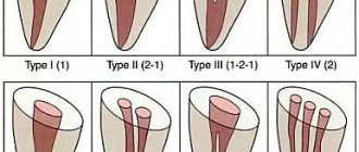
In the edematous form, the enlargement of the gingival papillae occurs due to their chronic persistent swelling, which is practically untreatable. This form can occur not only in pregnant women, but also in patients with long-term catarrhal gingivitis (in the absence of adequate treatment). In the fibrous form, gum enlargement occurs due to the proliferation of fibrous tissue (Fig. 4). You can read more about the forms of catarrhal and hypertrophic gingivitis and their treatment regimens at the link below.
→ Treatment of gingivitis in adults
Preventive measures
To keep your gums healthy, you need to follow the following rules:
- brush your teeth regularly and do it correctly;
- eat a balanced diet;
- promptly treat existing malocclusions;
- undergo preventive examinations at the dentist annually;
- Do professional dental hygiene once a year.
It is also important to quit smoking. Tobacco smoke constricts blood vessels, causing poor blood supply to the gums.
Content:
- The most common gum diseases
- Why gum disease occurs
- How diseases manifest themselves
- Treatment
- Preventive measures
4.1. Oral hygiene 4.2. Physiotherapy 4.3. Anti-inflammatory drugs 4.4. Curettage 4.5. Surgery
Worried about the whiteness of their teeth and the aesthetics of their smile, people often forget about the health of their gums. Meanwhile, among other dental diseases, their pathologies provide significant competition to caries. Moreover, premature loosening and loss of incisors and molars is very often associated with infection of the apparatus that holds them.
Causes of gum inflammation?
After each meal, a small amount of food debris remains on the teeth and between the teeth, which forms a soft plaque. It is difficult to see, but oral microorganisms immediately settle in it. If plaque is not regularly removed from the surface of the tooth, it turns into tartar as a result of the deposition of salivary salts - a substrate for bacterial colonies.
The accumulation of bacterial plaque and waste products of microorganisms in the area of the gingival sulcus leads to gingivitis, subsequently to the destruction of the sulcus, the formation of periodontal pockets, the development of periodontitis and damage to bone tissue. Inflammation of the gums develops faster when the general defenses of the body are weakened, with dental caries, after a tooth injury (for example, with a blow, microtrauma when biting a thread), after unsuccessful prosthetics.
Diseases such as hypertension, diabetes, diseases of the thyroid gland and adrenal glands lead to serious changes in the entire body, including the periodontium. Patients with this pathology should be under constant supervision of a periodontist and therapist. Regular visits to these specialists allow you to successfully treat gum inflammation and save teeth in the presence of general diseases of the body.
Plaque, which is the main cause of gum disease, contains a huge variety of bacteria that can spread to other organs. In this regard, periodontal pockets are centers of chronic infection. It has been proven that the long-term presence of infection in such a formation leads to the development of rheumatoid arthritis, infective endocarditis, gastritis and enterocolitis.