I. K. Lutskaya Head of the Department of Therapeutic Dentistry BelMAPO (Minsk)
HIV infection is a disease caused by the human immunodeficiency virus, which occurs with damage to the immune and nervous systems and is manifested by the development of severe infectious (parasitic) diseases and/or malignant neoplasms, as well as signs of encephalomyelopathy. AIDS (acquired immunodeficiency syndrome) is the final (terminal) stage of HIV infection.
The infectious process in the human body (from the moment of infection with the human immunodeficiency virus until the death of the patient) is characterized by a long incubation period (from several months to 5 or more years), a slow course, and selective damage to T-lymphocytes and neuroglial cells.
General information
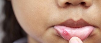
The problem of mycotic diseases in general, including fungal diseases of the throat (pharynx and larynx), becomes important due to the wide distribution and severity of the disease. The risk of dissemination of the process and even the development of fungal sepsis increases. Thus, over the past 10 years, the incidence of candidal pharyngitis ( pharyngomycosis ), laryngeal mycoses ( laryngomycosis ) and candidal tonsillitis have increased sharply - they account for 30 to 45% in the structure of all infectious lesions of the tonsils and pharynx.
Below you can see what a fungus in the throat looks like (photo). The growth of fungal infections of the throat is facilitated by massive antibiotic therapy, taking immunosuppressive and glucocorticoid drugs over a long period of time.
These and a number of other factors create favorable conditions for the development and spread of deep mycoses caused by opportunistic fungal flora, which is widespread in the external environment, can saprophyte the mucous membrane of the throat and was previously considered non-pathogenic.
Pharyngomycosis / laryngomycosis is not a highly contagious disease. In the vast majority of cases, inflammation of the mucous membranes of the throat caused by fungi of the genus Candida is secondary, developing against the background of predisposing factors. Fungal diseases of the throat are easily chronic (especially in cases of unreasonable/inadequate/adequate, but not aggressive enough therapy) and are prone to frequent recurrence. In most cases, they become stubbornly persistent and therapeutically resistant, and in cases of absence/incorrect treatment can lead to severe complications. The symptoms and treatment of fungal diseases of the throat are practically the same when the throat is affected by one or another type of fungus.
Pathogenesis
The pathogenetic mechanisms of fungal infection of the pharynx are based on a restructuring of the body’s immune reactivity (accumulation in the blood and circulation of fungal antibodies), which causes immediate/delayed reactions. The development of throat lesions by fungi is also facilitated by changes in cellular immunity . In the pathogenesis of pharyngomycosis, an important link is specific/nonspecific sensitization of the body and allergy . The factor of traumatization of the mucous membrane of the oropharynx, which contributes to the development of the fungal process, also plays an important role. Tissue reactions are manifested by the formation of granulomas, atypical hyperplasia of the epithelium, necrotic changes of varying severity. Fungal elements in the tissues of the mucous membrane of the throat and the stroma of the tonsils are located predominantly subepithelial. Most often, mycotic foci in the pharynx are localized on the palatine tonsils, posterior wall, arches, velum and tongue.
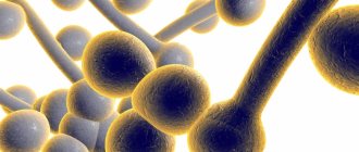
Why are Candida mushrooms dangerous?
The fungus contains several substances that are harmful to our body:
- endotoxins - poisons released during the breakdown of a microorganism after its death;
- aspartyl proteinases and phospholipases are enzymes that cause tissue necrosis;
- oligosaccharides in the cell wall that suppress immune reactions [1].
In addition, the fungus is capable of transforming into a thread-like form (the so-called pseudomycelium), which penetrates into the intercellular spaces and destroys tissue.
Causes
The main causative agents of fungal infections of the oropharynx are fungi of the genus Candida (97%) and molds of the genus Aspergillus (in 3% of cases). Among the fungi of the genus Candida, the yeast-like Candida albicans (39%), C. tropicalis (12%), and C. krusei (9%) were most often isolated in patients. Almost all of these types of listed fungi belong to saprophytes (opportunistic microflora), which are activated when the body’s reactivity decreases and acquire pathogenic properties.
The leading reasons for the decrease in the body's resistance are immunodeficiency states caused by frequent and long-term administration of immunosuppressive/glucocorticoid drugs and massive antibacterial therapy. The development of pharyngomycosis is promoted by systemic diseases of the gastrointestinal tract and blood, diabetes mellitus , bronchial asthma and especially often intestinal dysbiosis , in which the lack of bifidobacteria leads to a decrease in the synthesis of B vitamins and, as a result, the unhindered and rapid colonization of fungal flora not only in the intestines, but also in all other cavities organisms in contact with the external environment (oral cavity, nose).
Occupational hazards include constant contact with gases, dust, and elevated temperatures. Often, due to diagnostic errors and inadequate treatment, an acute mycotic process in the throat becomes chronic.
Risk group and causes
In order for a fungus of the genus Candida to trigger symptoms of inflammation, it is necessary to reduce the protective properties of the body. Therefore, children with reduced immunity are at risk.
- Premature newborns.
- Children with intrauterine growth retardation and congenital defects.
- Birth trauma helps to reduce defense mechanisms.
- Severe infectious diseases.
- An unbalanced, monotonous diet leads to hypovitaminosis and decreased immune status.
- Long-term antibiotic therapy and chemotherapy directly suppress the immune system.
- Metabolic disorders.
- Endocrine diseases.
- Poor hygiene of newborns. The fungus reaches the baby from the mother through dirty nipples, hands, and care items.
The listed reasons for decreased immunity are the most studied. In reality, there are many more such factors. That is why the diagnosis of oral thrush is very common in pediatric practice.
Symptoms of fungus in the throat
Symptoms of fungal diseases of the throat (pharyngomycosis) are variable and are determined primarily by the general/antimycotic activity of the body, and then by the type of fungus that caused the disease. The clinical manifestations of candidiasis , aspergillosis and penicilliosis are practically the same. The acute form of the disease is characterized by a rise in temperature to a subfebrile level, a feeling of dryness/scratching and pain in the throat, hyperemia of the oropharyngeal mucosa, the appearance of necrosis of the epithelium, first white small and then extensive plaques that arise under the influence of a fungal infection, which persists when treated with conservative traditional methods .
Upon examination, multiple, difficult to remove, grayish-white plaques of irregular shape are detected on the mucous membrane, which are located on the palate, tonsils, arches and other places. After their removal, as a rule, erosion remains on the mucous membrane.
In the chronic form, patients' complaints appear cyclically every 2-3 weeks, that is, with frequent alternation of new exacerbations and unstable remissions, depending on the current immune status and other determining factors.
In cases of superficial damage, unexpressed hyperemia of the mucous membrane is determined with areas of translucent/dense deposits of a grayish-white color, having a predominantly lumpy, curd-like character, which are easily removed, exposing the hyperemic smooth mucous membrane. Less often, plaques become denser and merge, and when they are removed, the mucous membrane that bleeds with any damage is exposed (like diphtheria).
With the ulcerative-membranous variant of the lesion, localization of the pathological process is characteristic of the tuberous infiltrated tonsils and adjacent formations. At the same time, the ulcers are covered with an easily removable white coating. The submandibular and submandibular regional lymph nodes are in most cases slightly enlarged and slightly painful. Often lesions are found in other parts of the digestive/respiratory tract ( candidiasis , cheilitis , glossitis ).
Classification of oral candidiasis
The infection can occur in several forms with different symptoms. All of them are collected in a pivot table.
| Form of candidiasis | Preferential localization | External signs | Patient's feelings | At-risk groups |
| Acute pseudomembranous (thrush) | Cheeks, palate, tongue | White grains appear on the mucous membranes, which can merge into a cheesy film. | Burning and dryness in the mouth, pain while eating. | Usually affects children, almost never occurs in adults. |
| Acute atrophic | Cheeks, tongue | The lesions are redness with a smooth, varnished surface without plaque. | Severe pain, the mucous membrane becomes extremely sensitive to any irritants. | After thrush, a course of antibiotics or corticosteroids. |
| Chronic hyperplastic | Cheeks, tongue | White spots and plaques that, without treatment, turn into rough gray films. | As a rule, the course is painless, only some patients complain of pain when eating spicy and sour foods. | Smokers, patients with tuberculosis or blood pathology. |
| Chronic atrophic | Tongue, areas under or adjacent to dentures. | Redness and swelling of the mucous membranes. In later stages, the papillae of the tongue atrophy, and its surface becomes smooth. | Dry mouth, burning, secretion of viscous viscous saliva. | Patients with gastritis or diabetes, elderly people with dentures [2]. |
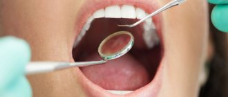
Tests and diagnostics
The diagnosis of mycotic lesions of the oropharynx is based on complaints, examination of the patient’s oral cavity and laboratory data. If upon examination there are changes in the mucous membrane and a curdled white coating is detected, then a diagnosis can be made at this stage. If the picture of the pharynx (with an erosive-ulcerative/catarrhal process) is not specific, then additional studies are prescribed:
- Microscopy of the native preparation (examination of plaque under a microscope in a smear from the oropharynx allows you to quickly visualize fungal cells, spores, pseudomycelium).
- Bacteriological method (inoculation of the contents of the mask on a nutrient medium). Allows you to identify a specific type of fungus and determine its sensitivity to antifungal drugs.
- Immunological examination (detection of pathogen antigens in the blood).
Stages of infection development
The fungus can penetrate into the oral cavity in three ways: airborne, with food, and through contact with a contaminated surface (for example, cutlery) [2]. Microorganisms then go through the following stages:
- attachment to the surface of the mucous membrane;
- emergence of a colony;
- violation of the barrier properties of the mucous membrane;
- penetration into the underlying tissue.
This process occurs most simply with mucous membranes, the epithelium of which is not prone to keratinization—the oral cavity is covered with these [1, 2].
Fungus in a child's throat
The incidence of FM in childhood is significantly higher. Candidiasis of the mucous membrane of the mouth and throat is especially common in newborns (thrush) and in children under 1 year of age. This is due to the incomplete formation of the child’s immune system from exposure to a mycotic infection. Pharyngomycosis also often affects older children, in whom the onset of the disease is caused by fungal infection at an early age and the lack of complete elimination of the source of infection in the oropharynx. Treatment of fungus in the throat in a child is identical to the treatment of adults, taking into account the age-specific dosage of drugs. It is also necessary to take into account the complexity of local treatment in children.
Prevention
Prevention of pharyngomycosis is aimed at increasing immunity and involves the rational and justified use of antibiotics and glucocorticoid drugs. The duration of antibiotic therapy should not exceed the period sufficient to eradicate the pathogen. Antibiotics should not be prescribed for prophylactic purposes, especially for viral respiratory infections. If repeated courses of antibiotic therapy are indicated, mandatory antifungal therapy is required.
Adequate treatment of endocrine pathology ( thyroiditis , diabetes , obesity ) is necessary. Hardening, good nutrition, healthy lifestyle.
List of sources
- Kryukov A.I., Kunelskaya V.Ya., Shadrin G.B. Epidemiology of fungal diseases of the upper respiratory tract and ear. Problems of medical mycology. St. Petersburg, 2011. T. 13, No. 1. — P. 28-31.
- Sergeev A.Yu., Sergeev Yu.V. Fungal infections. Guide for doctors. 2nd ed. -M.: BINOM Publishing House, 2008. - 408 p.
- Kunelskaya N.L., Izotova G.N., Kunelskaya V.Ya., Shadrin G.B., Krasnikova D.I., Andreenkova O.A. Pharyngomycosis. Diagnosis, prevention and treatment. Medical advice. 2013. No. 2. P. 42-45/
- Kunelskaya V.Ya., Romanenko S.G., Shadrin G.B., Krasnikova D.I. Laryngomycosis. Modern approach to diagnosis and treatment. Materials of the XIV Scientific and Practical Conference “Pharmacological and physical methods of treatment in otorhinolaryngology”, Russian Otorhinolaryngology. 2021. 3(82). pp. 38-39.
- Kunelskaya V.Ya., Shadrin G.B., Krasnikova D.I., Andreenkova O.A. Rational methods of treating UDP candidiasis. Advances in medical mycology. 2013. 11. P.99-102.
Prevention of candidiasis
A few simple rules can significantly reduce the risk of developing an infection:
- use of antibiotics strictly according to indications;
- regular sanitization of children's pacifiers, toys, household items;
- brushing your teeth 2 times a day with high-quality toothpaste - for example, Colgate Total 12;
- sanitation of the oral cavity: timely treatment of caries and periodontal diseases, removal of the roots of damaged teeth;
- dental assistance: rational prosthetics, correction of occlusion pathology;
- careful adherence to the rules of care for removable dentures [1].
Candidiasis can cause serious harm to the oral cavity if the pathological process is not eliminated at an early stage. If you notice any symptoms of the disease, you must consult a doctor, who will make an accurate diagnosis and prescribe effective treatment.
List of sources:
- Sakharuk N. A. Candidiasis: etiology, clinical picture, diagnosis, treatment: Monograph. Vitebsk: VSMU, 2010. // URL: https://elib.vsmu.by/bitstream/123/10746/1/Sakharuk-NA_Kandidoz%20etiologiia%20klinika%20diagnostika%20lechenie_2010.pdf (access date 12/10/2020) .
- Molokov V.D., Galchenko V.M. Oral candidiasis. Irkutsk: Irkutsk State Medical University, 2009. // URL: https://ismu.baikal.ru/src/downloads/5eb06a90_kandidoz_polosti_rta.pdf (access date 12/10/2020).
- Weisgeim L. D., Dubacheva S. M., Gavrikova L. M. Complex treatment of oral candidiasis // International Journal of Applied and Fundamental Research. 2014. No. 2. pp. 48-51. // URL: https://applied-research.ru/ru/article/view?id=4692 (accessed 12/10/2020).