- Signs of tonsillitis
- Diseases associated with chronic tonsillitis
- Treatment methods for chronic tonsillitis
- Surgical methods for treating chronic tonsillitis
- Conservative methods of treating chronic tonsillitis
- Physiotherapeutic methods of treating chronic tonsillitis
Chronic tonsillitis is a chronic inflammation of the tonsils.
The palatine tonsils are an organ that takes an active part in the formation of the body’s immunobiological defense mechanisms. There are two forms of chronic tonsillitis:
- compensated
- decompensated.
What are tonsils, structural features
The immune system consists of many components. They participate in the formation of the body’s immune response to pathogenic agents when attempting to introduce them. The tonsils are considered an important part of it. They are accumulations of lymphoid tissue. The tonsils are located in the mucous membrane lining the pharynx. Formations become the first filter on the path of infection into the body.
Together with other accumulations of lymphoid tissue, they form the pharyngeal ring. Tonsils have a complex structure. They are pierced by winding deep channels called crypts. Branching reaches the third or even fourth order. Their number is usually from 16 to 18 pieces. The epithelium covering the walls of the crypts is associated with lymphoid tissue over a large area.
These formations are most developed in the area of the upper pole of the tonsil. In their lumen, desquamated epithelial cells, food debris, leukocytes, and lymphocytes accumulate. Bacteria actively multiply in this environment. This structure promotes the development of chronic inflammatory processes inside the crypts.
Diseases associated with chronic tonsillitis
There are quite a lot of them. Such diseases may be directly or indirectly associated with chronic inflammation of the tonsils. First of all, these are collagen diseases (rheumatism, systemic lupus erythematosus, periarteritis nodosa, scleroderma, dermatomyositis), a number of skin diseases (psoriasis, eczema, polymorphic exudative erythema), nephritis, thyrotoxicosis, peripheral nerve damage (plexitis, radiculitis). Long-term tonsillogenic intoxication can contribute to the development of thrombocytopenic purpura and hemorrhagic vasculitis.
Chronic tonsillitis often causes a prolonged increase in low temperature (low-grade fever), pathological auditory sensations (tinnitus), worsens the course of vasomotor dysfunction of the nose, vegetative-vascular dystonia, vestibular dysfunction, etc.
Chronic tonsillitis has serious complications - these include difficulty breathing due to constant swelling of the nasal cavity and its mucous membrane. It has been established that during the night, about 200 ml of pus enters the stomach, which disrupts the normal functioning of the stomach and intestines. Such patients are at risk of developing a sore throat, which often leads to a peritonsillar abscess. A peritonsillar abscess often develops into cellulitis of the neck, a disease that can be fatal. In total, up to 55 diseases can be identified that arise as a complication of chronic tonsillitis.
Methods for treating inflammation of the tonsils
Chronic tonsillitis is usually treated using conservative and surgical methods, which are used only as a last resort.
The following types of therapy are used:
- antibiotic therapy prescribed by otolaryngologists taking into account the sensitivity of bacteria; treating the throat with antiseptic solutions (“Octenisept”, “Miramistin” and others); antihistamines to relieve swelling of the mucous membrane; immunomodulators to stimulate the immune system; painkillers.
The patient must strictly follow a diet. Exclude solid foods, cold or hot dishes and drinks, alcohol, soda.
When should you wash your tonsils?
The fact is that pathogenic microorganisms and dead leukocytes not only accumulate inside the tonsils, but they also cannot leave their location, since inside the tonsils there are many voids and tortuous “passages”. If you influence them with the same drug, then sooner or later addiction occurs - microorganisms (streptococci and staphylococci) become resistant, adapt to the antibiotic, and it is no longer powerless to cope with them. This is where a special procedure comes to the rescue - washing the tonsils.
Washing the tonsils is prescribed to a person who has long suffered from chronic tonsillitis with constant exacerbations, who feels constant weakness, soreness in the throat, in the cervical and submandibular lymph nodes.
What methods of hardware washing of tonsils are there?
Washing the tonsils is a procedure that is performed only by an ENT doctor. This can be done in several ways:
- Using a special syringe. During the procedure, the doctor uses a syringe with a curved metal tube attached to it. It is introduced into the lacuna. This breaks the traffic jams. The syringe contains a solution of an antiseptic - furacillin, potassium permanganate and others. Liquid is pumped into the lacuna. It pours out, squeezing out pus. Vacuum washing method. Removal of purulent plugs occurs under the influence of ultrasound. Radiation promotes the penetration of drugs into deep lacunar passages. This type of washing can produce almost complete cleansing of lacunae. Cavitation method combined with ultrasound. When ultraacoustic vibrations pass through a liquid, cavities with air are formed in it. When they collapse, a shock wave is formed. It damages the cell membranes of bacteria.
Using these techniques, together with drug therapy, it is possible to achieve a significant reduction in exacerbations of chronic tonsillitis.
Types of pathologies
Tonsils are the first barrier that traps pathogenic microorganisms. Therefore, inflammatory and infectious lesions of the tonsils are quite common, especially among children from 5 to 10 years old.
Angina
The term tonsillitis (acute tonsillitis) refers to acute inflammation of the palatine or pharyngeal tonsils. The main signs of the disease are dry, sore and sore throat, hyperemia and enlargement of the tonsils, increased temperature, and symptoms of intoxication.
There are several forms of angina, most often diagnosed:
- Catarrhal. The symptoms are mild and, with appropriate treatment, disappear within 3-5 days.
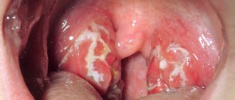
- Follicular. Follicles are affected. A characteristic symptom is pain when swallowing, radiating to the ear. The temperature can rise to 38 degrees or higher; upon examination, enlarged tonsils with whitish-yellow dots are visible.
- Lacunarnaya. The manifestations are similar to follicular, however, the symptoms of intoxication can be more pronounced. A yellowish-white coating covers most of the inflamed tonsils.
Treatment of sore throat involves the use of antibiotics, frequent gargling, and lowering the temperature if necessary. In the first days of acute inflammation, bed rest is necessary; compliance with it reduces the risk of complications.
Chronic tonsillitis
Most often it is a consequence of an untreated form of acute tonsillitis. It occurs with periods of relapses and remissions. A chronic focus of infection often becomes the cause of the development of dangerous diseases such as rheumatism, lupus erythematosus, and nephritis.
The treatment is complex. Antibiotics and drugs that enhance immunity are selected for the patient. If there is no effect from conservative therapy, partial or complete removal of the tonsils is recommended.
Hypertrophy
Tissue proliferation in most cases is characteristic of the nasopharyngeal tonsil. The disease is otherwise called adenoiditis and young children who often suffer from ARVI are susceptible to it. Primary signs are a frequently recurring runny nose, noisy breathing and snoring during sleep, a cough that gets worse at night, and mucous discharge from the nose.
Advanced forms of adenoiditis lead to chronic hypoxia, which causes lethargy and inattention. Lack of timely treatment can cause improper formation of the bones of the facial skull. Depending on the severity of hypertrophy, treatment can be medicinal or surgical.
Abscesses
They are characterized by the formation of cavities filled with pus near the tonsils. Most often, abscesses occur against the background of a sore throat. Symptoms are severe pain on the affected side, fever, high temperature, intoxication of the body. Abscesses are opened surgically.
Neoplasms
Cysts can form on the tonsils. A malignant lesion cannot be excluded, which at an early stage is manifested by tissue enlargement and periodic pain.
There are many reasons for the development of tonsil diseases. The main ones are infection with staphylococci, streptococci, and E. coli. Inflammation can be caused by hypothermia, mechanical injury, or chemical burn. The likelihood of pathologies increases with hypovitaminosis and low immunity. Pathogenic microorganisms can enter the tissues of the lymphatic ring not only directly, but also through the blood during chronic and acute diseases of other organs.
Indications and contraindications for washing the tonsils
Most often, the procedure is performed to treat chronic forms of tonsillitis in adults. The doctor decides whether this procedure is necessary for the patient. The main indication is the presence of purulent plugs in the lacunae of the tonsils.
Contraindications for treatment are as follows:
- state of exacerbation of inflammatory processes; oncological processes; pathologies of the retina; severe heart defects; decompensation of hypertension.
This manipulation is not recommended for pregnant women and children under three years of age.
What is the reason for this difference in approaches in the practice of Russian doctors and their colleagues from other countries?
First of all, the criteria on the basis of which the diagnosis of chronic tonsillitis is made are very different. In Russian medical practice, this diagnosis is often made based on an examination of the throat, even without assessing the patient’s history. In the practice of doctors from European countries, Japan, and the USA, such a diagnosis is rarely made, and based on a number of signs.
Patients who have been diagnosed with this can often hear about a “locus of chronic infection”, the risk of developing other diseases due to the fact that dangerous bacteria spread from the palatine tonsils to other organs and systems. Today this medical concept is outdated. Numerous studies and meta-studies have shown that only one disease has a risk of spreading beyond the tonsils - tonsillitis or, in scientific terms, “streptococcal acute tonsillitis”.
Sore throat is a disease caused by certain bacteria that are easily transmitted from person to person. If a patient suffers from tonsillitis more than 2-3 times a year for several years, the tonsils need to be removed. This is the only measure that, from the standpoint of modern science, can help the patient in this situation and negate the risks of developing other diseases.
Rinsing the tonsils does not reduce the risk of developing tonsillitis.
How is the tonsil rinsing procedure performed?
No special preparation is required before performing this procedure. Washing on this device occurs in several stages:
- first, the tonsils are treated with lidocaine; a vacuum attachment is fixed to the surface of the formation; the device creates negative pressure; the nozzle pulls out purulent plugs and removes them into a special container; the lacunae are washed with an antiseptic solution. Ultrasound injects a drug into the tonsil.
The procedure relieves inflammation, activates tissue regeneration, and removes swelling.
What to do if your throat constantly hurts? Is this an infection?
No, persistent sore throat is most often caused by non-infectious causes. The most common cause is acid from the stomach entering the back of the throat through the esophagus. This condition is medically called gastroesophageal, or extraesophageal, reflux. This is a very common illness that can occur without abdominal pain or heartburn. If heartburn occurs constantly and your throat often hurts, then the connection between these events is obvious and you need to deal with this particular problem, and not try to cure your throat.
Histological analysis of the condition of the palatine tonsils in chronic tonsillitis
Magazine "Medical Council" No. 20, 2021
DOI: https://doi.org/10.21518/2079-701X-2019-20-68-71
V.T. Finger
,
A.V.
Gurov , ORCID: 0000-0001-9811-8397, e-mail,
T.K.
Dubovaya , ORCID: 0000-0001-7936-180X,
A.G.
Ermolaev , ORCID: 0000-0003-2642-5173,
Russian National Research Medical University named after N.I.
Pirogova The article is devoted to the problem of chronic tonsillitis. The purpose of the study was to assess changes in the morphofunctional state of the palatine tonsils under conditions of chronic inflammation. The study presents comparative data from an immunohistochemical study of the palatine tonsils in normal conditions and in cases of chronic tonsillar pathology. We used the clinical classification of chronic tonsillitis developed by B.S. Preobrazhensky and V.T. Finger. In accordance with this classification, histological changes in the palatine tonsils were described in chronic tonsillitis of toxic-allergic form II. The control group consisted of tonsil tissue from patients without chronic tonsillitis. The analysis was carried out using markers of apoptosis, anti-apoptosis, cell proliferation, as well as various types of lymphocytes. The method of working with tonsil tissue, the morphofunctional state in each study group are described, the studied parameters are compared in the two groups, and the dynamics of changes in the tonsils in chronic tonsillitis is reflected. An increase in apoptotic and anti-apoptotic activity in combination with a decrease in cell proliferation, an increase in the number of T-helper cells and B-lymphocytes indicates a significant decrease in the functional activity of the palatine tonsils under conditions of chronic inflammation. These changes are correlated with the clinical picture to develop the most optimal treatment tactics.
For quotation:
Palchun V.T., Gurov A.V., Dubovaya T.K., Ermolaev A.G., Histological analysis of the condition of the palatine tonsils in chronic tonsillitis. Medical advice. 2019;(20):68-71. https://doi.org/10.21518/2079-701X-2019-20-68-71
Conflict of interest:
The authors declare no conflict of interest.
Influence of vitamin D deficiency on progression of inflammation and reparative healing in patients with orofacial region diseases
Vladimir T. Pal'chun, Aleksandr V. Gurov
, ORCID: 0000-0001-9811-8397, e-mail,
Tatyana K. Dubovaya,
ORCID: 0000-0001-7936-180X,
Vladimir G. Ermolaev
, ORCID: 0000-0003-2642-5173,
Pirogov Russian National Research Medical University
The article is dedicated to the problem of chronic tonsillitis. The aim of the study was to assess changes in the morphofunctional state of the palatine tonsils in chronic inflammation. The study presents comparative data of immunohistochemical examination of the tonsils in normal and chronic tonsillar pathology. The authors used the clinical classification of chronic tonsillitis developed by BS Preobrazhensky and VT Pal'chun. Histological changes in the palatine tonsils of patients with toxic-allergic form 2 chronic tonsillitis were described in accordance with this classification. The control group represented palatine tonsils tissue taken from patients without chronic tonsillitis. The examination was carried out using markers of apoptosis, antiapoptosis, cell proliferation, as well as various types of lymphocytes. The authors describe a tonsil tissue handling technique, morphological and functional state in each study group, compared the studied parameters in two groups, reflected the dynamics of changes in the palatine tonsils with chronic tonsillitis. Increased apoptotic and anti-apoptotic activity combined with decreased cell proliferation, increased number of T-helpers and B-lymphocytes gives evidence of significantly decreased functional activity of the palatine tonsils in chronic inflammation. These changes are correlated with the clinical picture to develop the most optimal management of the disease.
For citation:
Pal'chun VT, Gurov AV, Dubovaya TK, Ermolaev AG Histological examination of the palatine tonsils in chronic tonsillitis. Medical Council. 2019;(20):68-71. (In Russ.) https://doi.org/10.21518/2079-701X-2019-20-68-71
Conflict of interest:
The authors declare no conflict of interest.
Introduction
The high frequency of visits to outpatient doctors has persisted for many years, including to otolaryngologists, and a significant part of the visits are repeated. In turn, among ENT pathologies, a significant share is occupied by pathology of the pharynx in general and the palatine tonsils in particular. A large percentage of repeat visits are caused by chronic tonsillar pathology.
Chronic tonsillitis (CT) is a well-studied nosology with developed diagnostic criteria and treatment algorithms. At the same time, some issues of classification of tonsillar pathology and approaches to treatment remain controversial.
In this study, we used the classification of chemotherapy developed by B.S. Preobrazhensky and V.T. Palchun [1]. According to this classification, a simple form of the disease is distinguished, which is characterized by only local manifestations of the disease and conservative therapy is recommended. In the toxic-allergic form of degree I (TAF I) with more severe pathology, in addition to the local signs characteristic of the simple form, there are general toxic-allergic manifestations. For this form, conservative therapy is also recommended, and if there is no effect, surgical treatment. The toxic-allergic form of degree II (TAF II) is characterized by even more pronounced symptoms compared to TAF I and the presence of associated diseases (having a common etiological basis with β-hemolytic streptococcus of group A). For this form of chemotherapy, surgical treatment is recommended.
Considering that CT is a focal infection, timely diagnosis of the form of the disease and the choice of treatment tactics are of great importance, which helps not only to improve the patient’s quality of life, but also to prevent the development of metatonsillar pathology, as well as to reduce the economic costs of treatment associated with repeated visits to the doctor and a decrease in the number of days of disability [2].
There are a large number of methods for conservative treatment of chemotherapy, but the most controversial issue is the need for surgical treatment, namely the moment when it is worth abandoning conservative therapy and performing a tonsillectomy. To answer this question, an in-depth study of the condition of the palatine tonsils is necessary. In this regard, the goal of our work is to conduct a morphofunctional study of the state of the palatine tonsils in normal conditions and in chronic tonsillar pathology.
In the literature available to us, there are a large number of descriptions of the histological structure of the palatine tonsils and the dynamics of the development of pathological changes during chemotherapy. However, in the vast majority of cases, standard histological methods are used, which provide only a general idea of the structure of the palatine tonsils in normal conditions and in chronic tonsillar pathology [3, 4].
Currently, more informative methods are used in histological and pathological studies, in particular immunohistochemical (IHC). There is data in the literature on the use of these methods in the study of chemotherapy, and when they were obtained, no more than 2-3 markers were used simultaneously in one study, which did not allow obtaining a complete picture of the condition of the object of study [5, 6].
In this work, to assess the morphofunctional state of the palatine tonsils, two study groups of patients were formed and, accordingly, two blocks of biomaterial (tonsil tissue samples) were obtained from these patients.
The first group, the control group, consisted of patients who did not have clinical signs of chemotherapy and had a negative microbiological test result for Streptococcus pyogenes. Material for the study was collected during tonsillotomy in patients with papillomas of the palatine tonsils (n = 3) and tonsillectomy during uvulopalatopharyngoplasty (n = 2). The total number in this group was 5 people (of which two men and three women) in the age range from 27 to 46 years, average age 37.4 years.
The second group, experimental, included patients with a clinically diagnosed and laboratory confirmed diagnosis of CT TAF II. Tissue samples for research were obtained during planned and emergency tonsillectomies. The total number of patients included in this group was 42 people (24 men and 18 women) in the age range from 18 to 58 years, average age 35.57 years.
The criteria for inclusion in both groups of the study, in addition to those described above, was the absence of severe concomitant diseases in patients that could directly or indirectly affect the results of the study (diabetes mellitus, angiopathy, immunocompetent conditions, endocrine diseases, cancer, tuberculosis, hepatitis, HIV).
Tissue samples of the palatine tonsils, obtained directly from their removal, were placed in a 10% solution of neutral buffered formalin for fixation in the operating room.
Laboratory stage of the study.
Fixation of the material in a formaldehyde solution for 3-5 days -> dehydration in alcohol solutions of increasing concentration -> clearing in xylene -> embedding in paraffin -> production of paraffin blocks, followed by the production of sections 3-4 microns thick. The sections were placed on glasses coated with an adhesive composition.
The next stage of the work was deparaffinization of the sections (slides were placed in a thermostat at a temperature of 60 °C for 30 minutes, then treated twice with xylene for 3 minutes each, then kept in a 100% alcohol solution twice for 3 minutes, then placed in 95% alcohol solution and gently washed with running water). Wet slides were stained with appropriate IHC dyes. A light optical microscope was used to analyze the obtained sections. Appropriate morphometric assessment methods were used to quantitatively and semiquantitatively assess responses to markers. The area of areas occupied by labeled cells was assessed, which was expressed in points from 1 to 5, as well as by the intensity of staining from 1 to 3 points. The points were summed up. The next stage was statistical processing of the results obtained and their comparison with clinical data.
When staining, monoclonal antibodies were used, which are characterized by uniformity in different batches. This method has significant advantages over the use of polyclonal antibodies.
Each study included positive and negative controls.
The IHC study assessed the biological characteristics of lymphocytes and their subpopulations in the palatine tonsils, the level of their proliferative activity and apoptosis, as well as elements of the microenvironment (Table).
Table. Monoclonal immunohistochemical antibodies used and labeled objects
| Marker name | Index |
| Ki-67 | Cell proliferation protein |
| Bcl-2 | Marker of anti-apoptotic activity |
| Caspase-3 | Marker of apoptotic activity |
| CD-4 | Marker of T-lymphocytes, helpers |
| CD-19 | B-lymphocyte marker |
| CD-20 | Pro-B lymphocyte marker |
Research results
Results obtained in the first group (control). Labeling for Ki-67 according to their localization in lymphoid nodules was identical in both palatine tonsils. High expression of 6-7 points was noted in the dark zones of the germinal centers, and moderate expression of 4-5 points was noted in the light and mantle centers. At the same time, lymphocytes in the interfollicular spaces were negative for the test marker, with the exception of single, diffusely located cells.
When carrying out a reaction to detect the Bcl-2 protein, stained cells were recorded only in the lymphocytes of the mantle zone of lymphoid follicles (2-3 points) in the absence of staining in the germinal center.
A weak IHC reaction of 2-3 points was visualized for caspase-3 in the germinal center and mantle zones of lymphoid follicles.
Low CD4 expression of 2-3 points was noted in the lymphocytes of the germinal center of lymphoid follicles.
CD19 was expressed by lymphocytes of the mantle zone of lymphoid follicles at 3-4 points.
When studying with the antibodies to CD20 used, weak staining of the cells of the germinal center of the lymphoid follicles was noted, 1-2 points.
Results obtained in the second group (experimental). Labeling for Ki-67 in lymphoid nodules showed moderate expression of 3-4 points in lymphocytes of the germinal centers of lymphoid follicles.
A moderate positive reaction to Bcl-2 of 3-4 points was noted only in the mantle zone of lymphoid follicles.
Marking for the detection of caspase-3 is positive in the germinal center and mantle zones of lymphoid follicles with a score of 5-6.
CD4 was expressed mainly by cells of the germinal center with scores of 6-7 and to a lesser extent by lymphocytes of the mantle zone of lymphoid follicles with scores of 4-5.
CD19 was expressed at 3-4 points by lymphocytes of the mantle zone of lymphoid follicles.
In a study using antibodies to CD20, pronounced staining of 7-8 points in the light zone of the germinal center of lymphoid follicles was noted. In addition, high expression was observed in single lymphocytes of the stratified squamous non-keratinizing epithelium of the crypts.
Analysis of the results obtained.
High values of the proliferation index (Ki-67) of lymphocytes indicate pronounced mitotic activity (87.0 ± 0.34%, p < 0.01) in tonsil tissue samples obtained from patients in the control group, and a decrease in this indicator by almost half with chemotherapy (41.0 ± 0.1%, p < 0.01).
Analysis of labeling for Bcl-2 indicated that anti-apoptotic activity prevails in the lymphocytes of the mantle zone of lymphoid follicles in patients of group 2 (43.2 ± 0.44%, p < 0.01) compared to the control group (21, 0 ± 0.1%, p < 0.01).
The indicator of the termination phase of programmed cell death (caspase-3) was most intensively demonstrated by lymphocytes during chemotherapy (53.1 ± 0.1%, p < 0.01) in contrast to cells of biomaterial obtained from patients in the control group (26.2 ± 0 .3%, p < 0.01).
The number of helper T lymphocytes was significantly higher in patients with chemotherapy compared to controls (62.2 ± 0.3 and 28.1 ± 0.1%, respectively, p < 0.01).
The proportion of B-lymphocytes in the biomaterial obtained from patients was almost at the same level in both groups (second group - 38.4 ± 0.1%; control - 42.5 ± 0.2%, p < 0.01).
The number of B-lymphocytes (lymphoblasts), according to CD-20 labeling, was increased in patients of the second group (78.0 ± 0.14%, p < 0.01) compared to the first control (44.0 ± 0.21 %, p < 0.01). This indicates the intensification of lymphocytopoiesis in the territory occupied by lymphoid follicles, as well as the processes of differentiation and proliferation under conditions of antigenic stimulation.
The data obtained indicate a pronounced change in the cytoarchitecture of the palatine tonsils during chemotherapy TAF II. The immunocompetent and barrier functions are significantly reduced, which leads to the formation of a focus of chronic infection in the tonsil.
When comparing the data obtained by immunohistochemical study with the clinical picture of the disease and the corresponding treatment tactics used according to the classification of chemotherapy according to B.S. Preobrazhensky and V.T. Palchun, it is clearly seen that surgical treatment is the most justified for CT TAF II.
Bibliography
- Palchun V.T. Classification and treatment tactics for chronic tonsillitis. Bulletin of otorhinolaryngology. 2013;78(3):8-11. Access mode: https://www.mediasphera. ru/issues/vestnik-otorinolaringolog ii/2013/3/030042-4668201332.
- Bhattachatyya N., Kepnes LJ Economic Benefit of Tonsillectomy in Adults with Chronic Tonsillitis. Annals of Otology, Rhinology & Laryngology. 2002;111(11):983-988. https://doi.org/10.1177/000348940211101106.
- Palchun V.T., Gurov A.V., Guseva O.A. Pathogenetic features of the formation of chronic tonsillar pathology. Bulletin of otorhinolaryngology. 2018;83(2):30-33. https://doi.org/10.17116/otorino201883230-33.
- Preobrazhenksky B.S., Popova G.N. Sore throat, chronic tonsillitis, and associated common diseases. M.: Medicine; 1970.
- Lange MJ, Lasiter JC, Misfeldt ML Toll-like receptors in tonsillar epithelial cells. International Journal of Pediatric Otorhinolaryngology. 2009;73(4):613-621. https://doi.org/10.1016/j.ijporl.2008.12.013.
- Avramović V., Petrović V., Jović M., Vlahović P. Quantification of cells expressing markers of proliferation and apoptosis in chronic tonsilitis. Acta Otorhinolaryngologica Italica. 2015;35(4):277-84. Available at: https://www. https://ncbi.nlm.nih.gov/pmc/articles/PMC4731890.
Are there any contraindications?
Despite the simplicity and apparent safety of washing, the procedure is not allowed for everyone. Contraindications to its implementation are:
- children under 3 years of age;
- pregnancy in the 1st and 3rd trimesters;
- oncological diseases;
- any acute infectious processes in the oropharynx;
- retinal detachment;
- heart defects;
- pathologies of blood vessels and heart in the stage of decompensation;
- intolerance to used antiseptics.
A relative contraindication is hypertension. The patient's condition is assessed individually. If the likelihood of a crisis developing is low, then washing is allowed.
An absolute prohibition is not only acute inflammation of the tonsils. Any inflammatory processes, even caries, when washed, can cause infection of nearby tissues and provoke complications.
In case of chronic tonsillitis, washing the tonsils can be an alternative to removal. Washing the lacunar folds is an unpleasant procedure. But sometimes this is the only alternative to tonsil removal. Regular sanitation cleans cavities, prevents exacerbation of the chronic process and helps restore lost organ functions.