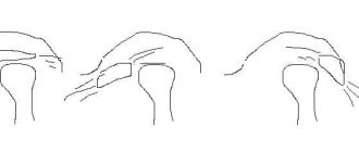
Limited mouth opening, inability to chew food, missing teeth
Before
Stages
After
Specialists:
Nazaryan David Nazaretovich
Description:
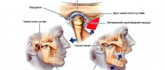
The patient came to the clinic with complaints of pain in the TMJ area, more pronounced on the left side, limited mouth opening, inability to chew food, missing teeth in the lower jaw, and destruction of some teeth. During the diagnosis, an input MRI of the TMJ revealed displacement of the articular discs of the temporomandibular joint without the possibility of reduction. Due to the absence of chewing teeth on the lower jaw on the left side, a decrease in the height of the crowns of the chewing teeth on the right side, and TMJ disease, the head of the joint fell deeper into the articular fossa. Due to the displaced articular disc, the head of the lower jaw began to contact areas not intended for load (hence the pain syndrome) and change its shape in response to contact with the bone tissue of the temporal bone fossa. The displaced disc blocked the opening of the mouth and the ability to move the jaw forward and to the sides.
The first stage of treatment was the production of a therapeutic and diagnostic distraction mouthguard - Splint, which made it possible to move the head of the lower jaw away from the articular fossa. The second stage involved surgical correction of the position of the displaced discs. You can notice changes in the height of the bite in the lateral sections with the correct position of the discs on the heads of the joint. The third stage involved orthodontic correction to ensure support for the TMJ structures using the patient’s own teeth. At this stage, implantation of missing teeth and temporary prosthetics were performed. While wearing the braces system, we were able to regain the lost bite height. After removing the brace system, local prosthetics will need to be performed for long-term stability of the result.
Treatment
Treatment for TMJ can range from conservative dental and medical treatments to complex surgical procedures. Depending on the diagnosis, treatment may include a short course of anti-inflammatory, painkillers and drugs aimed at muscle relaxation, special mouthguards/plates/splint therapy, consultation with medical specialists on psychological problems and stress.
If non-surgical treatment is unsuccessful or if there is obvious joint damage, surgery may be recommended. Surgery may involve arthroscopy and arthrolavage (a technique identical to orthopedic procedures used to examine and treat large joints such as the knee) or removing damaged tissue and replacing it with implants.
After diagnosing and making the correct diagnosis for problems with the temporomandibular joint, our specialists will provide the appropriate treatment necessary for a particular case.
Complaints of headaches, pain in the TMJ area, clicking when moving the lower jaw
Before
After
Specialists:
Nazaryan David Nazaretovich
Description:
The patient came to the clinic with complaints of headaches, pain in the TMJ area, clicking when moving the lower jaw, and lack of stable position of the lower jaw.
From the anamnesis: about 6 years ago, prosthetics were performed in the lateral sections of the upper and lower jaw in another medical facility. Tooth 4.6 was removed for therapeutic reasons (exacerbation of chronic periodontitis).
At the time of examination, the patient had no lateral bite support on the right side and the 8th teeth on the lower jaw stood above the occlusal plane and created traumatic nodes during chewing. This could provoke a forced displacement of the lower jaw.
According to the conclusion of MRI of the TMJ, displacement of the articular discs with reposition was noted. (with restoration of position when opening the mouth)
At the rehabilitation stage, teeth 3.8, 4.8 were removed, the bridge structure on the upper jaw was removed and single composite crowns were made, implantation was performed in the area of the missing tooth 4.6, and a therapeutic and diagnostic mouth guard (Splint) was performed with preliminary diagnosis of the dental system and electrical myostimulation of the masticatory muscle group for fixation true position of the lower jaw.
During the active wearing of Splint and its corrections, the patient’s complaints resolved and subsequently required orthodontic treatment to ensure correct bite and support of the TMJ.
The temporomandibular joint (TMJ) (articulatio temporomandibularis) , formed by the head of the mandible and the mandibular fossa of the temporal bone (Fig. 1-24).
Rice. 1-24. Temporomandibular joint (TMJ) . A: 1 - zygomatic arch; 2 - zygomatic bone; 3 - coronoid process of the lower jaw; 4 - maxillary bone; 5 - second molar; 6 - lower jaw; 7 - third molar; 8 - masticatory tuberosity; 9 - branch of the lower jaw; 10 - stylomandibular ligament; 11 - condylar process of the lower jaw; 12 - anterior (outer) part of the lateral ligament of the temporomandibular joint; 13 - posterior (internal) part of the lateral ligament of the temporomandibular joint; 14 - mastoid process of the temporal bone; 15 - external auditory canal.
B: 1 - sphenoid sinus; 2 - lateral plate of the pterygoid process of the sphenoid bone; 3 - pterygospinous ligament; 4 - spine of the sphenoid bone; 5 - neck of the lower jaw; 6 - sphenomandibular ligament; 7 - styloid process of the temporal bone; 8 - condylar process of the lower jaw; 9 - stylomandibular ligament; 10 - opening of the lower jaw; 11 - wing-shaped hook; 12 - pterygoid tuberosity; 13 - angle of the lower jaw; 14 - mylohyoid line; 15 - molars; 16 - premolars; 17 — fangs; 18 - hard palate; 19 - medial plate of the pterygoid process; 20 - inferior nasal concha; 21 - sphenopalatine foramen; 22 - middle turbinate; 23 - superior nasal concha; 24 - frontal sinus
The head of the lower jaw is a roller-shaped thickening of an ellipsoidal shape, elongated in the transverse direction. The axes, extended along the length of the head, converge at the anterior edge of the foramen magnum, forming an obtuse angle. In front of the head, in the pterygoid fossa, the lateral pterygoid muscle is attached. The posterior surface of the head is slightly convex, triangular in shape, with the base facing upward. The articular surface of the mandibular fossa is 2-3 times larger than the head of the mandible. It has an elliptical shape. The fossa is divided into two parts: anterior - intracapsular and posterior - extracapsular. The incongruity between the head and fossa is leveled by the articular disc and the attachment of the joint capsule on the temporal bone. The intracapsular part of the articular fossa is limited in front by the slope of the articular tubercle, and in the back by the petrotympanic fissure. From the outside, the fossa is limited by the root of the zygomatic process, from the inside by the angular spine of the sphenoid bone. The shape of the mandibular fossa is different and depends on individual development factors, as well as the nature of dental occlusion. There are two extreme forms - deep and flat.
One of the characteristic features of TMJ is the presence of an articular tubercle, which is unique to humans. The articular tubercle, limiting the fossa in front, is a bony prominence of the zygomatic process. There are two extreme forms of the tubercle: a low and wide tubercle corresponds to a flat mandibular fossa, a high and narrow one corresponds to a deep fossa (Fig. 1-25).
Rice. 1-25. Shape of the articular tubercle : a - flat; b - medium-convex; v - cool
The articular disc (discus articularis) consists of fibrous cartilage tissue. It divides the joint cavity into two isolated slits - upper and lower. The disc has the shape of a biconcave lens, in which anterior and posterior sections are distinguished. Between the latter there is a thinner and narrower middle part of the disk. The anterior part of the disc is thicker than the posterior part. Its thickness depends on the shape of the articular fossa: the deeper and narrower the fossa, the thicker the disc, and, conversely, the flatter and wider the fossa, the thinner the disc (Fig. 1-26).
Rice. 1-26. Differences in the structure of the articular surfaces of the TMJ : a - ovoid shape of the condylar process and deep mandibular fossa; b — flat shape of the condylar process and the mandibular fossa: 1 — mandibular fossa, 2 — articular disc, 3 — condylar process; 4 — mandibular fossa (bottom view), 5 — isolated condylar process
Therefore, two extreme forms of the articular disc are distinguished: with one of them, the articular disc is flat and thin, with the other, narrow and thick. The purpose of the disc is to equalize the discrepancy between the articular fossa and the head and, due to its elasticity, to soften chewing impacts. The superior joint space is located between the glenoid fossa and the articular tubercle and the superior surface of the articular disc. The lower articular space is limited at the top by the concave surface of the disc, and at the bottom by the articular head of the mandible. The articulated surfaces in the lower joint gap fit one another more tightly, so it is narrower here than the upper one. The tendon fibers of the lateral pterygoid muscle are woven into the anteromedial edge of the articular disc, thanks to which it can move down the slope of the articular tubercle down and forward.
The articular capsule of the TMJ is extensive and flexible, allowing significant movements of the lower jaw. At the top, the capsule is attached anteriorly along the edge of the zygomatic arch, posteriorly along the fissura petrotympanica, medially along the spina angularis and sutura petrotympanica, then it turns outward and captures the articular tubercle in front. On the lower jaw, the capsule runs along the neck of the articular process, leaving the fovea pterygoidea outside the capsule. At the back, the capsule is thickened, and the extracapsular part of the mandibular fossa is filled with loose connective tissue, forming a maxillary cushion. The TMJ ligaments are divided into intracapsular and extracapsular . The intracapsular ligaments include the anterior and posterior discotemporal ligaments, running from the upper edge of the disc upward and forward and backward towards the root of the zygomatic arch; lateral and medial disconomandibular, located from the lower edge of the disc down to the attachment of the capsule at the neck of the lower jaw. Three ligaments are extracapsular. 1. The lateral ligament (ligamentum laterale) starts from the base of the zygomatic process and the zygomatic arch, goes down to the neck of the articular process. The ligament has the shape of a triangle, with the base facing the zygomatic arch, and consists of two parts: the posterior one, in which the fiber bundles go from above and forward, and the anterior one - the fiber bundles go from top to bottom and back. This ligament inhibits the lateral inward movements of the lower jaw. 2. The sphenomandibular ligament (ligamentum sphenomandibulare) originates from the angular spine of the sphenoid bone, extends downward, attaching to the lingula of the lower jaw. The ligament delays the lateral and vertical movements of the lower jaw.
3. The stylomandibular ligament (ligamentum stylomandibular) runs from the styloid process of the temporal bone down to the posterior edge of the ramus of the mandible. This ligament inhibits the forward movement of the lower jaw. The TMJ is a combined joint. Its articular surfaces are covered with fibrous cartilage. According to the nature of the movements, the joint is classified as block-shaped. The lowering and raising of the lower jaw is possible in the joint. With a slight lowering of the lower jaw, movement occurs around the frontal axis in the lower joint gap, while the head of the lower jaw produces rotational movements along the lower surface of the disc. The forward movement of the lower jaw occurs in the upper gap of the joint. In this case, the head together with the disc is one piece and slides forward and down the slope of the articular tubercle. Simultaneously with this movement, the head of the jaw makes rotational movements in the lower gap of the joint. Lateral movements of the lower jaw occur due to unilateral contraction of the lateral pterygoid muscle and the anterior bundles of the temporal muscle of the opposite side. The angle of deviation towards the lower jaw is 15-17°. The head of the jaw on the side of the contracting muscles makes its way down and forward onto the articular tubercle along with the disc, while turning inward. The movement occurs in the upper gap between the upper surface of the articular disc and the slope of the articular tubercle. In the joint of the opposite side, where the lower jaw has advanced, the head remains in the articular fossa, performing rotational movements around the vertical axis. In addition, the head shifts backward and inward. Movement occurs in the lower chamber of the joint between the lower surface of the disc and the articular head (Fig. 1-27).
Rice. 1-27. Sagittal section of the temporomandibular joint (TMJ)
In the joint cavity there is a biconcave Z-shaped curved cartilage disc. Since the lower jaw anatomically has two joints, it is classified as combined, complex, or biaxial. The movements in it are complex. The structure of the joint allows the lower jaw to perform rotational movements around the frontal axis - lower the jaw (open the mouth) to a distance of up to 5 cm between the front teeth in an adult. Further lowering leads to dislocation. If the mouth opens excessively, the condyle of the lower jaw can slip forward through the tubercle and be fixed in this position by muscle contraction. All this causes dislocation of the lower jaw, which can be on one or both sides. In this position, movements of the lower jaw are impossible, there is no speech, only inarticulate sounds are made. The dislocation must be reduced, and as quickly as possible, otherwise the stretched capsule creates conditions for repeated phenomena. But this must be done by a doctor, since inept reduction can be complicated by a fracture of the neck of the condylar process of the mandible. Since the joints are separated from each other, movements in them can be separate. This is facilitated by the wide capsule and ellipsoidal shape of the condyle of the jaw, i.e. presence of a vertical axis. More precisely, it is possible to move the jaw forward in one joint, but not to do this in the second joint, thus, in a non-adjustable joint, the condyle rotates around a vertical axis. The chin part of the jaw moves in a circle around the center. This displacement is limited by the joint of the opposite side and, above all, by the depth of its fossa of the temporal bone, the severity (height) of the articular tubercle and the strength of the articular ligaments. From the average position, the chin can move to the sides by no more than 15-17°, i.e. by 4.5% of the circumference. In addition to these movements, the jaw can move forward and backward simultaneously in both joints: this movement is called translational. Thus, the TMJ is the only joint that allows translational movements. Combinations of the described movements create the ability to chew, not only squeezing, but also crushing food by the shear type (move the jaw forward, sideways). The relief of the teeth also serves this purpose.
Materials used : Anatomy, physiology and biomechanics of the dentofacial system: Ed. L.L. Kolesnikova, S.D. Arutyunova, I.Yu. Lebedenko, V.P. Degtyareva. — M.: GEOTAR-Media, 2009
You might be interested in:
- Incisors (dentes incisivi). There are 8 incisors located in the middle of the dental arches, they are called the front teeth. There are upper and lower incisors, as well as medial...
">Structure of incisors
- Human teeth are an integral part of the masticatory-speech apparatus, which is a complex of interacting and interconnected organs...
">General structure of teeth
We recommend reading:
- General information about the structure of the human skull. The skeleton of the head is made up of paired and unpaired bones, which together are called the skull, cranium. ...
">Structure of the human skull
- Lower jaw The lower jaw (mandibula) is unpaired, horseshoe-shaped, the only movable bone of the skull. It consists of two symmetrical...
">Structure of the lower jaw
- Upper jaw The upper jaw, maxilla, paired, is located in the center of the face and connects with all its bones, as well as the ethmoid, frontal and…
">Structure of the upper jaw
Constant clicking in the TMJ area when chewing
Before
Frontal photograph of the bite. There is an inclination of the occlusal plane of the upper jaw to the right side
Lateral photograph of the bite (right). Incorrect inclination of the maxillary incisors is noted. (Lower jaw lock). Stamped crown on tooth 4.6
Lateral photograph of the bite (left). Missing tooth 2.6
Photograph of the teeth of the upper jaw. Crowded position of the anterior group of teeth. Rotations of the molar and premolar groups. Erupted teeth 1.8, 2.8 (as one of the causes of traumatic nodes in occlusion). Asymmetrical dentition
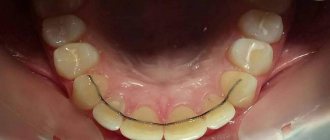
Photograph of the teeth of the lower jaw. Secondary caries under old restorations on the molar group of teeth. lack of correct anatomy of the stamped crown of tooth 4.6. erupted teeth 3.8, 4.8
MRI slice of the left TMJ in the mouth opening position of 54 mm. The head of the joint extends beyond the articular tubercle, but to a lesser extent on the right side. (joint hypermobility)
MRI slice of the left TMJ in the open mouth position 43mm. Reposition of the disc is noted (the disc is in the correct position)
MRI slice of the Left TMJ in the closed mouth position. Anterior displacement of the articular disc is noted
MRI slice of the right TMJ in the open mouth position 54 mm. The head of the joint extends beyond the articular tubercle (hypermobility of the joint)
MRI slice of the right TMJ in the open mouth position at 43mm. The normal position of the articular disc is noted
MRI slice of the right TMJ in the closed mouth position. The normal position of the articular disc is noted
Stages
Frontal photograph of the bite. Beginning of correction of the inclination of the occlusal plane of the upper jaw
Lateral photograph of the bite (right). There is a rise in the bite due to orthodontic onlays
Lateral photograph of the bite (left). There is a rise in the bite due to orthodontic onlays
Photograph of the teeth of the upper jaw
Photograph of lower jaw teeth
After
Frontal photograph of the bite. There is no inclination of the occlusal planes of the upper and lower jaw
Lateral bite photograph (right)
Lateral photograph of the bite (left). The contact density in the molar area will be restored by replacing the temporary crown on the 2.6 implant
Photograph of the teeth of the upper jaw
Photograph of the teeth of the lower jaw. The temporary crown of tooth 4.6 needs to be replaced
Specialists:
Nazaryan David Nazaretovich
Description:
The patient complained of constant ringing clicks in the TMJ area when chewing, opening the mouth and moving the lower jaw. Entry MRI revealed joint hypermobility and disc dislocation with recovery.
After wearing Splint, positive dynamics, and analysis of the results of the control MRI, no indications for joint surgery were identified. The task was to rebuild the patient's bite to support the joint. Treatment with braces lasted 17 months. At this stage, implantation was carried out in the area of 2.6 teeth, removal of all 8 teeth, temporary prosthetics on an implant, as well as restoration of the erased cutting edges of the central incisors. After removing the braces, local orthopedic work will be performed to stabilize the bite.
Temporomandibular joint dysfunction (TMJ)
Causes of joint dysfunction.
The temporomandibular joint (TMJ) is a complex apparatus
consisting of muscles, ligaments, cartilaginous discs, bones. This system provides a movable articulation of the temporal bone of the skull with the lower jaw, facilitating its movement in three planes, back and forth, up and down, left and right. At the same time, the temporomandibular joint is considered one of the most active structures of the human body; it is involved in such important processes as chewing food, swallowing, breathing, yawning, and speech. Any problem that results in a disruption in the normal functioning of a joint is called TMJ dysfunction. According to statistics, such violations are by no means rare. According to various sources, they occur in 30-70% of cases of dental diseases. The dysfunction of the TMJ is based on a multifactorial process. It is believed that the development of symptoms is caused by pathology of the joint itself or damage to the masticatory muscles.
More specifically, the development of negative signs may be due to:
- Anatomical features, discrepancy between individual elements of the articulation, the articular fossa and the head of the articular bone and others
anomalies of the dental system. - Trauma to the joint, facial bones and jaw, their improper fusion after a fracture.
- Malocclusion due to tooth loss, increased wear
, or other factors. - Bruxism, involuntary jaw clenching and teeth grinding;
- overstrain of the jaw muscles due to their overload (professional activity of lecturers, teachers, habit of biting nails, incorrect position of the telephone, long-term, more than three hours, dentist appointment without rest breaks).
- Stress and mental disorders that also affect joint movements.
- degenerative-dystrophic disorders (arthrosis).
- Inflammatory process in the joint (arthritis).
- Endocrine disorders, infectious diseases.
IMPORTANT:
A fairly common cause of TMJ dysfunction is professional errors by dentists. The violation can be caused by incorrect placement of a filling on a chewing tooth or incorrectly performed prosthetics.
Characteristic symptoms.
Diagnosis and treatment of TMJ dysfunctions is difficult, since this type of disorder is characterized by many different manifestations.
At the same time, TMJ dysfunction must be differentiated from a number of other diseases. It can be difficult for patients to understand that the root of the problem is a malfunction of the joint. Specialists at the Dentist clinic often encounter the fact that before getting an appointment with a gnathologist, people underwent long-term and ineffective treatment from neurologists, therapists, and ENT doctors. The following symptoms are considered typical for temporomandibular joint dysfunction:
- Clicking and noise effects in the joint. Joint clicking and other sounds
occurring when moving a joint, chewing, or yawning are so loud that others can hear them. They may be accompanied by pain that is limited to the joint or radiates to the face and neck. - Headache concentrated in the temples, back of the head, radiating to the forehead, neck, shoulder. Such sensations can be so intense that the patient undergoes examinations to rule out dangerous brain diseases.
- Bite problems, manifested by a violation of jaw closure.
- Facial asymmetry.
- Change in the amplitude of mouth opening.
- Jaw jamming.
- Soreness and tension in the jaw muscles.
- Toothache in the absence of manifestations of caries and other dental damage.
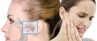
- Signs of damage to the ear area, manifested by pain and ear congestion, hearing loss, ringing and noise in the ears, which is associated with the anatomical proximity of both structures.
Less typical complaints should also be treated with the utmost care:
dry mouth;
burning sensation in the nose, throat, tongue; photophobia; insomnia; snore; apnea (holding your breath during sleep). IMPORTANT:
Because TMJ dysfunction is difficult to diagnose and many dentists are not adequately trained to care for these patients, people may not always be able to get the help they need. To avoid this, if questionable symptoms appear, contact the dentists of the Dentist clinic.
Which doctor treats TMJ dysfunction.
Treatment of TMJ dysfunction is the responsibility of a gnathologist dentist
. Exactly
This specialist, through his actions, helps set the patient’s lower jaw in the correct position, ensuring balanced operation of the entire system.
After conducting a diagnosis, the doctor will determine the causes of failures and suggest the most optimal ways to solve existing problems. Having outlined the correction methods, he will monitor the progress of treatment.
Despite the importance of this dental specialty, not every center is ready to provide a consultation with a gnathologist in Moscow. The issue of conducting diagnostic studies is not always fully resolved. In search of the most suitable center, pay attention to the Dentist clinic. Our center offers a full range of services in this area.
Diagnosis of the disease.
Diagnosis of TMJ dysfunction
is multi-stage and includes a whole
range of procedures
:
- The patient's complaints and anamnesis data are studied;
- The patient's posture, head position, facial symmetry, as well as functions such as breathing, swallowing, and chewing are assessed.
- A study of the closure of teeth in different positions of the lower jaw is carried out; the condition of the masticatory muscles, the tension of the muscles of the head, neck, and back are studied by palpation.
However, purely objective examination and functional tests are not enough. Additional studies are needed
for a complete clinical picture :
- X-ray diagnostics (panoramic image, survey X-ray in different projections, cone-beam and magnetic resonance imaging);
- Functional analysis, which involves taking impressions, making diagnostic models, and analyzing occlusal contacts. Electromyography is prescribed to determine the strength of muscle compression.
All techniques necessary for reliable diagnosis can be fully used by the specialists of the Dentist clinic. In their work, doctors use modern expert-level equipment that provides the most accurate research results. Diagnosis and processing of the results of diagnostic studies takes about a week, after which the dentist-gnathologist prepares a conclusion regarding the nature of the disorders, ways to solve the problem, possible treatment options, and prognosis are discussed.
Treatment methods used.
The goal of treating TMJ dysfunction is to reduce pain, expand the functional activity of the joint, and therefore improve the patient’s quality of life.
and depending on the nature of the detected disorders, conservative, reconstructive and surgical tactics are distinguished. Typically, treating TMJ pain and dysfunction requires a comprehensive approach. It is carried out through the interaction of a number of techniques and the efforts of dentists of different specialties. However, not in all cases, sound phenomena require corrective procedures. Treatment will be required if clicking in the joint is combined with limited mobility and accompanied by pain. Main areas of treatment:
- Elimination of pain, restoration of the normal process of opening the mouth and movements of the lower jaw.
- Bringing the masticatory muscles to normal tone.
- Improvement of the bite, which is ensured by the formation of correct closure of the teeth, restoration of the number of teeth.
Conservative methods include:
- Myogymnastics, when with the help of special exercises it is achieved
improvement of muscle tone; - Psychotherapy, during which a specialist teaches the patient how to properly close the jaws;
- Drug therapy, including non-steroidal anti-inflammatory drugs, muscle relaxants, corticosteroids, botulinum toxin injections;
- Deprogramming of the masticatory muscles.
- Tire installation. This removable device made of polymer material is used to prevent the dentition from closing during sleep, as well as to relax the masticatory muscles and protect against tooth decay during bruxism.
- The use of devices aimed at limiting the mobility of the lower jaw. This is also facilitated by recommendations such as changing the diet in favor of softer foods and reducing speech load.
Reconstructive tactics
in the treatment of these disorders consist of restoring the height of the bite, the number of teeth, and the formation of correct closure of the dentition.
This task is achieved by installing dentures, inlays, using orthodontic techniques, as well as grinding the surface tissues of teeth that prevent the correct closure of the dentition. IMPORTANT:
Treatment for TMJ dysfunction in Moscow can be done at the Dentist clinic. If necessary, some of the activities will be entrusted to dentists of other specialties, orthopedist, orthodontist, periodontist, and surgeon.
Forecast and prevention.
If signs of TMJ dysfunction are detected, it is imperative to
consult a gnathologist and carry out the corrective procedures prescribed by him. If treatment is started in a timely manner, the prognosis is favorable and a lasting improvement in the situation will be achieved.
Ignoring treatment will invariably cause the condition to worsen. Over time, functional disorders will lead to the development of organic degenerative processes, accompanied by immobilization of the joint.
IMPORTANT:
Prevention of damage to the temporomandibular joint consists of taking care of the health of the oral cavity, preserving natural teeth, their timely treatment, and, if necessary, prosthetics.
Advantages of dentistry "Dentist".
In Moscow, a highly qualified consultation with a gnathologist dentist is available
get it at the dental clinic "Dentist". Using the services of our center, each patient can count on high-quality diagnostics using modern techniques, as well as a full range of treatment procedures.
The cost of gnathologist services in Moscow is in a wide price range, but often does not correspond to the professional training of the specialist or the level of diagnosis. If you make an appointment with a gnathologist at the Dentist clinic, then during the free consultation you will be able to verify the professionalism of the doctor and the excellent diagnostic and treatment capabilities of our center. Since doctors use the most advanced technologies in their medical activities, this allows them to achieve success in the most complex cases of pathology.
Dissatisfaction with facial aesthetics, limitations in mouth opening
Before
Frontal photograph of the bite. Cross occlusion is noted on the right side. Absence of contacts of the premolar group on the left side. Tilt of the occlusal plane of the lower jaw to the left
Lateral photograph of the bite (right). Incorrect inclination of the frontal group of teeth in the upper jaw is noted. The lower jaw occupies a posterior (retracted) position
Lateral photograph of the bite (left). The incorrect inclination of the anterior group of teeth in the lower jaw is clearly expressed. Lack of closure of teeth in the lateral region
MRI slice of the right TMJ in the closed mouth position. The normal position of the articular disc is noted. Articular head of regular shape
MRI slice of the Left TMJ in the closed mouth position. There is a pronounced structural deformation of the articular head
MRI slice of the right TMJ in the open mouth position at 34mm. The normal position of the articular disc is noted
MRI slice of the left TMJ in the open mouth position 34mm. There is a limitation in the movement of this deformed joint. The articular disc is not in the correct position
After
Frontal photograph of the bite. Patient with a splint in the postoperative period
Lateral photograph of the bite (right). Ensures uniform contacts in the lateral sections
Lateral photograph of the bite (left). Ensures uniform contacts in the lateral sections
Specialists:
Nazaryan David Nazaretovich
Description:
The main complaints of the patient in this clinical case were dissatisfaction with facial aesthetics (the lower jaw is shifted to the right side) and limitations in mouth opening with pain in the area of the left TMJ. The entry MRI of the TMJ shows a pronounced deformation of the head of the left condyle with unreducible displacement of the articular disc.
The patient was recommended to undergo surgical treatment of the TMJ with preliminary production of Splint in preparation for the operation and subsequent use of this mouth guard in the postoperative period to support the joint structures during the healing period. The second stage of treatment for this patient will be orthodontic treatment in preparation for orthognathic surgery aimed at changing the position of the jaw bones and eliminating asymmetry.
Pain syndrome and restrictions in movements of the lower jaw
Before
Frontal photograph of the bite
Lateral photograph of the bite (right). Absence of tooth 4.5, absence of tooth 1.4 are noted with replacement of the defect with a removable partial apparatus
Lateral bite photograph (left)
MRI slice of the left TMJ. There is a structural change in the shape of the joint head. Anterior disc dislocation without reduction (without restoration)
After
Frontal photograph of occlusion after joint surgery
Lateral bite photograph (right)
Lateral photograph of the bite (left). There is a rise in the height of the bite on the operated side
MRI section of the left TMJ after surgical reduction of the articular disc. The correct position of the disc is noted
Frontal photograph of the bite in the mouthguard (Splint)
Lateral photograph of the bite in the mouth guard (Splint) (right view)
Lateral photograph of the bite in the mouthguard (Splint) (left view)
Specialists:
Nazaryan David Nazaretovich
Description:
A typical complaint for patients with TMJ problems is pain and restrictions in the movements of the lower jaw. Such complaints were also present in this clinical case. The entry MRI of the TMJ shows a pronounced structural change in the shape of the head of the left joint and an articular disc displaced forward without the ability to restore its position.
We carried out preliminary Splint therapy in preparation for joint surgery (we made a transparent rigid mouthguard for the lower dentition) and assessed the result over time with a series of control MRIs of the TMJ. Using the example of a section from one of these studies, one can clearly compare the picture before and after surgery. The patient also noted positive dynamics regarding pain symptoms already at the stage of wearing a mouth guard. In the postoperative period, pain was no longer observed. The range of movements of the lower jaw was restored. Of course, the closure of the teeth changed after placing the disc in the correct position. We will require orthodontic and orthopedic treatment to support the stability of the TMJ.