Teeth go numb quite often. The cause of the symptom is the trigeminal nerve (nervus trigeminus). More precisely, its damage or other violations associated with it. Pathology of the trigeminal nerve most often develops due to serious errors made during dental procedures - installation of dentures, tooth extraction, and their treatment. Numbness can be caused by neuroses, inflammation, or trauma. To eliminate this symptom, various types of treatment may be needed:
- Medicines
- Ultrasonic
- Using laser
- Surgical
- Physiotherapeutic
Next, methods for treating dental numbness will be described in more detail.
Causes
There are a large number of factors that provoke loss of sensitivity in the lower or upper teeth. These include:
- mechanical injuries;
- periodontitis and other tooth root pathologies;
- malocclusion , which disrupts the functioning of the temporomandibular joint;
- errors during implantation and dental treatment;
- damage to the canal in the area of the mandibular nerve;
- stretching, partial or complete rupture of the nerve itself;
- severe emotional stress;
- outflow of blood from the vessels of the facial area;
- syndrome of an allergic reaction to a sharp decrease in ambient temperature.
All of these problems can provoke, in addition to jaw numbness, pain or, conversely, loss of sensitivity in the tongue, cheeks or gums. Excessive salivation may be an accompanying symptom.
Paresthesia is a condition of numbness of the teeth. The pathology is accompanied by burning, tingling, and loss of sensitivity of the lower or upper row of teeth.
Skull tomography, x-rays, and vascular Doppler sonography help identify the cause of the disease.
Treatment includes surgical correction of the nerve, electrophoresis, laser therapy, exposure to diadynamic currents, and UHF procedures.
Any movement of the jaw can be accompanied by unpleasant sensations, including severe pain. If paresthesia is due to the body's reaction to cold, swelling and rashes or blisters may form.
Main causes of nerve damage during implant placement
Damage to the maxillary or mandibular processes of the trigeminal nerve, which is the main cause of long-term numbness of the lips, tongue, and chin after dental implantation, can be caused by an unintentional mistake by the dentist or negligence. Damage to the mandibular nerve, which provokes paresthesia, can occur in the following cases:
- Incorrect selection of implants (chosen too long).
- The specialist has not studied the anatomical features of the jaw and the implanted implant touches the nerve ending.
- Injury to the nerve ending by a needle during the administration of anesthesia or dental instruments during bone preparation.
If numbness in the lower part of the face is caused by injury to the nerve endings during the administration of anesthesia, the discomfort will go away on its own after the nerve fibers heal. Typically, the healing period ranges from 3 months to six months. You can understand that numbness of the lip and chin after implantation is associated with damage to the trigeminal nerve by the presence of the following signs after surgery:
- salivation increases;
- difficulty eating;
- facial expressions and articulation are impaired;
- the sensitivity of the tongue and lips decreases;
- there is pain and discomfort in the implant area;
- numbness is felt on the side where the titanium root is implanted.
Chief physician, Salatsky Dmitry Nikolaevich
Sign up for a free consultation
+7
If the feeling of numbness in the lower jaw does not decrease within several hours after implantation, then an urgent consultation with the doctor who performed the operation is necessary. Only after conducting diagnostic studies will a specialist be able to identify the extent of nerve damage and, based on this, prescribe appropriate treatment.
Diagnostics
In most cases, the dentist determines the cause of the condition visually or after interviewing the patient. However, sometimes additional research is required to clarify the diagnosis:
- tomography of the cranium;
- Dopplerography of blood vessels;
- X-ray of the jaw.
Competent treatment is impossible without establishing the exact cause of the syndrome, so if a doctor prescribes diagnostic procedures, you should not refuse them, otherwise the effectiveness of therapy can be noticeably reduced.
The most serious causes of paresthesia are injuries to the trigeminal nerve as a result of blows, falls and dental implants.
After such injuries, a preventive examination by a dentist is mandatory, even if the patient seems to be fine.Early diagnosis will help eliminate the consequences quickly and with a high degree of success.
Dentist
Novikova Olga Alexandrovna
8 years of experience
Anesthesia during surgery
Numbness prevents you from feeling pain during surgery. Achieved by administering anesthetic drugs. If sensitivity returns and the procedure has not yet come to an end, the drug is added to prolong the effect of local anesthesia.
In rare cases, a substance used for pain relief or improperly administered anesthesia can cause longer-term numbness - up to six months.
Basic treatment methods
Tooth numbness is usually treated by a dentist. First of all, you should find out the cause of the syndrome. If you notice signs of paresthesia, you should make an appointment with a dentist.
Self-medication is dangerous with complications!
Attention
Despite the fact that our articles are based on trusted sources and have been tested by practicing doctors, the same symptoms can be signs of different diseases, and the disease may not proceed according to the textbook.
Pros of seeing a doctor:
- Only a specialist will prescribe suitable medications.
- Recovery will be easier and faster.
- The doctor will monitor the course of the disease and help avoid complications.
find a doctor
Do not try to treat yourself - consult a specialist.
In some cases, the syndrome is corrected without surgery. Sometimes it will be necessary to perform an operation using one of the methods of jaw microsurgery.
The main goal of therapy in both cases is to restore sensitivity to the jaw nerve. It is extremely important to take into account that if the disease continues for three months, the likelihood of successful treatment is noticeably reduced, and if paresthesia persists for more than one year, it becomes almost impossible to completely restore tooth sensitivity.
Why can our articles be trusted?
We make health information clear, accessible and relevant.
- All articles are checked by practicing doctors.
- We take scientific literature and the latest research as a basis.
- We publish detailed articles that answer all questions.
If the face is hypothermic, doctors recommend taking antihistamines and trying to increase body temperature to increase blood flow.
During treatment in a dental office or during the rehabilitation period after surgical correction of the mandibular nerve, physiotherapeutic treatment procedures are of great benefit:
- electrophoresis, which is usually used together with painkillers;
- UHF procedures;
- laser therapy;
- exposure to affected areas with diadynamic currents.
All these procedures are not mandatory and are prescribed based on the doctor’s opinion of their appropriateness.
Helping the patient with numbness after implantation
During a consultation regarding the loss or disappearance of sensitivity after installation of an implant, the dentist first of all finds out the cause. To do this, a diagnostic study is carried out, which makes it possible to determine the type and degree of damage to the nerve fibers. Then a set of therapeutic measures is prescribed.
If therapy begins at an early stage of development of disorders, sensitivity can be restored more quickly. The rehabilitation program for each patient is drawn up individually, with the participation of a dentist, neurologist and physiotherapist.
Treatment usually includes medications and physiotherapeutic procedures, such as acupressure, acupuncture, electrophoresis, and ultraphonophoresis.
Numbness during dental implantation
Incorrectly performed dental implantation is one of the most common factors leading to numbness of the front teeth. Sometimes the main reason is unsuccessful anesthesia, since during the injection process the needle may well damage the jaw nerve and lead to loss of its sensitivity.
The syndrome occurs as a result of erroneous determination of the length of the implant. In this case, after the procedure of its implantation, the nerve is either injured or pinched.
There are several stages of numbness, which can be caused by a dentist’s mistake during installation of implants.
At the stage of neuropraxia, only partial numbness is observed, which usually lasts several hours and disappears either on the day of surgery or the next morning.
At the stage of axonotmesis, symptoms persist for about a month and are accompanied by pain.
The most severe form is neurotmesis, in which numbness is accompanied by the appearance of a scar and disappears no earlier than two months after implantation.
All of these listed forms are not the norm, so the appearance of any stage of jaw numbness should be discussed with your doctor and undergo an additional course of treatment if necessary.
Causes of numbness of teeth and gums
Experts believe that the causes of numbness or paresthesia of the teeth, jaws, and face are: impaired blood supply (ischemia), trauma, inflammation, hypovitaminosis, and intoxication.
Not only the teeth, but also the tongue, cheek, and lips
can become numb, causing burning, pain and discomfort. This unpleasant sensation can spread to the tongue and lips; it is often accompanied by burning, aching pain, and discomfort when touching these areas. Jaw numbness can be caused by:
- Complications after installation of implants. This is a difficult surgical operation, after which various complications, including neuro-dental ones, can occur. Numbness of the teeth in this case can be caused by nerve damage during implantation of the rod, infection of the postoperative wound and resulting inflammation, implant rejection
- Poor quality treatment. When extraction of one or more teeth is difficult - a deformed or twisted root - the result is often numbness. It is caused by the fact that in the area of tooth extraction there is an alveolar plexus or a branch of the nerve, which can easily be affected during complex manipulation. Removing wisdom teeth is always associated with a high risk of nerve damage, which can cause numbness not only in neighboring teeth, but also in part of the gums, lips, and areas of the face. Errors during treatment, such as root perforation, poor quality of materials used, can contribute to the appearance of numbness of the teeth
- Injuries. Injury to the branches of the trigeminal nerve can occur due to hard contact with a hard surface, strong blows to the face, or during ENT or dental operations. In case of accidental perforation of the canals, during the treatment of caries in wisdom teeth, the third branch of the nervus trigeminus may be damaged, which is why throbbing pain and a pins and needles sensation are added to the symptom of numbness
- Anesthesia. If the dose of anesthetic administered before a dental procedure is calculated incorrectly, then the result may also be damage to the nervus trigeminus branch. Such errors are considered medical errors and should not be allowed.
- Neuroses. Neurological symptoms – convulsions, muscle spasms, bruxism. If numbness is caused by neurosis, then an indirect confirmation of this may be the periodic contraction of the jaw in sleep, when nothing affects it and there is no load
- Inflammatory processes. Teeth may become numb due to developing trigeminal neuropathy. The causes of the disease can be otitis media, sinusitis, inflammation of the middle ear, chronic pulpitis during an exacerbation, osteomyelitis, periodontitis, and gum disease. Neuropathy is accompanied by acute pain, with a slight increase, often paroxysmal. Other signs of the condition appear depending on the location of the pathology. Arthritis of the temporomandibular joint, problems with the infraorbital canal can cause numbness
- Lack of vitamins and microelements. In order for the nervous system to function normally, food must be nutritious and rich in essential vitamins and minerals. A sufficient amount of vitamin B intake is especially important. It is its deficiency that can cause disruptions and neurological disorders. Therefore, you need to take care of taking complex vitamins and eating foods containing this element
Trigeminal neuritis, leading to numbness of the teeth, can be caused not only by dental reasons
In addition to the listed reasons, trigeminal neuritis, leading to numbness of the teeth, can be caused by a hypertensive crisis, migraine, allergic manifestations, tumors, osteochondrosis of the cervical spine, and circulatory disorders.
Numbness of the facial muscles can be caused by certain infectious diseases.
Prevention
Since the cause is usually mechanical trauma, there are no special preventive methods to protect against the syndrome. However, to minimize the risks of this problem, you should keep an eye on the following things:
- qualifications of the treating dentist and prosthetist;
- availability of free time for proper rest;
- face protection from low temperatures.
Teeth numbness is an unpleasant syndrome that does not go away on its own. If you delay in contacting a doctor, the changes will develop into an irreversible stage. Moreover, paresthesia can act as a symptom of a serious illness. That is why it is worth taking this pathology seriously and starting to eliminate it in a timely manner.
Why does numbness in the upper or lower jaw persist after implant installation?
Often, after placing an implant, temporary complications arise in the form of loss of sensitivity, aching pain, bleeding and redness of the gums, swelling - all this is a normal reaction to surgery. Usually paresthesia goes away without a trace after 3-5 hours, all other symptoms - after 4-6 days.
There is no need to delay a visit to the doctor if numbness lasts more than five hours . It can occur either due to the effect of anesthesia or due to:
- poor preparation for surgery, incorrect selection of implants, the site of their integration;
- injuries of the mandibular trigeminal nerve.
Solutions to the problem
Treatment of dental stiffness should be carried out taking into account the cause of the disease. Damage to the jaw nerve can also be eliminated non-surgically, it all depends on what factors caused it. The main condition for successful treatment is timely provision of assistance. Other treatment options are microsurgery, physiotherapy, and taking special medications.
If your teeth have been numb for more than three months, it will be difficult to restore their sensitivity. Therefore, the sooner you seek professional help, the better.
In addition to taking special medications, the doctor may prescribe physiotherapeutic techniques to the patient. Main options:
- electrophoresis of teeth with calcium (it is applied to the affected part of the face);
- UHF;
- dynamic currents;
- laser therapy. A similar technique can be used to eliminate caries. Find out more about laser treatment for caries here.
The treatment program in each case is drawn up individually, the doctor considers the advisability of using certain therapeutic techniques for a particular patient.
At the moment, different methods of treating the trigeminal nerve are used, and they all have their own characteristics. In the initial stages, anti-seizure medications are often prescribed to prevent the seizure from progressing. Doctors may prescribe antispasmodics, sedatives, and vascular medications.
Sometimes trigeminal neuralgia is treated with folk remedies - for example, amphibian knotweed decoction or yarrow infusion. You can target the affected area with a laser - this method is applied across the fields in the area where the nerve branches exit the skull, cutaneously.
Surgical treatment of trigeminal neuralgia is quite complex, and doctors do not agree on its effectiveness. The first method of surgical therapy consists of trephination of the cranial fossa from the back and restoration of the correct location of the roots relative to the arteries and veins.
During compression, they are separated - a gasket is placed between the spine and the vessels, which prevents the vessels from affecting the spine. But there is a nuance here - the effect of blood vessels on the nerve is not always the main cause of the disease, so such actions may simply not bring the desired results.
The second method of surgical therapy is percutaneous radiofrequency destruction of the roots. It involves the impact of thermal energy when high-frequency currents pass through biomaterials. This method has a number of advantages compared to the first - the main ones are low trauma and the use of local anesthesia, which means quick recovery.
It is precisely because of the high degree of trauma that surgical methods of treatment are used relatively rarely.
Treatments for tooth numbness depend on the cause. If the jaw nerve is damaged, the problem can be solved with conservative methods, but sometimes surgery will be required. Much depends on how long this situation lasts. For example, if numbness of the teeth lasts longer than 3 months, then it is difficult to eliminate this phenomenon even with the use of modern medications.
For neuropathy, the doctor prescribes anticonvulsants (carbamazepine or its analogues - Tegretol and Finlepsin). For allergic reactions, antihistamines are used, but they are also needed to enhance the effect of anticonvulsants. For this purpose, agents are used in the form of a solution for intramuscular injection, for example Diprazine.
The doctor may prescribe various physiotherapeutic procedures. Options such as ultraphonophoresis (hydrocortisone is administered simultaneously) and ionogalvanization, which requires the use of novocaine, have proven themselves to be effective.
Sometimes surgery is performed to relieve the trigeminal nerve from decompression caused by compression of the vessels.
If the reason is improper dental procedures, you need an anti-inflammatory drug, for example Nimesil. Regardless of the cause of pain, local application of lidocaine or anesthetic ointment is indicated.
If the problem is caused by poor nutrition, vitamin complexes are prescribed, but only based on the results of the examination, when it is determined which vitamins are missing. A special diet is also prescribed, this also applies to allergic reactions.
We suggest you read: Causes of white coating on the tongue of an adult
For injuries, in addition to pain relief, chondroitin and glucosamine are prescribed for faster restoration of cartilage and bone tissue. This is also useful in cases where numbness is caused by osteochondrosis.
Numbness of the jaw is a rare symptom that occurs mainly with damage to nerves, blood vessels and due to dental problems. Most often the lower jaw goes numb, and the patient may experience both minor discomfort and difficulty eating and speaking. If a problem arises, first of all you need to contact a therapist and undergo an examination.
Nerve damage during implantation: diagnosis, treatment, prevention
Damage to branches of the trigeminal nerve (eg, inferior alveolar, lingual, mental, or infraorbital) is a potential complication that can develop during the dental implant procedure.
Direct damage to the nerve fiber can be caused by injury, inflammation, or the result of an infectious factor. Most often, the branches of the trigeminal nerve are affected during anesthesia, flap separation, bone augmentation, osteotomy and direct installation of a titanium intraosseous support. Since the restoration of damaged nerve fibers is quite problematic, the best tactics for treating such complications is prevention. Therefore, it is extremely important for the doctor to understand the features of the histology and anatomy of the nerves of the maxillofacial region, and to be informed about the symptoms that most often accompany their lesions. Also, the clinician must take into account aspects of differential diagnosis in order to correctly establish the cause of the development of certain symptoms, based on which in the future he will have to carry out appropriate treatment. Treatment options for trigeminal nerve branch lesions include the use of various pharmacological agents, monitoring with physical therapy, or even removal of the problematic dental implant.
In this article we will discuss approaches to the treatment of dental patients with damage to the nerves of the maxillofacial area associated with the dental implantation procedure, as well as the main aspects of the etiology and pathogenesis of such pathologies in general.
Anatomy and histology of the trigeminal nerve
The trigeminal nerve is the fifth and largest pair of cranial nerves, which consists of the following branches: the ophthalmic nerve (V1), the maxillary nerve (V2), and the mandibular nerve (V3). The mandibular nerve is the largest branch and innervates the lower lip, chin area, teeth, adjacent soft tissues, lower jaw and part of the external ear. The motor fibers of the mandibular nerve are not damaged during the implantation procedure because they arise from the main branch of V3 before exiting the mental foramen. The main structural unit of a nerve is the nerve fiber. The structure of V3 is dominated by myelinated nerve fibers. Each axon and Schwann cell is covered in connective tissue called the endoneurium. Groups of nerve fibers form bundles that are surrounded by epineurium. Damage to any part of the nerve bundle can lead to neurosensory impairment. The trigeminal nerve consists of 7000-12000 axons, and the number of bundles varies in different parts of the maxillofacial region. The inferior alveolar nerve (IAN) is polyfascicular (consisting of more than 10 fascicles), while the lingual nerve contains only a few similar nerve structures. Since the NAN is characterized by a large number of nerve bundles, its regenerative abilities are also significantly higher compared to the lingual nerve.
Types of nerve lesions
Lesions of the trigeminal nerve can be caused by compression, stretching, complete or partial disruption of the integrity of the nerve fiber. Damage may result in neurosensory changes in touch, pressure, temperature, and pain. Such pathologies significantly affect the patient’s comfort and ability to talk, eat, kiss, shave, apply makeup, brush teeth and drink normally. In addition, neurosensory disorders also affect the patient's ability to interact normally in society. Signs of these pathologies can be identified directly during surgery (if there is a pain symptom), or during long-term monitoring of the patient’s condition. To describe traumatic lesions of axons of varying degrees of complexity, the following terms are used:
- neurapraxia - a lesion in which the integrity of the nerve fiber is preserved, and the mechanism of injury is associated with stretching or impact such as blunt trauma; Sensitivity usually returns to normal within a few days or weeks.
- axonotmesis - damage to the nerve, in which the processes of its degeneration and regeneration develop, but the axon itself does not lose its integrity, and sensitivity is normalized over 2-4 months; however, sensitivity after recovery may be slightly less than before the intervention, and in some clinical cases it is characterized by accompanying dysesthesia.
- neurotmesis - damage to the nerve, in which there is a violation of its integrity, and the prognosis for the restoration of normal sensitivity is unfavorable.
The International Association for the Study of Pain has standardized nomenclature regarding traumatic nerve injuries. In particular, the definition of the term paresthesia, which was previously used to refer to loss of sensation, was changed. Current terminology provides the following definitions:
- paresthesia - a change in sensitivity without accompanying discomfort;
- dysesthesia - a change in sensitivity, which is accompanied by unpleasant sensations;
- anesthesia - loss of sensitivity.
To describe changes in neurosensory functions, terms such as allodynia (the occurrence of pain to stimuli that normally do not provoke pain), causalgia (the presence of persistent burning pain), hypoesthesia (decreased sensitivity to the action of stimuli), hyperesthesia (increased sensitivity to action) are also used. irritants).
When nerves are stretched or compressed, the perineurium protects the bundles from damage. However, lengthening the nerve by more than 30% can provoke structural damage. When the integrity of the nerve is completely disrupted, symptoms of anesthesia and a decrease in certain sensory functions develop. When the integrity of the nerve fiber is partially disrupted, various symptoms of damage, including dysesthesia, may be observed. It should be noted that the presence of persistent pain after surgery is not a criterion for determining the potential for complete restoration of the function of the affected fiber.
After damage to the peripheral nerve, Wallerian degeneration begins to develop, which continues for several weeks and even months. Axonal necrosis develops distal to the site of traumatic intersection. Degeneration in such cases is progressive and irreversible and lasts for up to 18 months. The ability of the affected nerve area to heal is influenced by factors such as the patient's general health, age, and type of injury. A key point in the process of nerve recovery after damage is the formation of scar tissue in the area of endoneurial tubules.
Evaluation of traumatic lesions of the trigeminal nerve
NAN is most often affected during the installation of dental implants. Signs of inferior alveolar nerve involvement include anesthesia, paresthesia, or dysesthesia in the skin, lower lip, cheek, and gums up to the second molar site. Patients with damage to the lingual nerve are characterized by uncontrolled salivation, biting the tongue, a feeling of heartburn, loss of taste, changes in speech and swallowing function, numbness of the mucous membrane and tongue. Both during and after surgery, all potential symptoms of sensorineural impairment should be documented. Areas of altered sensitivity are mapped (both by location and area of the affected area). Thus, it is possible to monitor changes in all parameters in the future, and determine whether the patient needs microsurgical intervention or not. To identify and determine the extent of disorders, both objective and subjective diagnostic tests are used, which are conventionally divided into mechanoceptive (response to mechanical stimuli and compression) and nociceptive (sensation of pain).
Mechanoceptive tests include static touch with a soft brush, two-point discrimination, and determination of the direction of brush movement. The sensation of a needle prick and the recognition of thermal stimuli are classified as nociceptive diagnostic procedures. To compare indicators, not only the affected area is always diagnosed, but also a symmetrical area, thus accurately identifying the fact and degree of neurosensory impairment. If the patient complains of loss of taste, a cotton swab moistened with salt or sugar is used for diagnosis.
Prevalence of traumatic nerve injuries
After implantation, permanent loss of sensitivity in the lip area due to traumatic damage to nerve fibers is observed in 0-36% of clinical cases. However, these data can be considered somewhat outdated and do not correspond to the approaches of modern implantological practice. After all, earlier during operations, dental surgeons more often used vestibular incisions, which caused sensitivity disorders to develop. Today, during the installation of dental implants, midline mucosal incisions are made along the top of the residual ridge, and the entire procedure is pre-planned, taking into account the data obtained after a computed tomographic examination. Thus, it can be assumed that the prevalence of nerve fiber damage due to implantation is significantly less than 36%.
Dannan et al reported that the incidence of nerve damage with implantation was as high as 2.95% (5 of 169 patients treated) in cases of temporary neurosensory changes, and 1.7% in cases of irreversible implant-associated neuropathies. Another study found that the incidence of nerve damage after maxillofacial surgery was 2.69% (42 of 1559 patients), with an even lower percentage of irreversible neurosensory damage, but the exact number was not specified in the study. . In the author's opinion, however, even such rates of implant-associated damage to neural structures are too high for clinical practice. Transient loss of sensation in the lip can often be associated with swelling, which is observed during the first two weeks after surgery.
Traumatic damage to the lingual nerve during surgical procedures
The lingual nerve in the region of the mandibular molars passes through the soft tissues on the lingual side of the jaw. Sometimes the nerve is located coronal to the surface of the bone tissue and is tightly adjacent to the cortical bone plate on the lingual side. Therefore, any surgical interventions must be carried out very carefully in this area. After removal of the third molars of the mandible, lesions of the lingual nerve are observed in 0.5-2.1% of clinical cases. Traumatic disorders of the lingual nerve during dental implantation are not a common phenomenon and are recorded quite rarely. To prevent such complications when installing dental implants, the following rules should be followed: only intrasulcular incisions can be made without releasing incisions and flap separation from the lingual side; During flap separation, it is necessary to avoid overstretching it and maintain a safe distance when performing osteotomy. 90% of all recorded cases of neurosensory changes associated with lesions of the lingual nerve resolve within 8-10 weeks after surgery.
Preoperative planning: prevention of traumatic nerve injuries
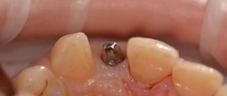
To prevent most complications associated with the installation of dental implants, it is necessary to ensure careful planning of the surgical intervention. Using the capabilities of computed tomography and surgical templates allows you to avoid unexpected outcomes of iatrogenic intervention. When installing a dental implant, a minimum of 2 mm of bone thickness must be left between the apical part and the coronal part of the mandibular nerve canal. In addition, it is important to adhere to the specified osteotomy length and strictly follow the bone preparation protocol. The presence of 2 mm of bone thickness also avoids excessive bone compression in the area of the nerve after installation of a titanium intraosseous support (photos 1 - 2).
Photo 1. The implant was installed in the area of the 30th tooth. After the anesthesia wore off, the patient began to complain of parasthesia in the area of the right lip and chin. On an x-ray taken immediately after implantation, there are no signs of implant penetration into the mandibular nerve canal.
Photo 2. The implant was installed 10 years ago, and during this time the patient was able to adapt to changes in sensitivity. The CBCT image shows that the implant in the area of the 30th tooth is much closer to the nerve canal than previously thought.
If necessary, to ensure the safety of the intervention, short dental implants can be used. It is also important for the doctor to be familiar with the absolute length of all drills that are used during the manipulation, since failure to take these parameters into account can provoke excessive deepening by more than 0.4-1.5 mm relative to the selected safe boundary. To control the deepening into the bone tissue, it is also recommended to use special stoppers. However, the physician must understand that neither the thickness nor the density of the bone tissue over the area of the nerve ensures the safety of its condition during the osteotomy procedure, therefore applying too much force and pressure during the preparation of the bone tissue is strictly prohibited. Finally, it should be noted that up to 50% of lawsuits related to nerve damage after implantation are caused by the lack of informed consent on the part of the patient, which the doctor must obtain before surgery. It is also a good idea to assess the patient’s neurosensory parameters before the intervention in order to compare them with the data that will be obtained after implantation.
Local anesthesia: a potential cause of nerve damage
Traumatic lesions of the mandibular and lingual nerves can occur during anesthesia due to needle trauma, hematoma, and exposure to the components of the anesthetic solution. The mechanisms of such lesions are still quite unexplored. One retrospective study estimated the incidence of nerve injury during anesthesia to be between 1/26,762 and 1/160,5716 cases, while Haas and Lennon predicted the incidence of such complications to be 1/785,000. Other data suggest that the prevalence of short-term transient lesions of the mandibular and lingual nerves as a result of anesthesia ranges from 0.15% to 0.54%. Whereas cases of the development of permanent changes in sensitivity of the same etiology are quite rare, with a prevalence of 0.0001-0.01%. After performing mandibular anesthesia, 3-7% of patients experience electrical sensations that resolve spontaneously over time. However, when the clinician notes that the patient has overreacted to the needle insertion, the needle may need to be withdrawn and repositioned slightly. Methods for the treatment or prevention of nervous complications associated with the anesthesia procedure have not yet been developed. Between 70% and 89% of anesthesia-related neurosensory lesions develop in the region of the lingual nerve. This trend can be explained by the fact that the lingual nerve consists of only a few bundles, while the lower alveolar nerve consists of a huge number of them, which, in turn, increases its potential for regeneration. From a geometric point of view, everything is explained much more simply: the size of the needle is on average 0.45 mm, while the diameter of the lingual nerve is 1.86 mm, and the diameter of the inferior alveolar nerve is 3 mm.
Anesthesia-associated nephropathy most often develops after anesthesia with 4% solutions of articaine or pilocarpine. Compared to lidocaine, pilocarpine and articaine cause 7.3 and 3.6 times more neurosensory impairment. Garisto et al reported that in 4 of 9 studies, the complication rate was higher with prilocaine or articaine 4% solutions than with lower concentration anesthetic injections. According to the authors, local anesthesia with these drugs should be avoided in order to reduce the incidence of associated neuropathies after iatrogenic interventions. However, according to Malamed, the cases in which Arcticaine has demonstrated a greater association with neuropathies than lidocaine are anecdotal, and do not have sufficient evidence-based argumentation. Similarly, in 2013, after an extensive review of the literature, Toma and colleagues concluded that studies suggesting extreme neurotoxicity of articaine were retrospective in design, the data presented were at high risk of bias, and the findings should not be regarded as sufficient evidence. . The authors concluded that direct trauma to the nerve fiber is the predominant cause of neurosensory impairment, and the latter has little to do with the chemical toxicity of the anesthetics used. In general, there is disagreement in the literature on this issue; Therefore, clinicians should make decisions regarding the use of higher concentrations of anesthetics based on the conditions of each individual clinical case, while interpreting prior information and taking into account the recommendations of the drug manufacturers.
Osteotomy procedure for implantation
The osteotomy procedure should be performed using well-sharpened drills and abundant irrigation. Hypothetically, overheating of the intervention area during osteotomy can provoke traumatic nerve damage. The extent of bone necrosis caused by overheating is directly proportional to the preparation temperature under which iatrogenic intervention was performed.
Eriksson and Albrektsson summarized that performing an osteotomy at 47°C for 1 minute may provoke subsequent bone resorption. In cases of progressive resorption, given the shift in the position of the mental foramen, a transcrete incision is contraindicated; instead, it should be shifted to the lingual side. When placing implants anterior to the mental foramen, careful analysis of CT images should be performed, which may help detect the presence of a loop of mental nerve.
Flap separation procedures
As a rule, flap separation does not provoke any neurosensory disturbances, however, the doctor should still pay great attention when performing this iatrogenic intervention in the chin area. The doctor must clearly understand where the mental nerve exits the mental foramen, so that when separating the flap it does not cause damage to the nerve fiber.
Tooth extraction
Before extracting molars and premolars in the lower jaw for subsequent installation of dental implants, the relationship between the position of their roots relative to the course of the inferior alveolar and mental nerves should be carefully studied. Care should also be taken to curettage the sockets after resection, since periapical lesions may often be close to neural structures (Figures 3–4).
Photo 3. The patient sought help for pain in the area of the 31st tooth. The X-ray image shows signs of acute apical periodontitis.
Photo 4. The results of CBCT diagnostics indicate that the pathological focus is located near the nerve canal. The tooth was carefully removed, followed by careful curettage of the defect area.
Pharmacological therapy of neuropathies associated with the installation of dental implants
There is no clear opinion regarding which drugs are best to use in diagnosing traumatic lesions of the nerves of the maxillofacial area. Some authors prefer corticosteroids and nonsteroidal anti-inflammatory drugs (NSAIDs). It is worth remembering that the use of various pharmacological agents is relevant only if the integrity of the nerve fiber as a whole is preserved. In cases of sensory impairment after injection, the patient can be given dexamethasone 4 g/ml directly to the area of the injury, and after 3 days the dose of steroids can be increased. If a nerve is compressed or a fiber is injured during surgery, 1-2 ml of dexamethasone is administered intravenously, after which dexamethasone is prescribed orally for 6 days (4 mg, 2 tablets after meals for three days, then 1 tablet after meals for another three days). If the integrity of the nerve is compromised, steroids may be prescribed for a period of 5 to 7 days. Impaired sensitivity can be relieved by 800 mg of ibuprofen, which should be taken three times a day for 3 weeks. If neuropathy develops after removal of the implant, patients can also be prescribed 800 mg of ibuprofen three times a day and with the same frequency 500 mg of amoxicillin for 5-7 days. In parallel, 50 g of prednisolone is prescribed, reducing the dose by 10 mg every day (for 5 days). Post-traumatic neuropathy can be treated with low doses of antidepressants and antiepileptic drugs.
In combination with a pharmaceutical approach, Renton and Yilma recommend physical therapy interventions for patients with chronic neuropathy. The goal of such a treatment algorithm is to reduce the patient’s discomfort and help him get rid of pain. Bagheri and Meyer generally question the effectiveness of corticosteroids in the treatment of traumatic lesions of the inferior alveolar nerve, since the density of the bone tissue forming the nerve canal is so high that it is simply impossible for medications to reach their space. At this time, there are no results from clinical studies that confirm the feasibility and success of using corticosteroids or NSAIDs for traumatic neuropathies associated with the installation of dental implants.
When to refer a patient to a microsurgeon
Universal recommendations regarding when to refer patients with traumatic trigeminal neuropathies to a microsurgeon have not yet been developed. Some authors argue that the patient should be referred to a specialist immediately after noticing sensory disturbances after the implantation procedure. Other researchers recommend referral to a microsurgeon after 2, 3, 4, or even 6 months of monitoring if signs of traumatic neuropathy are observed during these months. Ziccardi and Zuniga recommend microsurgical intervention for up to 1 year after the implantation procedure, since the effectiveness of microsurgical correction noticeably decreases one year after the intervention. When determining the fact of traumatic neuropathy after implantation, the physician must determine whether the patient needs to be referred to a microsurgeon, or whether the situation can be improved with the use of various pharmacological drugs.
Sometimes explantation, or slight “unscrewing” of the implant from the bone, can help resolve the symptoms of traumatic sensory dysfunction. The results of individual studies indicate that the advisability of referral to a microsurgeon is justified only by stating the fact of a complete violation of the integrity of the nerve. When the doctor is sure that he did not violate the integrity of the bone canal of the nerve, then the disturbance in sensitivity can be caused by compression of the nerve fiber or an inflammatory effect. In such cases, a pharmacological approach to treatment can also help. Meanwhile, the doctor must understand that even if he did not perforate the main nerve canal, the nerve often has additional branches, the course of which is quite difficult to predict. The latter is possible only with a careful analysis of the data obtained after CBCT scanning, because two-dimensional radiographs provoke superimposition of individual anatomical structures, therefore, the prediction of possible traumatic nerve damage with their help is ineffective (photo 1-2).
In cases of discomfort after implant installation, if it can be confirmed that the intraosseous support is not located near the nerve canal, treatment algorithms are variable. Bagheri and Meyer suggest monitoring for 3 to 4 months to see if sensory improvements are observed before referring the patient to a microsurgeon. The authors also claim that if the position of the implant is too close to the nerve, the level of compression of the latter can be reduced by a slight reverse movement of the implant. Other researchers suggest explanting the intraosseous construct within 36 hours and immediately prescribing steroids if the patient exhibits signs of neurosensory impairment after surgery.
Khawaja and Renton described four clinical cases of the development of sensory symptoms after the installation of dental implants, in which the intraosseous structure was not located near the nerve. However, upon explantation, the symptoms of neurosensory disorders resolved quite quickly in two of the four patients. Interestingly, positive dynamics were observed in patients whose implants were removed within 18 and 36 hours, and residual symptoms were observed in patients whose explantation was performed 3 and 4 days after surgery.
Renton, Dawood and colleagues suggest that patients with such symptoms should be monitored for no more than 3 months, since after this period the risk of developing neural changes outweighs the potential success of microsurgery. Ziccardi and Steinberg in their review article recommend initial monitoring for 1 month, and if symptoms improve, continue to monitor the patient. If the symptoms do not improve or worsen, it is necessary to refer the patient to a microsurgeon. Research results indicate that patients operated on for neurosensory impairment 6-8 months after implantation have the same level of success as patients operated on in a shorter period after iatrogenic intervention. According to the authors, earlier intervention is possible and more effective, but there is no evidence to support this assumption. In addition, microsurgery is not indicated in cases of only minor neurosensory changes, since the risk from the intervention itself in such cases exceeds the predicted success rate. There is also a 12-week waiting period - on average, this is the amount of time a doctor needs to decide whether to refer a patient for microsurgical surgery.
It is logical that in cases where patients have acute pain, it is not necessary to wait 12 weeks. Before arguing for certain approaches to treatment, the doctor must objectively compare X-ray data and clinical symptoms, and then take into account all possible medico-legal aspects of the violations.
Surgical restoration of damaged trigeminal nerve
There are specific reasons that justify the need for microsurgical restoration of the damaged trigeminal nerve, and contractual factors that determine the prognosis of this manipulation. Ziccardi and Zuniga formulated the following list of reasons that justify referral of a patient to a microsurgeon: sensory impairment that persists for more than 3 months and interferes with the patient’s normal life activities, confirmation of the fact of nerve transection, lack of improvement in signs of hypoesthesia, development of pain after implantation. After microsurgical correction is performed, a number of factors influence the success of it: the waiting time after implantation, the type and volume of the lesion, the degree of vascularization of the affected area, the experience of the surgeon, the method of harvesting and preparing the graft, the presence of tension in the repair area, the age and general health of the patient.
In fact, microsurgical restoration of the branches of the lingual and inferior alveolar nerves is possible. However, the success rate of such manipulations is very variable - on average, researchers indicate only 50-59.4% effectiveness of such treatment, and in only two studies the results of the intervention reached levels of 81.7% and 63.1%. In all the studies conducted, the number of subjects is quite small, so it is methodologically impossible to compare the final results of such studies. However, 50-60% of patients after microsurgical correction show signs of improvement in neurological impairment. Ziccardi and Zuniga warn that patients with severe sensorineural impairment should be informed that full rehabilitation is virtually impossible. Also, many researchers indicate that the effectiveness of microsurgical procedures in cases of treatment of anesthesia, dysesthesia and spontaneous pain is exaggerated. To summarize, microsurgical correction can help individual patients, but the level of predictability of the results of such treatment is ambiguous. Thus, the best treatment for neurosensory disorders associated with implantation is their prevention.
Conclusion
A common dilemma in clinical practice is: if an implant is successfully osseointegrated but causes mild paresthesia without pain, what should be done with it? After all, explantation may not always help resolve symptoms, and retention of the implant in the bone with actual nerve damage can trigger the development of neuroma. The latter is formed as a result of excessive healing of the area of the damaged nerve and hyperplasia of adjacent tissues, and very often requires subsequent surgical removal. The decision to choose a possible treatment method should be made together with the patient after a thorough discussion of all possible options, and before starting rehabilitation, the patient must formally confirm his consent by filling out a special written form.
Authors: Gary Greenstein, DDS, MS Joseph R. Carpentieri, DDS John Cavallaro, DDS