To understand postoperative sutures, you first need to understand what osteoplasty is and one of its types - sinus lifting. Osteoplasty is a surgical intervention in which bone tissue grows at the site where there was a decrease in bone volume and/or injury. Most often, osteoplasty is performed during dental implantation. And the sooner bone plastic surgery is performed after tooth extraction, the better the recovery effect will be and the easier the preparation for manipulation will be. The following types of osteoplasty are distinguished:
- Osteoplasty affecting the mandibular bone in its lateral parts;
- Osteoplasty affecting the maxillary bone and its alveolar process;
- Osteoplasty affecting the maxillary bone in its lateral parts, or sinus lifting
Osteoplasty of the lateral sections of the manidibular bone is used in cases where the width and length of the alveolar processes decrease and the distance to the mandibular canal decreases. If the amount of bone tissue there decreases significantly, this will lead to malocclusion, of the mesial type.
Osteoplasty on the alveolar process of the maxillary bone is associated primarily with the splitting of bone tissue and the subsequent formation of a sharp, fragile, thin alveolar ridge. The main difficulty of this operation is to achieve the most beautiful cosmetic effect.
Sinus lifting is currently the most popular type of osteoplasty, and is called so because the operation is performed in the area of the sinuses or maxillary sinuses. It is carried out according to the closed and open type, and the process differs only in the method of introducing the implant into the site of future tissue growth.
Classification of postoperative sutures
How quickly the sutures heal after surgery largely depends on the nature of their application and the materials used. In this regard, post-surgical procedures are usually classified as follows.
- Bloodless (the edges of the wound are glued together with a special plaster) and bloody (a classic suture that is applied manually with a medical instrument). In turn, the latter are divided into:
- simple knots (applied at a distance of 1–2 cm from each other, after which the knot is tightened until the edges of the incision touch);
- intradermal continuous (considered the most effective, since after their healing there are no traces left);
- mattress (applied after abdominal surgery);
- purse string (used in plastic surgery, as well as in operations to reduce the volume of the stomach);
- entwining (circular sutures that are used to sew together blood vessels and hollow organs).
- Manual (applied with a needle, thread and other special tools) and mechanical (performed with a medical stapler).
- Submersible (applied during operations on internal organs with threads that are absorbable or implanted into living tissue) and removable (they are used to stitch the skin, and after the edges of the wound have fused, the threads are removed).
Absorbable sutures are made in cases where long-term fixation of the edges of the incision is required, for example, when cutting the uterus during a cesarean section. As a rule, they are performed with threads from purified connective tissue, which is subsequently rejected into the organ cavity. To apply removable sutures, threads and other fasteners made of cotton, silk, metal and other non-absorbable materials are used (more than 30 varieties in total).
Hand stitching tools
Materials used in osteoplasty
Osteoplasty uses both natural implants and artificial ones (allosynthetic materials). Natural ones include:
- Autogenous grafts are those that were taken from the same patient and transplanted into him. This material survives better than all other grafts, but you need to understand that another operation will be performed to remove tissue from the donor site.
- Allografts are material that has been taken from a donor's dead body for medical purposes. The material is carefully processed, disinfected, it perfectly restores bone tissue and is used in osteoplasty with great success.
- Xenografts are material taken from certain species of animals and corals. It is also carefully processed, sterilized and prepared for further use in invasive dental procedures associated with implantation.
Synthetic (alloplastic) implants are made on the basis of calcium phosphate or hydroxyapatite; they are also widely used by all specialists around the world.
How to care for the suture after surgery?
Once the edges of the incision tighten, there will be no need for additional support. Removal of sutures in the head, face and neck area occurs already on the 5th day after the operation. If they were applied in the area of the torso or limbs, then it will take at least 10 days for the wound to heal. Daily dressings are necessary for the first few days. The patient usually spends this time in the hospital. After discharge, tight bandages are usually no longer needed. But if necessary, you can always change the dressing at the nearest hospital or medical center.
Caring for the suture after surgery consists of daily treating the incision area with an antiseptic and taking medications that accelerate tissue regeneration. All medications for home therapy are used strictly according to the doctor’s recommendation!
Treatment of sutures is usually carried out with ready-made pharmaceutical preparations or homemade antiseptics, such as solutions of iodine, potassium permanganate, brilliant green or hydrogen peroxide. To avoid getting a chemical burn when performing such procedures, the liquid for disinfection should be prepared only according to a prescription issued by a doctor.
To speed up regeneration processes, external agents with wound healing and antibacterial effects are used. These include balsamic liniment (better known as Vishnevsky ointment), levomekol, ichthyol ointment and many others.
Pain of varying intensity after surgery is absolutely normal. If discomfort is severe, analgesics approved by your physician may be used.
Techniques used in bone grafting
- Alveolar process resorption is used for horizontal bone splitting when it is necessary to increase the density of the alveolar process. It is performed on both jaws, and today is the most popular method for increasing the volume of the alveolar process, while having a low cost compared to more expensive procedures. Again, there are variations of this technique, but it’s worth paying attention to “Split Control”. The technique makes it possible to simultaneously expand the space and install an implant.
- To increase the volume and length of the alveolar process, a bone block transplantation technique is used. Autogenous material is more often used here. The principle is to screw a bone block taken, for example, from the maxillary region of the zygomatic-alveolar ridge to the bone, using medical mini-screws made of titanium alloy. Then it’s all covered with bone shavings and covered with a connective tissue membrane, which is secured. Subsequently, the surgical wound is tightly sutured with synthetic thread. The disadvantages of autogenous material were discussed above, which should be taken into account when choosing therapy.
- Guided tissue regeneration is a method in which it is possible to both increase the height and expand the volume of the alveolar processes. This technique also involves the simultaneous installation of an implant.
The guided bone regeneration technique includes two components:
- implanted bone material;
- a special barrier membrane that protects against the effects of various infectious agents and other unpleasant factors.
It is important to understand and take into account that the technique of directed bone regeneration does not always provide a sufficient effect. When using this method, bone material is “planted” on the outside of the cortical plate of the upper or lower jaw. The bone structure is very different from its own and is prone to incomplete splitting. Therefore, such an operation should be performed by an experienced doctor who knows all the nuances of the technique and can adequately assess further treatment tactics.
Additional recommendations for suture care after surgery
In addition to medical procedures, certain lifestyle adjustments are also necessary in the postoperative period. In particular, the following rules must be adhered to:
- Limit physical activity. Any high and medium intensity exercise (running, aerobics, etc.) during this period is strictly contraindicated.
- Avoid heavy lifting. As a rule, a person who has undergone surgery should not lift more than 2–2.5 kg. This rule is especially relevant when caring for a suture on the abdomen after surgery.
- Limit mobility in the waist area (avoid sharp and high-amplitude bends and turns of the body) after abdominal surgery.
- Carry out water procedures with great care. Before the stitches are removed, or better yet, before a scar forms, it is strongly recommended not to wet the wound.
- Avoid any pressure on the wound. When it comes to how to care for a suture after surgery, this is one of the main rules.
- In cases where surgery was performed on an arm or leg, the injured limb should be placed above the level of the heart (for example, placed on a pillow) during sleep. If the wound is above the level of the neck, then it is better to sleep with the head of the bed raised by 45° (two pillows are enough for this).
- During abdominal operations, observe bed rest until the doctor allows you to break it. To avoid bedsores and improve blood circulation, you can perform simple movements such as lifting your limbs and performing light self-massage.
- Strictly follow the diet prescribed by your doctor (especially after abdominal surgery).
- Protect the wound from exposure to direct sunlight. After the tissue has healed and until a full-fledged skin forms in this area, you can use sunscreen.
Recommendations for patients
To ensure that the recovery process takes little time and goes without complications, it is recommended:
- Follow all doctor's orders.
- Do not eat anything for the first few hours after surgery. In the first two to three days after therapy, chew food with the healthy side of the jaw and give preference to soft foods.
- Do not touch the unhealed hole with your hands.
- Do not try to remove a blood clot with your tongue or foreign objects.
- Rinse only if prescribed by a doctor.
- Avoid strenuous physical activity for the first two weeks.
- Do not attempt to remove stitches yourself.
By following these simple rules, a person can quickly return to their normal lifestyle after dental surgery.
In what cases should you consult a doctor?
Seeking help from a doctor during the healing process of a suture is a completely normal practice, even in the absence of serious problems. And in the event of adverse reactions due to a violation of the treatment regimen or any unforeseen circumstances, it is absolutely impossible to delay it. First of all, it is dangerous to ignore the following symptoms:
- bleeding that cannot be stopped by conventional means;
- high temperature (more than 38 degrees);
- weakness, chills;
- increasing pain or other progressive discomfort that cannot be relieved with medications;
- purulent discharge of a bright yellow or green color with a thick consistency and very often with an unpleasant odor;
- severe redness, swelling, or swelling in the wound area;
- the skin at the site of injury is hard and hot to the touch;
- the appearance of a rash or blisters;
- Suspicion of seam dehiscence.
The need for suturing in dentistry
Surgical intervention and, as a consequence, suturing are indicated for the following manipulations:
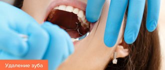
- Removal of wisdom teeth - third molars differ from the rest in the large size of the tooth crown and its root. Late eruption, the position of the “extreme” in the dentition very often leads to a violation of the physiological location of the tooth or even its impaction (partial or complete failure to erupt). In this regard, the procedure for removing a wisdom tooth is often accompanied by complications - extensive damage to the soft tissues of the oral cavity and alveolar process, ligaments, and blood vessels. To avoid this kind of consequences, dentists apply stitches to reduce the wound.
- Implantation - its implementation is always accompanied by the need for surgical intervention. A deep incision in the gum requires suturing.
- Maxillofacial operations - elimination of the consequences of traumatic injuries, congenital defects of the dentofacial apparatus.
- Removal of tumors of various etiologies - the integrity of the soft tissues of the oral cavity is damaged, often requiring layer-by-layer sutures.
How long does it take for a wound to heal after surgery?
The rate of healing of a postoperative wound depends on many conditions. Among them:
- age;
- body mass;
- state of immunity;
- state of the cardiovascular system.
On average, it takes about 3 months from the moment of surgery to the formation of a scar. Depending on the complexity of the operation and if there are complications, this period may last 12 months. Tissue regeneration takes place in 4 stages.
- Inflammation (5–7 days). The body's standard defense reaction to damage. During this period, there is an increased production of substances that stimulate blood clotting.
- Polyferation (from 10 days to 1 month). At this stage, the formation of young connective (granulation) tissue, penetrated by a dense network of microvessels, occurs. At first it is bright red in color and grainy in consistency, but as the wound heals it becomes pale and smooth, and its bleeding decreases.
- Epithelization (from 1 to 3 months). The connective tissue is finally formed. Skin begins to form at the site of the wound. The number of vessels decreases, a scar forms.
- Scar formation (from 3 to 12 months). Temporary vessels completely disappear. Fibers of collagen and elastin - elements of connective tissue - form the scar.
Sinus lift
Osteoplasty, which is performed in the lateral parts of the upper jaw, is called sinus lifting. The operation time in professional hands will be about 15 minutes. And here it’s not a matter of haste, but of the level of the specialist. The operation is carried out as follows:
- The membrane of the maxillary sinus is lifted with a special instrument;
- The doctor inserts a previously prepared graft into the newly formed space;
- End of surgery.
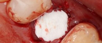
After surgery, the volume of the maxillary sinuses decreases slightly in size, but this does not interfere with breathing function. There are two types of sinus lifting: open type and closed type. Open involves cutting and folding back a flap of soft gum tissue, and subsequent drilling of the maxillary sinuses. Finally, stitches are placed on the gums for effective wound healing. The closed sinus lift method is a more gentle method that does not require subsequent stitches. But there is a condition that is important to comply with. The height of the bone must be at least 4 mm at the site proposed for implantation.
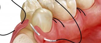
Sutures after bone grafting and sinus lifting are always placed tightly and firmly using special threads. They are divided into 2 types:
- Absorbable;
- Non-absorbable.
The first includes the legendary catgut, the sutures of which can remain in the oral cavity for up to 2 weeks. To prevent them from coming undone, they are tied with surgical and conventional knots. It happens that it can cause a local inflammatory reaction, as it is a foreign protein-containing material. But it is better not to use catgut threads if a local inflammatory reaction is possible. For example, this is bone tissue transplantation, the use of membranes that have tissue restoration properties in dental implantology.
Synthetic sutures for bone grafting and sinus lifting are made of Dexon and/or Vicryl. They are easier to use than catgut, but require knowledge in tying surgical knots. Resorption usually occurs after 1 month, they do not cause inflammation, but they need to be removed a maximum of 10 days after surgery.
The non-absorbable material primarily includes silk, which is produced from a protein secreted by the silkworm. This thread is easy to tie, it is strong and pliable, it is better to use surgical knots. But there is also a drawback - such threads often cause inflammation of the oral mucosa. It's all about a foreign protein that causes an inflammatory-allergic reaction. The next type of thread is braided polystyrene thread; its strength is comparable to silk, but does not cause tissue inflammation.
For fixation, a few knots are sufficient, which are tied on opposite sides and supplemented with surgical ones. Such threads are coated with silicone, polybugylate or polytetrafluoroethylene solutions, which reduce inflammatory reactions and give them elasticity. Monofilament sutures are another type of non-absorbable sutures that are a single thread made from polytetrafluoroethylene. They have good mechanical, anti-inflammatory and adaptive properties. They are often used in the application of periodontal membranes. But the ends of the threads must be covered with a bandage, as they have hard ends that can damage the mucous membranes of the lips and cheeks.
How to care for a wound when the stitches are removed after surgery?
It would be useful to remind you that stitches should only be removed by a specialist - a doctor or nurse. It is strictly forbidden to perform this procedure yourself, due to the high risk of causing infection in the wound or causing bleeding.
Treatment of the incision site after removal of the sutures is carried out using the same means as before. Treatment procedures last until the wound is completely healed. This usually takes about 1 week.
Precautionary measures
If you feel severe pain or see blood while removing a suture, stop immediately. This means that the wound has not yet healed and it is dangerous to touch it, otherwise the edges will separate. Pain, tissue inflammation, suppuration or increased temperature after removal of the threads are a dangerous sign. It indicates that the tissue has become infected and requires immediate medical attention.
We categorically do not recommend removing stitches at home, especially if you are not confident in your abilities. Contacting specialists Dr. Mos will relieve you of discomfort and unwanted consequences of the procedure.
Author: Ph.D. surgeon Samsonova G.S.
Professional care for postoperative sutures at Stoletnik MC
If in any matters related to taking care of your own health, you prefer to trust professionals, you are welcome at the medical office. To help those who are not sure how to properly care for a suture after surgery, a wide range of post-operative services is available. Among them:
- dressing large and small;
- removal of stitches up to 5 cm;
- removal of sutures 5–10 cm;
- removal of stitches more than 10 cm;
- sanitation of the wound surface;
- scar excision;
- and much more.
The responsibility and high professionalism of the clinic’s staff is the key to the safe and speedy recovery of our patients. And the affordability of the services provided will eliminate the need to waste time and nerves on trips to the clinic. Sign up for procedures by phone: +7 (8412) 999-395, 76-44-20. We are waiting for you at the address: Penza, st. Chaadaeva, 95 (Shuist microdistrict).
Healing time for external sutures on the perineum
It's no secret that all mothers who have just given birth are interested in the question of how long postpartum sutures last. The healing process is directly related to the size of the wound surface, the correctness of care during the recovery period, the general condition of the woman’s body, and the techniques and materials used when suturing. If self-absorbable threads (natural or synthetic) were used during suturing, healing will take about two weeks, and scar formation when using metal staples or non-absorbable threads takes longer - 15-30 days; in the latter case, the sutures are removed in the maternity hospital before discharge, approximately on the seventh day after birth.
Literature
- Belyaev A.N., Kozlov S.A., Taratykov I.B., Novikov E.I. Patient care in a surgical clinic: textbook. allowance. – Saransk: Mordovian University Publishing House, 2003. – 136 p.
- Buyanov V. M. Egiev V. N. Udotov O. A. Surgical suture. – M.: Antis, 2000. – 92 p.
- Zoltan J. Operating technique and conditions for optimal wound healing. – Budapest: Publishing House of the Academy of Sciences of Hungary, 1983. – 175 p.
- Mironova E. N. Fundamentals of physical rehabilitation. – M.: MOO “Academy of Safety and Survival”, 2021 – 310 p.
- Semenov G.M., Petrishin V.L., Kovshova M.V. Surgical suture - St. Petersburg: Peter, 2001. - 256 p.
Author: Korolev E. S.
Reviewer: reflexologist Kurus A. N.
How to remove suture threads from a tooth socket
Many people associate the removal of sutures with unpleasant pain. Pain occurs only in people with a low pain threshold, increased sensitivity, or when there is inflammation in the tooth socket. Usually the patient experiences only discomfort.
Stages of removing suture material:
- treating the patient's oral cavity with an antiseptic;
- examination of the mucous membranes for hyperthermia, swelling, suppuration;
- the use of topical anesthesia to prevent even minimal pain;
- cutting the threads with a special dental instrument - the stitches are divided in half, especially in the case of an intermittent suture line;
- removing pieces of thread with tweezers;
- checking the density of the scar and the quality of wound healing.
Next, the patient will be asked to rinse his mouth with an antiseptic composition.
The first three days after the stitches are removed, stiffness of movements and discomfort when opening the mouth are possible.
How do you take pictures on your face?
Facial surgeries are some of the most difficult, especially when surgical material is required. You always want to maintain a beautiful appearance, and scars are far from the best decoration.
If the wound is closed correctly and in a timely manner, then there are practically no scars left, so in this matter the main thing is to trust a good specialist.
On the face, stitches are usually left in place for about a week.
Plastic surgeon John Burns
How are sutures removed after eye surgery? Essentially, the removal technology is the same everywhere, as long as they are done superficially. If they are made specifically on the cornea, and they are done after transplantation, then they are removed no earlier than after 8 months.
The removal procedure is essentially painless, but quite unpleasant. In some cases, local anesthesia may be used if the patient experiences severe discomfort. In all other cases, anesthesia is not used.
Withdrawal deadlines and important factors
The sutures must be removed in time, when the wound has healed securely. If the procedure is performed early, the wound may open. If you remove the threads too late, inflammation will begin. Only a doctor can accurately determine whether or not the sutures can be removed.
The approximate timing for suture removal is given below:
- face/neck – 6-8 days;
- abdomen and chest – 10-14 days;
- after amputation – after 12 days;
- legs/arms – 9-11 days;
- after caesarean section – 7-11 days.
In the mouth they are removed after 3-6 days, and permanent sutures are placed on other mucous membranes.
When removing sutures, it is necessary to take into account a number of factors that will affect the entire process: the age and well-being of the patient, the characteristics of the disease and possible complications, the nature of the operation and the recovery characteristics of the body. For example, you are unlikely to be able to remove the sutures in your mouth without causing more harm.