There are plenty of reasons why you can lose a completely healthy tooth in our lives. In adults, the face, and along with it the jaws, are often injured in road accidents, as a result of falls, bad habits, and sometimes attempts to sort things out by force. Even more “opportunities” open up for children who love active games on playgrounds and sports grounds. In dentistry, this situation is usually called tooth dislocation.
Practice shows that many erudite adults get lost in such situations and don’t know how to behave. Meanwhile, you can save a tooth if you get an appointment with a dentist within an hour.
What to do if an adult or child knocks out a tooth?
Usually the front teeth that have one root and are easily torn off from the base are knocked out. The incisors of the upper jaw are mainly affected. Depending on the strength of the mechanical impact and the individual characteristics of the dental system, different symptoms may occur.
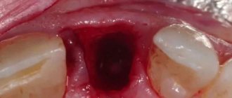
- With a strong blow to the dental crown, complete dislocation occurs: the tooth falls out of the socket and ends up on the floor or ground. In its place, a bleeding hole remains, in which a blood clot quickly forms. Soft labial tissue is often damaged.
- Incomplete dislocation is characterized by displacement of the crown from its normal position. It doesn’t go beyond the row, but it starts to hurt and stagger. In this case, the tooth root may become bent and stop developing. Hemorrhages and bruises are often observed in the soft tissue area.
- If mechanical impact occurs on the cutting edge of the tooth, the crown may be partially or completely immersed in the alveolus and embedded in the jaw bone tissue. Visually, there is a shortening of the tooth or its complete hiding in the bone.
Even if a child knocks out a baby tooth, only a specialist can assess the severity of the problem. You should not hope that a permanent one will soon grow in its place. The victim, along with the tooth found at the scene of the incident, must be taken to the dental clinic as quickly as possible. In this case, the clinic must admit the patient out of turn.
Should wisdom teeth be preserved?
We think yes.
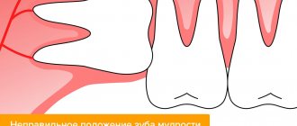
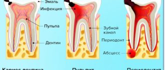
Although wisdom teeth are not involved in the process of chewing food, they can still benefit a person. There must be clear medical indications for their removal. And now we will provide you with another proof of the benefits of your own teeth-eights. Our patient Kim is about 20 years old, she is from Korea and lives in Moscow, as her parents work at the Korean embassy. I contacted the German Implantology Center due to inflammation of tooth 4.6 – the lower right six. The tooth was destroyed
, an abscess developed. There was a temporary filling on the tooth (placed in another clinic):
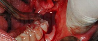
Where a temporary filling was placed, she was told to remove the tooth. That's why Kim came to us with one simple goal - to atraumatically remove the six, which began to greatly bother and hurt her. After doing a CT scan and looking at the tooth, we offered her an option, which, upon hearing, Kim at first did not believe her ears. Our proposal was to remove the six and immediately in its place... transplant its lower right wisdom tooth! This is called autotransplantation surgery.
What to do with a knocked out tooth?
A “lost piece” that has fallen into snow, mud or autumn slush can only be rinsed with water, but cannot be scraped and cleaned “to a mirror shine” so as not to damage the still living fibers. For the same reasons, you should pick up the tooth by the crown, and not by the root, which will be re-engrafted into the hole. Disinfection of living tissue with alcohol or hydrogen peroxide is also prohibited.
After dislocation, the tooth should be kept in a moist environment, which will significantly increase its chances of replantation and healing. How to achieve this?
- The best natural medium that supports the vitality of a lost tooth is saliva. Therefore, the tooth can be placed in the mouth, on the opposite side from the damaged side, tucked between the cheek and gum. When a baby tooth is dislocated, a child may scream and cry. To prevent the tooth from being swallowed, its transportation can be handled by an accompanying relative—preferably the mother.
- Perhaps the adult victim or those around him have a container for storing contact lenses. The tooth can be placed in the saline solution it contains.
- A small container filled with water or milk will also work.
- If there is a pharmacy in the neighborhood, you can buy any drug that represents a physiological environment - glucose, 0.02 percent furatsilin, novocaine, etc.
Did your child have a molar tooth removed? The orthodontist is already waiting...
Permanent teeth should ideally last a lifetime. But sometimes dentists are faced with the need to remove, as people say, a child’s “molar” tooth. This is done only as a last resort, when it is no longer possible to cure:
- advanced caries with tooth destruction of more than 70% and damage to the root;
- serious tooth trauma with root fracture;
- severe forms of periodontal pathology;
- untreatable purulent inflammatory processes.
Removing a permanent tooth in a child is a surgical procedure in which not just one of 32 teeth is removed, but a member that is very important for the balanced growth and functioning of the entire dental system. That is why dentists recommend not to go to extremes and regularly bring your child for preventive examinations at least 2 times a year.
Molars in children require more careful care than in adults. Frail enamel is susceptible to the effects of carious factors and the external environment, add to this a love of sweets and soda. Therefore, when children have their first permanent teeth (5
–
6 years), parents need to take special control over oral hygiene and diet, at least until the
14–15
, until the teenager begins to realize the importance of dental health.
Deleted and forgotten?
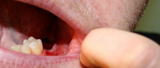
But no! Here everything is like in adults, a new tooth will not grow, and in place of the removed one there will be a gaping void. Many parents believe that they can leave everything as it is if the “hole” is not visible or the child is not embarrassed by it. And this is a fatal mistake, which in the future will result in expensive dental treatment and health problems.
This is exactly the situation faced by the girl Elena Badmaeva (name corrected), who had 2 chewing teeth removed at school age:
“I didn’t notice the problem for years, and I went on like this until I was 19 years old when I noticed that the neighboring teeth were leaning towards the missing ones, and the gums were sagging. My jaw began to click and pain radiated to my temple. The dentist immediately warned me that the treatment would not be easy or quick. As a result, I had to wear braces, undergo two surgical operations to restore the lost gum volume, and place implants. It took me 3 years and a tidy sum. But all this could have been avoided...
It is possible if you immediately contact an orthodontist after removal. Let us immediately warn you that you should not count on an implant, because they are placed only from the age of 18, after the end of active skeletal growth. Dentists have other methods for working with young patients.
For example, special spacer structures or temporary dentures that prevent teeth from shifting. As the bone tissue grows, the structures will be replaced so as not to disrupt the correct development of the facial skeleton. Well, when the child turns 18, he will be able to get implantation and prosthetics and will no longer remember about the ill-fated “lost” tooth.
– Observation by an orthodontist will eliminate the negative consequences of removing permanent teeth – displacement of adjacent teeth, uneven tissue atrophy, and other disorders of the dental system. In childhood and adolescence, maintaining or correcting minor changes in bite is much easier and faster. Timely treatment will allow you to avoid complex dental procedures in the future and save the family budget, warns Bato Vasiliev, chief physician of the Art Smile Dentistry Center.
Take care of the health of your children, monitor the condition of their milk and permanent teeth, so that in the future they only go to the dentist for preventive examinations.
And your children will thank you!
Art Smile Dentistry Center 69-777-0, 30-30-59 https://artsmile03.ru/ st. Klyuchevskaya 4/1
Author: Advertising INFPOL.RU
Do I need to insert a tooth myself?
In case of incomplete dislocation, the crown that protrudes strongly from the gums at the scene of the incident can be carefully inserted into place without applying significant effort. This will prevent complete tooth loss, the likelihood of which is quite high. But you shouldn’t touch a knocked-out baby tooth; it’s better to quickly go to the dentist to identify possible damage to soft tissue and bone.
If a piece of a tooth breaks off, you should rinse your mouth with clean water, try to find the fragment and rush to see a qualified doctor.
Why does a person's teeth change?
The structure and development of the dentition in different animals differs significantly from each other. In vertebrates and some species of reptiles, for example, teeth change several times in their lives, and over time, a new one grows in place of the lost tooth; in rodents, permanent teeth immediately grow. This is due to the characteristics of the body, residence and nutrition of representatives of the animal world.
There are several species of mammals whose teeth change once in their lifetime during childhood. This is necessary not only for the growth of the body, but also for survival - after the baby stops feeding on mother’s milk, its life largely depends on the capabilities of the dental apparatus. Humans can also be classified as mammals in this category, so changing teeth is an integral process of growing up for them.
Polyodontia (supernumerary teeth) - symptoms and treatment
For a successful prognosis in treatment, high-quality and timely diagnosis of patients is very important. The dentist-therapist must know the basic clinical manifestations and principles of diagnosing hyperdentia. This is especially important in the work of a pediatric specialist during periods of medical examination and planned sanitation of the oral cavity in children. This makes it possible to identify the presence of this anomaly at an early age and carry out appropriate treatment in the early stages.
Most often, “extra” teeth are located in the anterior part of the upper jaw. The question of the exact location of supernumerary teeth is very important, since the surgical stage of treatment depends on this. The more accurately the position of various anatomical formations is determined, the less surgical trauma (damage to the growth zones of complete teeth, the formation of large defects in the bone of the alveolar process) will result from the surgical stage of treatment. The volume and time of the procedure will be reduced, which is very important when it comes to treating children. The position of the causative tooth can be determined using instrumental examination methods:
- CT scan;
- orthopantomography;
- intraoral radiography;
- visiography;
- Magnetic resonance imaging.
Computed tomography allows you to obtain a cross-sectional, layer-by-layer image of any area of the human body, including the skull. Dental (dental) X-ray computed tomography allows you to make 4 types of images in different planes in one and a half minutes. The radiation dose with this examination method is less than other types of CT. The procedure is easily tolerated by patients and does not require special training, which is convenient when working with children. In addition, it is as informative as possible for the doctor.
Orthopantomography is a method of x-ray examination in which you can obtain an image of all the patient’s teeth, upper and lower jaws, adjacent tissues and areas of the facial skeleton. Conducted for children from 3-4 years old. This is a quick, simple, highly effective method that helps identify oral diseases in the early stages. It has virtually no clinical contraindications and is actively used by dentists.
Intraoral radiography is the simplest, most common diagnostic method in therapeutic dentistry. The picture shows from one to 3-4 teeth and adjacent formations. For the pathology under consideration, the method is not informative enough, but can be used if the above types of diagnostics are unavailable.
Visiography is a system for obtaining X-ray images without the use of film. In this type of research, instead of film, a special sensor is used, from which the image is transferred to a computer, after which it is processed and then saved. Advantages of this technique:
- The radiation exposure to the patient is tens of times lower compared to the most advanced film X-ray, which is extremely important for both the patient and the radiologist.
- Scalability and the possibility of mathematical image processing (measuring the size, density of objects, etc.).
- It is possible to store the received information and create your own database for each patient.
- There is the possibility of instant transfer of diagnostic data directly from the treatment room to the computer monitor of the attending doctor, wherever he is. This is very convenient if you need to consult doctors from different clinics or from different cities.
But, as with intraoral radiography, the image produces a low-informative planar image of the area under study, limited to 1-4 teeth.
Magnetic resonance imaging is a radiation diagnostic method that can be used to obtain an image of the layers of the human body in any plane. The method is simple and does not have any harmful effects on the body of the subject. A contraindication for MRI is the presence of metal foreign bodies (dentures, braces, surgical pins, etc.) in the patient’s body.
Sometimes, to determine the level of location of problem teeth, Yu.I.’s technique Zhigurta (1994). It consists of studying an orthopantomogram of the jaws and marking on it the four levels of occurrence of unerupted incisors and canines. However, this method is imperfect and does not allow an accurate assessment of where the supernumerary tooth is located. It is more advisable to use modern diagnostic methods described above.
To more accurately determine the relationship of the additional tooth with the roots and rudiments of the complete teeth, contact intraoral radiographs are taken in the preoperative period and 1, 3, 6 and 12 months after the operation. They are used to determine the degree of change in the position of the impacted tooth, the condition of the root of the complete tooth and the morphological structure of the bone of the alveolar process in the studied area [1][7][8].
If swelling takes a long time to go away after tooth extraction
Swelling of the gums persists for the first 2-3 days. Moreover, on the second day after removal it usually increases. If the swelling does not subside and new unpleasant symptoms arise, this may indicate that an inflammatory process has begun.
Symptoms of complications:
- aching pain followed by pulsation in the removal area;
- increased swelling;
- severe redness of the gums;
- socket suppuration;
- bad breath;
- temperature increase;
- general weakness.
If these manifestations are present, you should definitely contact your dentist to identify the cause of the complication. It could be an infection introduced into the wound, severe tissue trauma, a violation of sterility rules, a part of the tooth remained in the socket, or a cyst was not removed from the gum.
To ensure that your teeth are removed without complications, contact Neo-Dent dentistry. The clinic employs only highly qualified dental therapists and surgeons who will perform operations of any complexity with minimal consequences. We don’t even have to worry about removing wisdom teeth!
