It is a common belief that people with implants should not have an MRI. In fact, this was the case several decades ago, when patients were fitted with prosthetics made of steel, nickel and cobalt. In those years, magnetic resonance imaging could cause serious harm to human health.
TBS implant.
Let's be clear from the beginning that people with implants, pins, screws, retaining plates, breast implants, and dental implants CAN have an MRI.
What implants can be used for MRI?
MRI is allowed for people who have undergone hip or knee replacement. It is important that the endoprosthesis or fixation for osteosynthesis is made of metals or ceramics with low magnetic susceptibility. This avoids displacement or overheating of the structure during the examination.
Knee endoprosthesis.
People with hernia mesh, dental, breast and joint replacements are also allowed to have an MRI. All these implants are made from materials that do not interact with the magnetic field. This makes the study safe. However, you should consult a specialist before an MRI. The doctor will assess the possible risks and recommend the necessary precautions.
Minimally invasive endoprosthetics in the Czech Republic: doctors, rehabilitation, terms and prices.
Find out more
Examination with contrast
Contrast is used to improve visualization; in approximately 20% of all diagnostic cases it is necessary. Gadolinium-based drugs are used, which are safer than iodine-containing substances.
An intravenous injection is performed after routine imaging. For this, the patient is “rolled out” of the device and given a regular injection, after which the test is repeated.
Discuss with your doctor whether contrast is necessary, since in this case the cost of the examination doubles.
The procedure is indicated for suspected multiple sclerosis or for vascular examination. To obtain accurate information about myocardial viability or congenital pathologies, it is also advisable to perform an MRI of the heart with contrast.
Interaction of different metals with a magnetic field
Different metals tend to interact with magnets differently. Some of them are attracted to it, others are repelled, and others do not react at all. All three types of metals are used for the manufacture of endoprostheses.
Table 1. Metal classes.
| Class | Representatives | Description |
| Diamagnets | Copper Zirconium Silver Zinc | They have negative magnetic susceptibility. This means that when interacting with a magnetic field, they repel rather than attract. |
| Paramagnets | Titanium Tungsten Aluminum Tantalum Chrome Molybdenum | These metals are characterized by the presence of low magnetic susceptibility, independent of the magnetic field strength. Paramagnetic prostheses usually tolerate the MRI procedure well and do not move or heat up. |
| Ferromagnets | Iron Nickel Cobalt Steel | They have high magnetic susceptibility, depending on the magnetic field strength. Implants containing large amounts of these metals may become dislodged or become hot during an MRI scan. |
Composition of modern endoprostheses
All plates, pins and endoprostheses, which are used in modern traumatology and orthopedics, consist of a variety of alloys. Note that different implants contain different amounts of paramagnetic and ferromagnetic materials. The properties of each endoprosthesis, pin or plate depend on the composition.
Not all prostheses are 100% metal. Most of them contain ceramics or polyethylene. The latter does not interact with the magnetic field, therefore, it does not in any way affect the MRI results and the course of the procedure. However, ceramics most often contain aluminum oxide, which still has a certain magnetic susceptibility.
Destroyed components of the hip joint implant.
Possible combinations of materials in endoprostheses:
- ceramics + ceramics;
- ceramics + polyethylene;
- metal + polyethylene;
- metal + ceramics;
- metal + metal.
Fact! Plates and pins for fixing bone fragments are made of metal alloys. The same applies to external fixation devices (Illizarov type) and clips that are placed on vessels.
Composition of artificial joints:
- cobalt;
- chromium;
- molybdenum;
- titanium;
- zirconium;
- tantalum;
- niobium.
Having familiarized yourself with the composition, you can understand how it will behave in a resonant tomograph. The magnetic properties of each endoprosthesis are determined not only by the material from which it is made, but also by its shape and size. Steel pins and plates longer than 20 cm can heat up above the permissible limit.
Fact! Products containing large amounts of nickel and cobalt interact especially actively with a magnetic field. This means that diagnostics with such endoprostheses should be performed with extreme caution.
MRI making metal titanium plate leg metal titanium
MRI diagnostics often use a gadolinium-based contrast agent.
This method allows you to see changes in tissues or blood vessels and significantly expands the diagnostic capabilities of examinations. Through the vessels, the contrast agent penetrates the cells and changes their electromagnetic potential. The substance is a factory-made drug, completely safe and has passed the necessary certification.
It is administered intravenously by drip or as a whole immediately before the study.
Gadolinium chylate compounds are not involved in metabolism and are completely excreted, thus having very limited contraindications to MRI. They are as follows:
- Consequences of liver transplantation (postoperative period);
- Allergy to the drug;
- Decreased renal function (creatinine clearance less than 30 ml/min);
- Pregnancy and lactation.
- Individual intolerance to contrast media, because there is a risk of allergies.
- MRI with contrast should not be performed on pregnant or nursing mothers due to possible exposure of the fetus or child to the chemical.
- Chronic renal failure due to possible problems with removing contrast from the body.
Who should not have an MRI after endoprosthetics?
What to do if the patient has various types of implants right in his body? First of all, this must be reported to the specialist conducting the examination, since many metals are ferromagnetic and can shift in the body under the influence of a magnetic field.
For an MRI with a steel wire in the body, everything is not so simple. Iron causes the magnetic field to deviate from a given direction, which leads to distortion of the resulting images and the appearance of artifacts (defects) on them. In addition, the needle can heat up, creating unpleasant sensations for the patient.
Also, the answer to the question whether it is possible to do an MRI with an endoprosthesis depends on what material it is made of. If it is titanium, then there are no restrictions. If it is made of ferromagnetic materials, then this is a contraindication for research. You can find out exactly what metal the implant is made of in the design passport, which is issued to the patient after prosthetics.
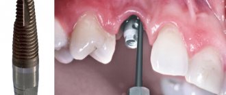
Is it possible to do an MRI with braces?
Modern brace systems are made of expensive and durable alloys that do not deform under the influence of magnetic-nuclear radiation and cannot move or injure the patient’s oral mucosa.
Small structures do not distort tomograph signals and do not heat up; their response to a magnetic field is very weak.
You cannot do an MRI if a fairly voluminous structure - more than 20 cm - is secured with ferromagnetic retainers. In this case, the bracket may become hot.
Is it necessary to do an MRI if a bracket is swallowed to accurately determine its position in the intestine? It is impossible to swallow a large brace, but a small one will come out naturally. To speed up this process, you need to eat more viscous porridge and drink fluids.
Braces do not affect the patient's condition during MRI, but they can result in insufficiently reliable results when scanning the brain, heart area, thoracic or cervical spine.
In cases where it is urgently necessary to screen the brain and cardiac system, and doctors do not see an alternative to MRI, you should contact orthodontists and remove dental implants. After tomography, they are reinstalled in the required volume.
Is it possible to do an MRI with iron crowns?
If you have old-style crowns made of iron, you cannot screen the brain and heart. The metal heats up significantly, which causes severe pain in the patient, deformation of the metal structure - the integrity of the implants may be compromised or they may fly off the teeth.
With crowns and dentures with metal-ceramics, screening of the brain and heart area is allowed, but there is a high probability of an unreliable result due to a distortion of the response to magnetic field signals.
Regardless of the type of alloys of crowns and prostheses, it is allowed to conduct MRI of the lumbar spine, abdominal organs and retroperitoneal space, pelvic region and extremities in closed-type devices.
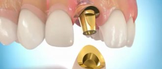
When installing pins, high-strength titanium implants are often used. Their presence does not affect the reliability of the examination results; moreover, the size of the pins is so small that the magnetic field does not have any significant effect on them.
Metal crowns made of polymer alloys also do not distort magnetic field signals, however, you should check with your dentist about the possibility of conducting an MRI. Some structures heat up, so the procedure will cause significant discomfort to the patient.
If a patient has dental bridges installed, then they probably contain separate parts - pins, plates, screws of various sizes. For their manufacture, diamagnetic, ferromagnetic and paramagnetic materials are used - cobalt, iron alloy and nickel, which react differently to magnetic field signals.
Is it possible to do an MRI with metal and titanium plates in the leg?
Be sure to tell your doctor before starting the study if you have at least one item from the list:
- General ferromagnetic devices and implants;
- hemostatic clips of cerebral vessels;
- fragments of bullets, shells, shot;
- nerve stimulators with metal elements;
- insulin pumps;
- intravascular stents;
- vascular clips;
- surgical braces;
- external fixation systems, wire splints or shunts for fractures;
- endoprostheses;
- stabilizing systems made of ferromagnetic alloys;
- non-ferromagnetic inner ear implants;
- ferromagnetic fragments;
- dental braces;
- steel intrauterine devices.
Depending on the relationship of any metal to the influence of a magnetic field, they are divided into diagmagnets (in the field they are subject to weak repulsion), paramagnetic (weakly attracted by the magnetic field) and ferromagnetic (strongly susceptible to the influence of the field).
In exceptional situations, the doctor may prescribe an MRI if the patient has metal plates.
If there is metal in the body, the examination can only be carried out if its immediate location is outside the range of the magnetic field, or the diagnosis will be performed using low-field equipment. However, in the vast majority of cases, metal prostheses are a contraindication to the procedure.
In the presence of titanium plates in the leg and other parts of the body, diagnostics are carried out without restrictions, since titanium is paramagnetic and is not characterized by strong attraction in a magnetic field. MRI with a titanium prosthesis is as informative and harmless as without it.
Obtaining MRI scans is based on recording a radiofrequency signal after exposure to a high-intensity magnetic field. Nuclear resonance of hydrogen protons allows you to obtain high-quality images. The effect operates in liquid tissues. Bones contain a low concentration of water, so it is better to examine them using computed tomography (CT).
Before scanning, you need to carefully analyze the contraindications:
- Presence of prosthetic knee and hip joints;
- Metal middle ear implants;
- Pacemakers, pacemakers;
- Vascular clips;
- Screws, pins.
A radiation diagnostics doctor can determine whether an MRI with vascular stents can be performed on a person with plates. Analyzing the physics course, all metals can be divided into 3 types with respect to magnetization - ferromagnetic, diamagnetic, paramagnetic.
Under the influence of a strong magnetic field, alloys of nickel, cobalt, and iron move. Pins, screws, and joint prostheses are made from metals. Steel is a cheap material widely used in surgery. The manufacturer's instructions determine the quality composition of the product. ferromagnets differ, so products behave differently in a magnetic field:
The danger of steel models is heating under the influence of a strong magnet. The likelihood of complications increases the longer the plate. Patients undergo supervised scanning. The person sits on the diagnostic table, holds a special button in his hand. When a feeling of warmth appears and a key is pressed, personnel are called and the examination is stopped.
Concentric structures such as the Illizarov apparatus pose a danger. The bulkiness and high metal content promotes heating. However, an MRI is performed while wearing the product. Concentric circles are located outside the leg and do not pose a health hazard.
Before deciding whether to do tomography of the joints after surgery, you should find out the composition of the structure. Endoprostheses are made of metal, titanium, chromium-molybdenum-cobalt alloys. The latest options do not move, they warm up. They do MRI with crowns and titanium implants.
Before scanning, you need to take an x-ray. The study will show the dimensions and number of artificial inserts (plates, prostheses, screws, screws). Even if a person has cobalt chrome endoprostheses, additional metal fragments may be a contraindication.
Attention! Magnetic resonance imaging of the area with a metal implant is not informative. The device distorts the signal. There is a lot of noise in the final tomogram.
MRI after stenting
After stenting, an MR examination is not only allowed, but also prescribed. Therefore, the answer to the question of whether it is possible to do an MRI after stenting is positive. But the specialist conducting magnetic resonance imaging must know exactly what material the stents are made of.
You can absolutely safely carry out examinations with bioabsorbable stents, since they consist of a biopolymer - after a given time they are absorbed, but the lumen of the vessel is preserved.
In other cases, stents are made of inert metal alloys: stainless steel, cobalt alloys, etc. Note that the patient is required to strictly follow the instructions for the stent, i.e.
if it states that MRI should not be done in the first few weeks after stenting, then this applies not only to the area where the stent is installed, but to the entire body.
Even if it is not directly located in the apparatus tunnel, the magnetic field works equally strongly in the room where the tomograph is installed.
Sometimes immediate diagnosis is necessary when the presence of a stent is not known before MRI is performed because the patient does not have time to report them. Practice has confirmed that the materials currently used for the manufacture of stents are not ferromagnetic and do not respond to external field influences, and, therefore, are MRI-compatible.
Traumatologists are assigned MRI scans to dynamically monitor the condition of the product. Even 3 Tesla field tomographs will not be able to move prosthetic joints due to strong fixation. Research with high-field devices with a power of 1.5 Tesla is affordable and characterized by weaker attraction.
The second important issue that needs to be addressed is the heating of the vascular clip installed during surgery. Practical studies show an increase in the temperature of metal wire up to 45 degrees (length about 20 meters).
Having an artificial prosthesis is not a contraindication. Before prescribing, you need to exclude clips and heart rate stimulants.
You cannot do an MRI with an endoprosthesis, pins, or screws for older people. Reduced bone density contributes to poor implant fixation. Moving metal can damage surrounding surfaces.
Steel structures are ferromagnetic and can be strongly attracted. Titanium is a paramagnetic material that does not heat up and is capable of moving.
General contraindications:
- Presence of metal objects in and on the body. We are talking not only about jewelry, hairpins and watches, but also about medical devices such as pacemakers, artificial valves, etc. It is also necessary to exclude cases with the presence of metal objects that entered the body accidentally or as a result of injury. A high magnetic field can disrupt the operation of the device and also heat up the metal.
- Contraindications during pregnancy: MRI should not be done in the early stages of pregnancy and if there is a threat of miscarriage;
- MRI with contrast is contraindicated at any stage of pregnancy.
Source: //biglionz.ru/mrt-s-metallom-v-tele/
Manufacturing companies
Over the past 20 years, medicine has mainly used implants made from chromium-cobalt alloys (as we have already found out, these metals actively react to a magnetic field). Many models made from better materials have appeared on the market. They are better tolerated by patients and do not cause allergies or MRI problems.
Table 2.
| Company manufacturer | Characteristics and Application | Behavior of implants during MRI diagnostics |
| Biomet | Produces high-quality implants that take root well and do not cause allergic reactions. | Due to their small size and low magnetic susceptibility, they do not interfere with MRI. |
| Zimmer | It produces products not from titanium, but from tantalum. The implants have a porous coating and fuse perfectly with the bone tissue. | They do not cause unexpected complications during magnetic resonance imaging and do not distort the results of the study. |
| Johnson&Johnson | The company produces implants using all available standards and technologies. | Do not interact with magnetic field. When available, MRI is absolutely safe. |
| Smith&Nephew | Manufactures endoprostheses from an alloy containing zirconium and niobium. | Smith&Nephew implants are hypoallergenic and practically do not interact with the magnetic field. |
| Stryker | World-famous company of beta-titanium endoprostheses and fixators for internal osteosynthesis. | Owners of Stryker implants can undergo MRI without any worries. Additional precautions may only be necessary if you have several large prostheses. |
| Aesculap | Produces endoprostheses from titanium, zirconium ceramics, chrome-cobalt alloys. | Most implants can easily withstand magnetic resonance imaging. |
If you have a prosthesis from one of the companies listed in the table, you can do an MRI without the slightest fear. However, in any case, you should not undergo the study without first consulting a doctor.
Operating principle of tomography equipment
First of all, you need to understand how exactly an MRI device works. The equipment is designed for targeted screening of soft tissues, bones and blood vessels. Advanced devices in diagnostic centers today have enormous technical capabilities. Thus, the power of the equipment is usually 1.5–3 Tesla, thanks to which doctors can examine anomalies whose dimensions are less than 0.2 mm.
MRI technology is based on the generation of radio waves and the ability to magnetize hydrogen elements present in different tissues of the body. The nuclei are able to respond to the influence of a magnetic field, that is, enter into resonance, and a computer program records all the data and converts it into detailed images. The research results are generated from many virtual sections of the selected department, for example, the brain, the vascular system, the abdominal cavity, and so on. Based on them, the radiologist draws up a medical report and a preliminary diagnosis.
Contraindications to the procedure
If prostheses, pins and plates are firmly connected to the bone tissue and cannot move, then implants of other locations can easily move under the influence of a magnet. Therefore, it is STRICTLY PROHIBITED to conduct magnetic resonance imaging if they are present.
Implants that cannot be used for MRI:
- artificial heart valves;
- stents and clips on vessels of any location;
- middle or inner ear implants;
- pacemakers;
- artificial lens;
- Illizarov apparatus;
- insulin pump;
- large metal implants.
Minimally invasive endoprosthetics in the Czech Republic: doctors, rehabilitation, terms and prices.
Find out more
Preparation
Many types of MRI examinations do not require special preparation, but there are exceptions.
Before an MRI of the abdominal cavity, it is advisable to follow the following diet:
- for two days, eliminate gas-forming foods, fresh fruits and vegetables;
- on the day of the procedure, avoid tea and coffee;
- It is advisable not to eat for at least 6 hours before the examination, and not to drink for 4 hours.
The doctor will recommend special medications for constipation and flatulence. To exclude artifacts from intestinal motility, two hours before the procedure you need to drink 2 tablets of No-shpa.
Before an MRI of the pelvis, it is advisable not to urinate for about two hours. If the study is carried out in women, it is recommended that it be carried out on days 5-12 of the cycle (immediately after menstruation), but this condition must be discussed with a doctor.
General recommendations
before MRI scan:
- remove all metal objects;
- leave jewelry and accessories at home;
- change into comfortable clothes;
- Leave valuables, money, and bank cards in a specially designated place or safe in front of the diagnostic room.
An approximate list of items that are not allowed in the diagnostic room:
- metal zippers on clothes, pins, hairpins;
- watch;
- Hearing Aids;
- pens;
- earrings, including piercings;
- glasses;
- removable dentures;
- pocket knives.
How to find out if you can have an MRI
Remember that an MRI can be done with the permission of a specialist. Only he will determine whether you need this research and whether it will harm you. Perhaps the doctor will make a diagnosis without magnetic resonance imaging. Spinal spondylosis and deforming osteoarthritis of stages II-IV can be detected using conventional radiography.
Comparison of visual diagnostic methods. MRI is on the right.
results
The study is interpreted by a diagnostician - a radiologist. The conclusion is issued on the day of the examination, but a description of the images may take the doctor several hours.
The doctor examines the obtained images and compares them with normal values:
- size and location of organs, symmetry;
- presence/absence of abnormal formations;
- presence/absence of an aneurysm, free fluid, blood clots in the blood stream;
- signs of infection and inflammation;
- stagnation in the ducts;
- traumatic injuries.
Possible complications and precautions
MRI in the presence of electronic implants can seriously harm a person or even lead to his death. Performing the study on persons with coronary walls and clips on cerebral vessels can provoke massive bleeding, which will lead to death. Endoprostheses made from some alloys may move out of place or heat up during an MRI, causing burns.
MRI installation before the procedure.
People with certain types of implants are strictly prohibited from undergoing magnetic resonance imaging. But for patients with implants made of “dangerous” alloys, you can still try to perform the study. As a precaution, a button is placed in the person's hand. If he feels a strong burning sensation, he presses it and the study is stopped.
Fact! Metal prostheses tend to “fade”, making the image of nearby tissues unclear. Therefore, it is pointless to try to obtain an MRI image of the replaced joint or bone held together by fonts or plates.
How preparation for research affects the result
It is important to remain still during the examination, otherwise the image quality may be somewhat distorted. Throughout the examination, the patient is under the supervision of a specialist. Inside, there is a button at hand to contact staff in case of an emergency. When examining children in the examination room, in exceptional cases (depending on age), an accompanying person may be present.
If you have a pacemaker, there are serious contraindications for MRI examination. You should consult your cardiologist as MRI may affect the implants and their function. When receiving a referral for diagnostics, you should have the implant passport with you.
Metal parts that are in the body (surgical screws, replaced joints, artificial heart valves, tattoos with metal particles) must be told to the doctor before the examination. Magnets can interfere with the normal functioning of pacemakers and can move pieces of metal that are present in the body.
A strong magnetic field can affect magnetic cards, electronic devices, and any magnetized metal body located inside or outside the body. Therefore, before entering the examination room, you must remove jewelry and watches. Personal belongings may be handed over to accompanying persons or placed in storage rooms located outside the examination room.