The treatment tactics for caries depend on the cause of the pathological process and its neglect. This is the most common dental disease, characterized by gradual tooth decay. The pathology goes through several successive stages; at first, the patient may not feel anything; in complex cases, complete loss of dental units is possible.
It is not difficult to detect the disease, because most of the symptoms are quite obvious. However, it is better to prevent the occurrence of carious lesions by following preventive recommendations, and then a snow-white smile will delight you for many years.
Types of caries localizations
Caries (lat. caries rotting) is a slow and hidden pathological process occurring in the hard tissues of the tooth. It is characterized first by focal demineralization of the enamel, then by the destruction of hard tooth tissues with the appearance of a cavity in the dentin. If left untreated, the tooth is lost or complications arise - pulpitis and periodontitis.
Cervical (circular)
The structure of a tooth is divided into a root, a neck and a crown - the latter is located above the gum. The neck and root are located in the gum and are protected by soft mucosal tissue.
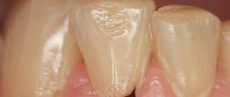
When the carious process begins to destroy the tooth in the area of contact of the crown with the mucous membrane, we can talk about the cervical form of caries.
This category is the most difficult to diagnose and treat. It most often affects the incisors. When biting food with incisors, the food is not only “cut off”, but also gets clogged into the gum pockets, moving towards the gums.
Crown caries
Caries has the ability to affect not only healthy, but also already treated teeth - under a filling or crown. If there is a crown, it is difficult to detect, because the tooth does not have a nerve and pain is not felt. There are no external signs for a long time.
The causes of this type of caries are:
- defect of the dental crown with the appearance of access to the tooth tissue underneath it
- poorly treated tooth before prosthetics;
- gum disease;
- poor oral hygiene.
The problem is that the process spreads into the deep layers and spreads to the bone structures, as a result of which the tooth is often completely removed. That is why preventive examinations and an orthopantomogram - a panoramic image of the entire jaw - are so important.
Radical caries
Basal and cervical caries are similar. Both incisors and chewing teeth are affected. Destruction begins with the enamel, gradually moving to the nerve in the root canal. When the process touches bone tissue, it threatens with osteomyelitis.
The main reason for the occurrence is a cariogenic situation in the oral cavity, when plaque accumulates especially quickly. The enamel in the root part of the tooth is much thinner and more easily destroyed. It is most often diagnosed between the ages of 30 and 60 years.
Root caries
Root or subgingival caries occurs hidden. It can be with or without a cavity. Along the flow - active, suspended, secondary. It most often affects molars.
Radiation diagnostics of dental injuries
After an injury, radiation diagnostics of the damaged and adjacent teeth, and sometimes antagonists, is almost always prescribed. The scope of emergency dental measures depends on its results. The most common injuries are:
- tooth fractures - isolated or combined, that is, in combination with a violation of the integrity of the jaw (the same symptoms are noticeable in the picture as with jaw fractures);
- dislocations - mainly in the area of the incisors (the x-ray shows how the tooth has come out of the socket, and with subluxation, the asymmetry of the periodontal gap is noticeable).
Radiation diagnostics is also used for other dental diseases if the benefits from it outweigh the expected harm from radiation.
What is the danger of this disease?
Caries is dangerous because there are no symptoms for a number of years. It is detected more often in an advanced stage, when tooth extraction becomes the solution.
Visual diagnosis is difficult because all destructive processes occur inside the tooth. Pronounced plaque or tartar hides any stains on the enamel.
The root is hidden under the gum and external irritants do not affect it until a certain time. On the other hand, the root walls are thin, so they are destroyed quickly and with complications.
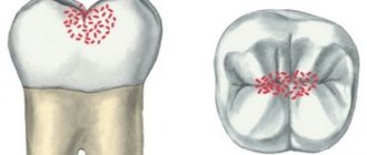
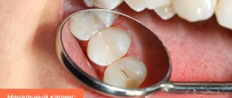
What does caries look like under a microscope?
Caries looks very interesting under a microscope, especially if you look at it on a tooth cut. Below you can see what superficial, medium and deep forms of caries look like. In addition, in each photograph in the center of the tooth crown you can see the cavity (pulp chamber) in which the neurovascular bundle of the tooth is located.
Causes of the disease
There are 3 main provoking factors that must act in a complex manner; none of them acts independently:
- Cariesogenic microflora - this includes Mutans streptococci, actinomycetes and certain types of lactobacilli. They should dominate the oral cavity. Bacteria produce organic acids from food carbohydrates, which cause demineralization of cement.
- Consumption of simple carbohydrates is the most cariogenic. Their breakdown produces glucan, which contributes to the appearance of plaque.
- Reduced caries resistance is a deterioration in the resistance of tooth tissue and the body as a whole. This is facilitated by a decrease in calcium content in hard root tissues, bad habits, and lack of saliva.
In addition, the anatomical features of the mouth can also have an effect - a small vestibule, a short frenulum, and bite pathology.
Gum disease, pocket formation
Gum pocket or otherwise periodontitis is a common occurrence in many patients. Many people do not treat it because it does not bother anyone for a long time.
The first manifestation of pathology is bleeding when brushing teeth, although outwardly they may look healthy. Dental pockets or depressions are normal, but their anatomical size should not exceed 3 mm.
Then the pocket is able to self-clean from food debris and epithelial particles. Reservoirs of bacterial accumulations form in deep gaps. This is accompanied by bad breath. Conventional treatment with antibiotics and brushing teeth with toothpaste have no effect.
Causes of periodontitis:
- insufficient hygiene;
- poor quality fillings;
- abnormal position of the dentition, malocclusion;
- hereditary predisposition;
- deficiency of vitamins and minerals;
- immunosuppression.
Features of pathology in children
It is common for children to replace baby teeth with permanent ones, but this does not mean that temporary teeth do not need to be treated - firstly, the baby will be bothered by pain, and secondly, it is important to eliminate the source of chronic infection.
Distinctive features of caries in children:
Children need help brushing their teeth; adults must constantly monitor the process, otherwise caries may occur.
Lifestyle
If you have learned about the risk group for root caries (older age), this does not mean that you can relax. Caries has many causes and can occur at any age if basic precautions are not followed.
Those at risk include smokers, diabetics, pregnant women and even children. The provoking factors in this case are:
- smoking and lack of oral hygiene;
- infrequent brushing of teeth and lack of fluoride;
- poor nutrition with preference for desserts;
- frequent stress;
- alternating hot and cold;
- structural features of the oral cavity;
- alcohol abuse - alcohol breaks down into sugars and acids;
- abuse of coffee and strong tea, which creates an acidic environment in the oral cavity;
- lack of water intake, and therefore lack of saliva;
- gum injuries.
Epidemiology of root caries
The incidence of root caries is constantly increasing in older people. This is due to the fact that:
- There are more people with periodontal diseases, since their prevention is ineffective
- Dental care has improved, and retirees now have many more teeth in their mouths.
- Well, life expectancy has increased, where would we be without it?
Be that as it may, in our country today people suffer from root caries
- 1.3% aged 25-29 years
- and 35.2% (aged 55-64 years)
Symptoms of the disease
The process usually occurs without symptoms, but pain may occur when brushing a toothbrush, eating sour, salty, sweet, cold or hot foods.
After eliminating the irritant, everything goes away. The patient can see a doctor if a stain is found on the front surface of the incisors, but often it is hidden under plaque or tartar.
The ongoing carious process gradually reaches the dentin junction, penetrating first into its surface layers and then further. The cavity deepens with bacteria and food debris. There is a smell from the mouth. Irritants cause pain more and more frequently.
With cement caries, teeth become mobile and lose their support, and bleeding gums occur. These are already symptoms of periodontitis. Now, even when chewing food or brushing teeth, severe discomfort occurs.
Digestion begins to suffer. Teeth become hypersensitive to hot or cold.
Next, the process follows the Leus classification:
- Active lesion - the edges of the cavity are undermined. The cavity is filled with softened tissues and tends to grow.
- Suspended caries - no increase in the affected area is observed. The cavity is clean, the bottom is shiny and smooth, the edges are even and dense.
- Secondary caries - occurs under a filling.
How to determine the presence of a carious infection
Often this disease is accompanied by such symptoms as:
- pain when cold or hot;
- increased sensitivity of the affected part;
- aching pain that increases at night;
- unpleasant odor from the mouth;
- change in tooth color.
In order to determine the presence of carious bacteria, it is best to contact trusted Dent Prestige dentists. Specialists diagnose the disease through a thorough examination and x-rays.
The presence of professional equipment contributes to the high-quality and quick implementation of the necessary procedures aimed at eliminating the problem.
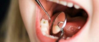
Diagnosis of the disease
The diagnosis is made in stages. Listening to complaints and visual examination make it possible to diagnose caries only in 13% of cases. Classic probing with a sharp probe and inspection with a mirror are informative. This allows you to examine the dentition.
Thermal diagnostics, electroodontometry and x-rays are also performed. If the patient has massive dental plaque, changes in the enamel will not be visible. In this case, professional teeth cleaning is first carried out to remove plaque and stone. All this is possible in a dental clinic.
At the dental clinic
The following types of studies can be performed at the clinic:
- Probing with a thin probe with a curved end - it is inserted under the gum, while the doctor has the opportunity to examine the structure of the tooth, its integrity, the presence of irregularities and chips. With rapidly progressing caries, the edges of the cavity are uneven and sharp. In the remission stage, the surface of the pathological focus is shiny, smooth, hard with smooth edges.
- Electroodontometry - can determine nerve damage and the depth of inflammation. The pulp reacts differently to the current strength.
- Thermal diagnostics - treatment of different areas of the tooth with a stream of water and heated wax. Unpleasant sensations that disappear after removing the irritant indicate caries.
- X-ray - a targeted photograph of one tooth or computed tomography. The presence and localization of obvious or hidden inflammation is determined with millimeter precision.
- Visiography is a special device - a visiograph scans the received data and transfers it to a computer, where the picture is studied from different angles and in detail, revealing the hidden process of inflammation.
Does genetics have an effect?
Some researchers attribute individual susceptibility to dental caries to genetic factors. By studying the development of caries in animal models, Japanese scientists suggested that loci on chromosomes 1, 2, 7, 8 and 17 contribute to susceptibility to caries. Genetics does not directly influence the onset of the disease, but determines its severity and duration. This is explained by the peculiarities of the structure of the oral cavity: genes involved in the development of enamel, in the formation and composition of saliva, and also those involved in the formation of the immune response are closely studied [4], [8], [9].
Treatment or removal - what determines the outcome
The doctor makes a decision on treatment or tooth extraction after determining the depth of spread and the area of localization of the inflammatory process. It is mandatory to first carry out professional cleaning using an ultrasonic scaler, an AirFlow water-abrasive device and hand tools.
Initial caries is treated with conservative retherapy. For hard tissue defects, preparation and filling are performed. In severe cases, the tooth is removed.
Treatment of the initial stage of root caries
Conservative retherapy includes:
- fluoridation of teeth;
- covering teeth with protective agents
- training in proper oral hygiene
Treatment of the disease
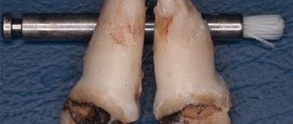
Caries between teeth is treated in the same way as regular one, with the only difference being that two units have to be “fixed” at once. In some special cases, in order to get to the affected part, the dentist is obliged to get rid of healthy tooth tissue by drilling. First, the specialist must prepare the cavity and then treat it.
It is important to be careful about the process so as not to touch or even damage the nerve. After which the doctor installs a plate that separates the teeth and inserts a filling. At the end of the process, it is put in order by grinding and polishing.
Cleaning in dentistry from stone and plaque
1243900247100
Professional teeth cleaning involves removing the accumulation of all pathogenic microflora around the root.
It involves not only removing plaque, but also stones. This elimination of negative factors protects teeth for a long time.
Antiseptic treatment of affected areas
Treatment of root caries involves drilling out all affected areas with a drill and treating the carious cavity with antiseptics and special preparations. A 2% chlorhexidine solution or a gel based on it is used as an antiseptic for the treatment of carious cavities.
Radiation diagnostics of chronic periodontitis
In childhood, chronic and acute periodontitis are localized in the area of the bifurcation of primary molars. The image shows a focus of destruction, which is characterized by fuzzy, uneven contours. Other radiological features include:
- predominance of granulating periodontitis;
- localization of destruction in the area of root bifurcation or around the root;
- pathological root resorption;
- spread of destruction to the follicle of the bud.
If the pathology has spread to the germ of a permanent tooth, usually a premolar, characteristic changes will appear on the x-ray - the end plate of the upper wall of the follicle will disappear, and the germ itself will take a different position or die altogether.
What to do to protect your gums
If the gum is not treated, inflammation will remain and all treatment will become meaningless. The gums are protected by diathermocoagulation and retraction.
Diathermocoagulation
Diathermocoagulation – excision of excess areas of periodontitis with a coagulation knife. The knife immediately cauterizes the bleeding edges. The procedure is performed when there are severe changes in the gums. In such cases, filling is postponed until the soft tissue is completely regenerated.
Retraction
If the condition of the gums is advanced, treatment of caries becomes impossible - the mucous membrane bleeds and interferes with tooth treatment. This gum is excised or a special device is applied to move the gum back. But more often, retraction is performed - lowering the gingival contour or moving apart the overhanging edges of the gums using special hemostatic threads. A temporary filling is placed until complete healing.
What symptoms appear with average caries?
To understand when a painful reaction to sweets occurs, you need to study the structure of the tooth a little.
In addition to enamel, the human tooth consists of dentin, and all dentin is penetrated by very thin dentinal tubules, which contain processes of odontoblast cells. These cells form the surface layer of the pulp and have their own sensitivity. They are the ones who react to sweets, they ate a piece of chocolate, the tooth ached sharply, but also quickly went away.
If only this sensitivity is present, then caries can be quickly cured, since the pulp has not yet been affected.
What preventative measures are there?
To keep your teeth healthy for many years, you need to brush your teeth twice a day, use floss and a tongue scraper. After sweets, you should rinse your mouth and chew gum for 5-10 minutes.
Prevention also includes the removal of tartar. A sufficient amount of saliva is also important, so it is necessary to maintain a water regime.
Like any other pathology, caries is better prevented than treated. Therefore, it is important to visit the dentist twice a year for preventive examinations. People at risk should approach this especially responsibly.
Other
A familiar diagnosis
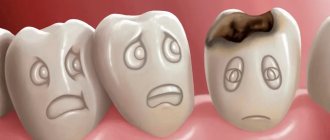
We are taught how to brush our teeth properly since kindergarten. However, more than 90% of people have faced and are still facing this problem.
The disease develops asymptomatically at the initial stage, affecting only the superficial layers of the enamel, and gradually penetrates deeper, reaching the nerve endings and blood vessels. As the process progresses, severe dental caries appears, which causes toothache and dentin damage.
Important! This is the insidiousness of the disease - the patient comes to the clinic when the pain becomes unbearable - which means complications, pulpitis and removal of the nerve.
Types of deep dental caries
The maximum penetration of carious infection can occur in different ways depending on the nature of the disease and its location (localization) on the crown of the tooth.
Types according to the nature of the flow
According to this principle there are:
- acute carious process - proceeds quickly, only slight destruction of the enamel can be visible from the outside, but with the help of a dental probe the doctor discovers a significant carious cavity inside;
- chronic - develops slowly; in a chronic course, a significant area of enamel and dentin is destroyed with the formation of a wide carious cavity.
If you suspect any type of caries, you should immediately contact your dentist. The earlier treatment is started, the greater the chance of saving the tooth.
Types by location of the source of destruction
According to localization, deep caries is divided into:
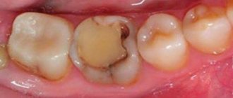
- fissure - develops in grooves on the chewing surface of the lateral teeth - fissures; the most common type, since food always gets stuck here; in some patients the fissures are shallow and easy to clean, in others they are narrow and deep, which increases the risk of infection;
- interdental (contact) – in areas of the crown in contact with adjacent teeth; food often gets stuck here, which is the cause of the disease; if teeth are closely spaced, food can only be removed using dental floss; It is difficult to detect and often this particular localization has to be treated at the stage of deep caries;
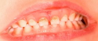
- cervical - in the area of the neck of the tooth - the place where the crown enters the root; the enamel and dentin here are thinner and the infection easily penetrates the tooth cavity, going down to the root through the root canals; and since the neck is covered by the gum, the pathological process is also not immediately detected, therefore it often reaches the stage of deep caries;
- circular - affects mainly baby teeth, is the result of a combination of genetic predisposition and improper feeding; develops as a cervical one, but not locally, but encircles the entire neck; affects several front teeth at once.
There is a more detailed classification according to Black:
Black classification
Factors provoking the development of caries
In addition to the above reasons, caries can be caused by:
- anatomical features of the structure of teeth (congenital dimples in the enamel in which plaque accumulates);
- malocclusion (crooked teeth lead to the fact that even good toothpaste and a high-quality brush are not able to fully cope with cleansing);
- increased viscosity of saliva (renewal of microflora does not occur properly due to this, resulting in various diseases);
- a state of constant stress and increased nervousness (this leads to a deterioration in the absorption of microelements from food).
Tooth hurts after deep caries - what to do
Some discomfort after treatment of deep caries may last for several days. Normally, the pain gradually subsides and is never acute. The doctor will definitely warn the patient how long the moderate pain will last. People sensitive to pain can take painkillers: Paracetamol tablets 500 mg, Ketorol 10 mg, etc.
Occasionally, the pain becomes progressive and is accompanied by other symptoms. This indicates some kind of complication. You should consult a doctor immediately if:
- Severe throbbing pain appears - a sign of pulpitis or periodontitis; Perhaps the pulp was not completely removed or the canals were not cleaned well enough.
- A reaction appears from eating sour and sweet foods - a sign of depressurization of the seal;
- Burning pain spreads to the entire jaw, the lip goes numb - the trigeminal nerve may have been injured;
- The mouth does not open, pain radiates to the ear and temple - a sign of injury to the temporomandibular joint;
- The cheek became swollen, a fever appeared - the untreated infection had spread to the soft tissues;
- The teeth do not close completely due to the high filling; it is easy to remove excess filling mass by grinding.
Complications are associated not only with medical errors, but also with the structural features of the patient’s tissues. To reduce the risk of complications to a minimum, you need to contact only trusted clinics. The St. Petersburg dental clinic “Yulistom” employs professionals, and their risk of mistakes tends to zero. If your tooth hurts after treatment of deep caries or after filling at an early stage, our clinic will definitely help you.