Caries (from the Latin process of decay) is a disease of dental tissues with their subsequent destruction. Dental caries account for 90% of all visits to the dentist. Caries develops as a result of the activity of pathogenic microflora. During the life of bacteria, acids are left on the surface of the teeth, destroying the enamel and deeper layers of the tooth.
Pathological processes occur slowly, and symptoms often appear only in the later stages of caries and are caused by exposure of the pulp capsule. If left untreated, caries also affects the pulp, leading to atrophy of the root system and early loss of teeth.
What is caries - main aspects
Dental caries is a long-term process of demineralization of enamel and destruction of hard tooth tissues. The disease is characterized by several stages of development. With a long course, a cavity defect is formed, which can be seen upon careful examination. A dental examination can detect caries at the chalk spot stage, an early stage of defect formation.
Symptoms of the early period of destruction of dental tissue are mild. On the surface of the enamel, spots ranging from pale yellow to gray are barely visible. Increased reaction to temperature and chewing of food. Medium and deep caries increases the intensity of symptoms, pain appears during exercise or at rest.
The disease can be complicated by the formation of retention cysts, pulpitis, and periodontitis. Complications from progressive caries are the main cause of tooth loss. According to WHO statistics, the incidence reaches 97%, with children accounting for 35% of the population. The largest number of cases are recorded in economically developed countries.
Classification and stages
The development of caries goes through several successive stages. The disease is classified not only by stage, but also by location and cause of occurrence. The stages of caries are of particular importance:
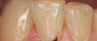
- Stage I. Formation of stains and clouding of the enamel layer. There are no visible destructions, the tissue structure is not changed. Sometimes the spot disappears on its own, which is associated with increased immunity.
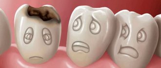
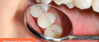
- Stage II. Superficial caries. The enamel undergoes pigmentation, and when examined with instruments, a softening of the structure is felt. Often, superficial damage to a tooth covers all layers of enamel, but is limited. Dentin is still not involved in the pathological process. Visually, a carious defect looks like a dark gray spot with a rough base.
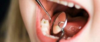
- Stage III. Average caries. The defect becomes pronounced, the lesion covers all layers of enamel and part of the dentin. Patients are increasingly noticing bad breath and discomfort when drinking or eating.
- Stage IV. Deep caries reaches the pulp membrane and is accompanied by tooth sensitivity, pain during chewing or at rest. The pain radiates to the temporal regions. The pain can be acute, throbbing, moderate, persistent. It intensifies and subsides involuntarily.
Typically, patients seek help from a doctor when symptoms worsen the quality of life and the dental-root system atrophies. When examining the oral cavity, caries is classified into:
- localized or generalized;
- acute or chronic;
- complicated or uncomplicated.
Carious lesions can be primary or secondary. In the first case, the disease occurs for the first time, in the second, caries develops again, under a filling, crown, bridge, or veneers. Clinically, there is another classification of caries according to Black:
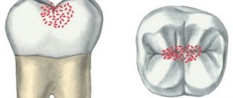
- Class I or fissure caries - characterized by deepening of the natural grooves of the enamel on the chewing surface;
- Class II or carious defect of molars - the formation of defects on the contact surfaces of premolars, molars;
- Class III - damage to the canines and incisors without the defect extending to the cutting edge;
- Class IV - the edges of the canines and incisors are involved in the pathological process;
- Class V - the formation of cervical caries in any group of teeth.
Other types of caries are described in the international classification of diseases. There are unspecified or other caries, odontoclasia (atrophy of the root part of milk teeth), stopped caries after hygienic cleaning, preventive sanitation of the oral cavity.
Diagnostics
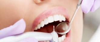
The dentist begins by examining the patient's mouth using a dental mirror. If there is a defect in the crown, it is examined using a probe. After this, the doctor prescribes:
- X-ray diagnostics; if cervical caries of only one tooth has been detected, targeted radiovisiography is prescribed to determine its depth and volume; if a general X-ray of the jaw is necessary, an orthopantomogram is prescribed;
- electroodontodiagnostics (EDD) – determination of pulp viability and pain sensitivity of the affected tooth;
- laser fluorescence – illumination of pathological foci using a laser device;
- painting the stain with water-soluble paints (methylene blue or magenta); in places where the enamel is damaged, it becomes permeable to paint and the pathological focus acquires clear contours;
- X-raying the crown using a special lamp to detect any hidden cavities.
Causes
Caries is the result of the activity of pathogenic microflora in accumulated plaque or tartar. A person’s immune status plays a major role in the development of carious defects. At particular risk are persons with autoimmune pathologies, AIDS, HIV, metabolic disorders, diabetes mellitus, and those taking immunosuppressive therapy for organ or tissue transplantation for a long time. The following factors can contribute to the development of caries:
- smoking, alcoholism;
- inadequate oral hygiene;
- natural aging (decrease in the body’s defenses, changes in the biochemical properties of saliva);
- metabolic disorders;
- endocrine pathologies;
- lack of food discipline (including prolonged fasting, poor nutrition, anorexia, bulimia);
- gum recession of various nature;
- excessive consumption of sugar and carbonated drinks;
- gastroesophageal reflux;
- bite pathologies;
- violation of hygiene rules when wearing braces and other orthodontic structures for the treatment of pathological occlusion.
Despite the variety of causes, eating disorders, bad habits and organ pathologies are the main etiological triad leading to the development of caries. Caries is not an independent disease. The pathogenetic link in the demineralization of enamel and destruction of the tooth body is a violation of the body's defenses.
The danger of decompensated form
Acute caries is dangerous for many reasons. The advanced form of the disease often leads to the following problems:
- development of pulpitis and periodontitis;
- development of periodontitis;
- tooth splitting;
- tooth loss.
In addition, the decompensated form is a signal of a disruption in the functioning of the entire organism. Untreated caries is often a consequence of decreased saliva production and a decrease in its bactericidal properties, which affects the general condition of the oral cavity as a whole. In pregnant women, this can affect the general physical condition of the expectant mother and the health of the fetus.
Symptoms
Symptoms of caries are determined by its stage and location. Patients practically do not notice the initial stage of the disease, especially if the affected area is outside the smile line or on the inner surface of the tooth. However, pay attention to:
- enamel roughness;
- change in shade;
- minor defects (deepening of fissures, cavity fragments).
As the carious process develops, defects become noticeable upon visual inspection, and the cavities deepen. Patients note an unpleasant odor from the mouth, even stench, mainly in the morning. Pain appears. When the first signs of caries appear, you should consult a doctor.
Acute caries affects several teeth at once. The enamel softens, crumbles, and the focal fragment has an irregular asymmetrical contour. Soreness accompanies a person almost all day. There is an acute form of generalized caries, when almost all teeth or several units in the jaw row are involved in the pathological process.
In chronic cases, enamel pigmentation comes to the fore. Destruction is sluggish, pain occurs rarely.
Lack of treatment for chronic caries naturally leads to the destruction of the pulpous membrane and the pulp tissue itself, and the formation of a cystic component.
General recommendations for treatment
Traditional preparation of the defect is carried out using a drill. If the carious cavity is located within the dentin and does not affect the pulp, then after thorough cleaning, a filling is applied. However, sometimes with a small cavity, applying a multi-layer filling (insulating pad + filling material) is impossible. This feature is taken into account when treating caries on the contact surface of premolars and molars.
Another way to treat shallow caries in dentistry is to use a composite material with adhesive properties. In this case, extensive preparation and creation of a wide cavity is not required. Another method involves polishing a carious defect with remineralization of the enamel with medicinal applications, electrophoresis with a 1% sodium fluoride solution and other approved medications.
Medium caries is treated only by deep preparation and filling. In this case, the cavity is cleaned until the affected tissue is completely removed. High-quality sanitation ensures reliable fixation of the filling and increases its service life.
Complications
Untimely treatment of caries leads to inevitable complications:
- development of pulpitis (the carious process gradually spreads to the deep layers of dentin surrounding the pulp of the tooth);
- if pulpitis is not treated, necrosis of the pulp occurs (i.e. it dies), as a result, a focus of inflammation appears at the apex of the tooth root;
- development of gum “flux” (a purulent process occurs between the jaw and periosteum);
- the appearance of a cyst at the apex of the tooth root (the bone tissue is resorbed, and inflammation forms in its place).
Features of the treatment of deep caries
Removing a carious cavity with deep damage to dental tissue is fraught with difficulties, and the choice of method depends on the degree of destruction of your own tooth. Treatment tactics:
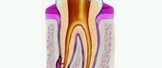
- With pulpitis. Pulp damage involves deep preparation and removal of the nucleus pulposus along with nerve endings from the root canals. An antiseptic is placed into the lumen of the root canal, and the cavity is closed with a temporary filling or medication. This is necessary for etching and eliminating a secondary infectious process. After 1-3 days, the root canals are filled, and the defect is covered with a composite material.
- No pulp damage. Sometimes, with deep damage, the pulp remains intact, covered by a small layer of intact dentin. Preparation is carried out precisely to this tissue in order to preserve the integrity and functionality of the root system. Often the therapist has to leave this part of the dentin, despite its softening, friability and pigmentation. In such cases, a pad based on paste-like calcemin is placed at the bottom of the defect for antiseptics and restoration of dentin. An insulating gasket and a permanent filling are also placed into the defect cavity.
In case of significant tooth decay, patients are recommended to restore the body of the unit with a composite material. This method is not considered durable due to abrasion, destruction, and the formation of new carious cavities. The only alternative in particularly difficult cases is implantation, microprosthetics or prosthetics with a bridge structure.
How is pulpitis treated?
Pulpitis can be treated biologically or surgically. The first is applicable if the disease is in its early stages. The dentist strives to preserve the pulp by eliminating only the inflammation. If possible, pulpitis of primary teeth is treated using a biological method, when it is especially important to preserve the tooth root. Treatment is carried out in several stages. First, the dentist prepares the carious cavity, opening it as wide as possible. Then he treats it with an antiseptic, puts a swab with an antibacterial agent and covers the tooth with a bandage. During the next appointment, if the patient has no complaints, the cavity is treated with medication, filled with a special composition that stimulates dentin production, and a temporary filling is placed for a period of 5 to 7 days. During the third appointment, the dental crown is filled.
When the biological method of treating pulpitis is ineffective, surgical treatment is used. In this case, the inflamed neurovascular bundle is removed completely or partially. Without a nerve, a tooth becomes unviable and its service life is reduced.
Surgical treatment is performed with local anesthesia. First, the carious cavity is prepared, the pulp is removed, the affected area is treated with an antiseptic, medication is injected, and first the root canals are filled, and then the dental crown. As with the biological method, surgical treatment is carried out over several visits to the dentist.
If pulpitis is not treated, it will progress and can lead to very serious complications:
- flux - inflammatory process in the periosteum;
- periodontitis - inflammation of the tissues surrounding the tooth;
- pulp necrosis - death of its cells;
- sepsis is a blood infection that can develop when an infection enters the bloodstream.
You can avoid such complications if you do not delay your visit to the dentist and begin treatment for the disease at the first symptoms.
Rules for selecting and making a filling
The key to success when applying a high-quality and durable filling is to follow the technology of its preparation. Even when using high-quality materials, violation of the manufacturing and application protocol sharply reduces its performance characteristics.
The choice of filling depends on the location of the tooth in the dental system, the strength of the chewing load, and the characteristics of the bite. So, if it is necessary to treat teeth along the smile line, the filling should not only be durable, but also aesthetically pleasing and inconspicuous. Dentists choose composite materials, silicates.
Filling a carious cavity as the final stage is performed only after complete cleansing of the prepared area, removal of dentinal fragments, and drying. The absence of moisture and organic sawdust increases the contact of the filling with the natural tooth tissue. After the filling is fixed and completely hardened, the doctor grinds and adjusts it to the anatomical shape (removes excess material, shapes the edges). Once the filling is comfortable for the patient when closing the jaws, it is polished.
Special attention is paid to polishing. The better the filling is polished, the lower the risk of developing recurrent caries. The uniformity and smoothness of the coating prevents corrosion, accumulation of plaque and tartar, and further destruction.
Modern methods of caries treatment
Modern technologies for the treatment of caries are based on reducing the invasiveness and aggressive effects of the drill and hard attachments and brushes. Minimally invasiveness and comfort during treatment are especially important in pediatric dentistry. Popular methods:
- ICON technology. Suitable for the treatment of caries at the chalk spot stage. Rehabilitation of the defect is carried out using the unique ICON resin, which has an antibacterial and regenerating effect. The component literally fills the pores in the enamel layer and prevents the further development of the pathological process. The procedure itself lasts no more than 20 minutes.
- Ozone therapy. Triatomic gas ozone is a natural oxidizing agent with high antibacterial properties. The substance removes pathogenic microflora and cleans the tooth surface of plaque. Ozone treatment is indicated for early stages of caries, as well as for superficial demineralization of enamel on molars and the cutting surface of teeth. In the treatment of complex cases, ozone treatment is auxiliary.
- Air abrasive processing. Dental sanitation is carried out using finely dispersed abrasive particles under pressure. This method eliminates the use of a drill and does not injure dead tooth tissue.
- Laser treatment. A minimally invasive treatment method that in most cases replaces a drill. The exclusion of traditional preparation of a carious cavity is possible only in case of superficial caries. Despite the fact that sanitation occurs with a laser beam, patients require anesthesia. During the removal of affected dentin, the laser somehow reaches the pulp and causes acute pain.
- Art methodology and artistic restoration. Non-traumatic cleaning of the defective cavity is performed mechanically, followed by filling with glass ionomer material. The method was developed in underdeveloped countries where it is not possible to open full-fledged dental clinics with a wide range of services. Manual cleaning helps to accurately remove affected tissue while preserving the thickness of the healthy part of the tooth as much as possible.
All methods of caries treatment are widely used in modern clinics, but are not advanced. Most often, dentists sanitize pathological cavities and remove dental defects using a drill.
Alternative medicine in the fight against caries
Caries can be eliminated at home only at the initial stage of destructive changes in the enamel. Non-traditional recipes can stop the progression of tooth decay, especially if treatment is currently difficult or impossible according to indications. Popular rinse recipes:
- Sage decoction. 1 tbsp. raw materials, pour 200 ml of boiling water, infuse until a bright amber-brown hue is obtained, filter and rinse your mouth throughout the day. The plant kills pathogenic microflora and is effective against stomatitis and periodontal disease. You can make lotions with a decoction of sage by applying a gauze pad to the affected area.
- Infusion of calamus roots. A steep tincture of calamus root helps relieve pain and slow down the development of superficial or medium caries. To prepare, you will need to pour 1 tbsp. crushed raw materials, 200 ml of vodka, alcohol solution of propolis. For greater effect, you can add 10 drops of tea tree ether. The resulting composition is placed in a dark place for 5-7 days. Before rinsing 1 tsp. alcohol infusion is combined with 100 ml of warm water.
- Mint based recipe. Fresh mint leaves are pounded in a mortar, poured with hot water (not boiling water!), and left for 30 minutes. Then add 1 tbsp. apple or wine vinegar and leave for 3 days. Mint essence prevents further destruction of dental tissue, removes plaque, softens tartar.
You can rinse your mouth with a solution of soda, sea salt, or an aqueous or alcoholic decoction of propolis. Camphor or fir oil, zinc-salicylic paste can be applied to the carious cavity.
Historical reference
It is believed that the “history” of caries begins with a change in the human diet, that is, from the time when coarse, unprocessed food began to be replaced by food subject to heat, steam and other processing, as well as from the moment when “sweets” appeared. Judging by archaeological finds, for example, by the teeth of a man who lived 13-14 thousand years ago found near the city of Lucca (Italy, Tuscany region), we can conclude that already then caries was encountered and attempts were made to treat it. In the teeth found near Lucca, cavities were found that reached the pulp, and on the walls of the teeth there were traces of drilling with some stone tools. Moreover, the teeth had fillings made of plant resin with plant fragments. This means that ancient healers tried to protect carious cavities from the penetration of food, and the plants added to the resin composition apparently served as an anesthetic.
The healers of the Mayan civilization, judging by other archaeological finds, made fillings from complex plant compositions with the addition of various minerals. In ancient Rus', caries in the initial stages was treated with honey, and in advanced stages - with propolis, which was applied to the tooth cavity, previously cleaned of decay products and affected tissues; to relieve pain, they applied a cloth soaked in a mixture of vodka and table salt. In ancient Rus', caries in the initial stages was treated with honey, and in advanced stages - with propolis, which was applied to the tooth cavity, previously cleaned of decay products and affected tissues; to relieve pain, they applied a cloth soaked in a mixture of vodka and table salt. In medieval Europe, where the development of medicine was hampered by the dogmas of the church, it was believed that caries developed due to the activity of “tooth worms”, and it was treated with poisons and hot metals introduced without anesthesia into the dental cavity affected by the disease. Moreover, these manipulations were carried out not by doctors, but by barbers. The poor people, having no means for “treatment”, received very strange, unscientific recommendations from the “doctors”: sit under the moonlight with your mouth open, tie a frog to your jaw, apply a mixture of oil and bird droppings, “rub” the affected area with the tooth of a deceased person , etc.
In the 15th century, healers began using metals, including gold, to fill teeth. In 1790, John Greenwood, the personal dentist of the first US President George Washington, came up with a primitive device for drilling teeth, where a foot drive from a spinning wheel was used to rotate the drill. More than 80 years later, in 1871, the American doctor James Morrison patented a dental drill he invented, which gave impetus to the development of therapeutic dentistry. Throughout the 20th century, therapeutic dentistry developed rapidly, adding more and more new techniques, materials and tools for the treatment of dental diseases, including caries. Today, caries treatment is the most common dental service, with high treatment effectiveness at any stage.
Preventive actions
Prevention of caries consists of high-quality removal of tartar and plaque using special means: toothpastes, rinses, flosses, irrigators. Prevention of caries in children comes down to early instillation of oral hygiene skills. Popular recommendations:
- annual dental examination;
- professional teeth cleaning to remove tartar and plaque at least once a year;
- taking vitamins and calcium-containing medications;
- normalization of diet;
- improving food quality, balanced diet;
- timely treatment of chronic diseases of internal organs and systems.
Persons with chronic diseases of the ENT organs, prone to colds and respiratory infections, should take immunomodulators in courses. In case of a burdened dental history or hereditary predisposition, special pastes with a high concentration of fluoride are recommended. For recurrent caries, periodic application of mineralizing, antiseptic and protective drugs and varnishes to the enamel is indicated.
If you have braces, orthodontic aligners or plates, you should use an irrigator and special brushes. However, even careful care does not exclude professional cleaning of the product and teeth from plaque 2-3 times a year.
Timely treatment of dental caries preserves the integrity of the unit, prevents its early loss and complications. Regular sanitation of the oral cavity improves the quality of life, the aesthetics of a smile, and self-confidence.
Prevention
To combat the development and recurrence of the carious process, you need:
- stop consuming large amounts of easily digestible carbohydrates - sweets, baked goods, sweet carbonated drinks;
- do not have frequent snacks;
- regularly include dairy products in the diet, which the body needs as a source of calcium and phosphorus;
- Brush your teeth twice a day with a soft toothbrush and use toothpaste recommended by your dentist (the choice of toothpaste depends on the condition of the teeth, heredity and the fluoride content of the water in the area);
- After each meal, rinse your mouth thoroughly and use dental floss or an irrigator to clean the interdental spaces;
- visit the dentist twice a year to identify and prevent the initial stages of cervical dental caries, and carry out professional cleaning.
If you regularly adhere to these requirements, you can maintain healthy teeth for a long time.