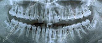
A panoramic photograph of the teeth (orthopantomogram or OTP) is an x-ray of the upper and lower jaw. Allows you to examine most dental problems in detail, conduct high-quality diagnostics and prescribe treatment.
A panoramic dental photograph shows not only the teeth, but also the jaw joints, paranasal sinuses and facial bones. All this makes it indispensable for orthodontic, surgical and reconstructive treatment. You can obtain OPTG using an orthopantomograph.
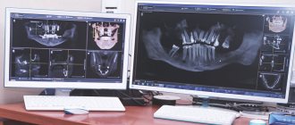
Panoramic photograph of teeth (optg) in Moscow
A panoramic photograph of the teeth is one of the most important and informative studies that must be performed before any orthodontic or surgical treatment.
This method allows you to determine with maximum accuracy the location of the teeth, their root system, and nerve endings. Also in the picture you can see cysts and foci of inflammatory processes formed for any reason. The more information the doctor has on hand before the operation, the more confident you can be in its favorable outcome. At the Kingdom of Smiles dental clinic, you can perform OPTG; the most advanced equipment is used for this. The company values its impeccable reputation, therefore it hires professionals with extensive experience. Highly qualified dentists and orthodontists will help any client become the owner of a perfect smile in the shortest possible time and without any risk to health.
What is a panoramic dental image (optg)
Otherwise, a panoramic image is called an orthopantomogram. This study is the most traditional and quite informative, so it is quite sufficient to obtain a complete picture of what is happening in the patient’s oral cavity. One picture clearly shows all the roots and teeth of the upper and lower jaws. They began to be called panoramic because the doctor receives maximum information about the entire dental system, which is incredibly convenient.
Some time ago, it was possible to see all the teeth at once only by taking many targeted photographs at once. In addition to hard tissues, the image contains maximum data about the maxillary sinuses and even the joints located in the jaw. OPTG has a number of advantages compared to alternative diagnostic methods. First of all, this concerns the relatively low cost, as well as convenience and the ability to get results in the shortest possible time.
Panoramic examination is prescribed in a variety of cases. The main indications are examination of the oral cavity in case of suspected periodontitis, jaw fracture due to trauma, oncological disease of hard tissues, as well as in the presence of partially or completely unerupted units. Unfortunately, X-rays do not provide data on the volume and density of bone tissue, so one such image before orthodontic treatment or implantation will not be enough. Root cracks, faults and minor deformations will also not be detected.
Orthopantomogram 3D
A three-dimensional panoramic x-ray of the jaw can be performed with a computed tomograph. He takes many sequential photographs in different projections and then completes the image in a computer program. The result is a three-dimensional image of the jaw without distortions, which are inevitably present on a conventional otopantomogram, since it conveys a curved object in a flat form. Only with the help of computed tomography does it become possible to look at an object from a different angle or in a different projection without taking new pictures. Also, this method of examination allows the doctor, in the absence of the patient, to see an image of his jaw and examine it from any side and even at any depth.
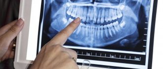
How to take a panoramic photograph of teeth (optg)
To conduct a panoramic examination, there is special equipment called an orthopantomograph. The person is asked to stand in the center of the machine, he must bite a small plate and hold it in his mouth. The latter is necessary to ensure that the jaws remain motionless throughout the operation of the technique. After this, the moving part of the device begins to move around the patient’s head. At the front, this part has a powerful source of X-ray radiation, and at the back there is a sensor and a film where the data is received.
All manipulations last no longer than 10-25 seconds, depending on the novelty of the device. Modern options make it possible to reduce irradiation time, which has a positive effect on the patient’s health. That is why you should not save on such important examinations; low prices can only be achieved if low-quality equipment is used.
The resulting images can be captured on film or in digital format, the latter being the most convenient and high-quality. All data can be studied in great detail on a computer; individual sections can be enlarged, cropped, etc. If necessary, the picture is printed on a regular printer, on regular or photo paper.
How is an orthopantomogram done?
No special preparation is required for the study. You will need to remove glasses, earrings, hair clips, other metal objects, and dentures. An orthopantomogram is usually done in a standing position. A special X-ray protective apron-cape is put on the chest. The position of the head is fixed with special clamps. As directed by the doctor, you will need to bite a special plastic mark (which is located in a disposable cover) - this will allow the image to be correctly centered. The tongue should be kept pressed to the palate. During an orthopantomogram, the module will move around the head, this will take 15-20 seconds, and during this time you must stand strictly still in order for the image to be of the proper quality.
You can get an orthopantomogram in Moscow at Family Doctor JSC.
Sign up for diagnostics Do not self-medicate. Contact our specialists who will correctly diagnose and prescribe treatment.
Types of panoramic dental photographs
Today there are two types of panoramic photographs: digital and film. Film machines work like a classic X-ray device, but specialists rarely use them these days. During the examination, there was an X-ray tube on one side of the person’s head, and a black film on the other. The information was immediately transferred to the film, but it had to be further developed.
A digital orthopantomogram involves replacing the film with a digital medium, that is, with a special cassette. Once all the data is received, it can be immediately transferred to the database on the computer. After this, the specialist begins processing the image, displaying it on the monitor or printing it. Modern technology has many advantages. The main ones are receiving a lower radiation dose and the highest quality images.
Previously, a new film had to be replaced in place of the used film, but the cassette did not need to be changed, since it was reusable. All data is convenient to store and transfer; it will not be lost over time, will not deteriorate or be lost due to moisture. In addition, if necessary, information can be easily sent by e-mail to a hospital in any country in the world. Now patients have the opportunity to have their teeth treated with the least harm to the body and at reasonable prices.
Digital or film orthopantomogram: what to choose?
Digital equipment today has practically replaced film analogues from practice. Although they cost clinics more, they do not require the costs of consumables and darkroom equipment.
In addition, film devices are characterized by a higher degree of radiation.
A panoramic image taken digitally is instantly transferred to the doctor’s computer and entered into the patient’s electronic record. If desired, the latter can receive the image on disk or in printed form.
Finally, the image quality with digital equipment is much higher than with film equipment. This means that the specialist receives better information for further work.
Contraindications
Like any medical procedure, an orthopantomograph examination has some contraindications. If a person, due to the nature of his work or due to undergoing radiation therapy for oncology, regularly receives small portions of radiation, then the possibility of research is considered on an individual basis. Compared to other devices, dental equipment is practically harmless.
If you only need to take one photo, then there is no point in worrying. It is important to understand that without it, it is very difficult to carry out full treatment, especially for complex pathologies. Lack of proper preparation can lead to the development of complications and medical errors. This is especially important before installing artificial teeth, which should serve their owner for many years.
Some doctors warn that it is not recommended to conduct such an examination during pregnancy and breastfeeding. However, as you know, the presence of an infection in the mouth, even due to a small carious cavity, can cause much more harm to the body of the mother and baby than its treatment. It is impossible to remove the affected areas without taking a picture; acting blindly can cause even more harm.
Painless procedure
The procedure itself is absolutely painless and quick. Unlike MRI, where a person is practically in a confined space, there is nothing to be afraid of. During one procedure, a person receives a minimum dose of radiation, which is incomparable to what can be obtained with radiation treatment (no more than 0.11 m3v). Fortunately, modern equipment allows us to reduce harm to minimal values, namely up to 0.04 m3v.
To better understand, you can compare a panoramic shot with a flight from America to Europe or vice versa. For 8-9 hours in the air, a person is exposed to radiation in the amount of more than 0.05 m3v, that is, a trip across the ocean and back can be easily compared with more than two studies on an orthopantomograph. The most dangerous device used by doctors is a CT scanner. The dose will be almost 30-40 times higher, so conscientious specialists do not prescribe a CT scan unless absolutely necessary.
If the patient needs to take several pictures in a year, then there is no need to worry either. Ultimately, the benefits of treatment will be much more noticeable than the harm from the device used. After completion of the procedure, there is no discomfort, discomfort, dizziness or pain. Immediately after leaving the clinic, you can return to the normal rhythm of life, drive a car, communicate with colleagues. If for any reason you feel unwell, it is best to immediately seek advice from your dentist. Only he will be able to understand the cause of what is happening and prescribe preventative therapy or medication.
Orthopantomogram on film or digital: which one to choose?
Today, electronic devices have almost replaced film devices. Their cost is higher, but they do not require a large amount of consumables, as for film orthopantomographs. To develop film, you need a darkroom, which is prohibited by law from being placed in residential buildings, which can become a problem for small private clinics. In addition, the irradiation exposure of film devices is several times higher than that of electronic devices. An orthopantomogram of the jaws, made on a digital device, is immediately transferred to the doctor’s computer, who can be located anywhere. It is much easier to store such an image: it will simply remain in the patient’s electronic record and can be viewed at any time. If desired or necessary, you will receive the result of the orthopantomogram on disk or printed on transparent film.
Unlike a panoramic photograph on film, a digital orthopantomogram will not be lost, which means that the patient will not have to undergo radiation again, which is present during dental x-rays, albeit in very small doses: only 0.002 - 0.003x mSv, whereas, according to Sanitary Regulations , a person can receive up to 1 mSv per year for research purposes, and from the environment we receive 2 - 3 mSv over the same period. In addition, the image clarity and image resolution on digital cameras is much higher than on film cameras.
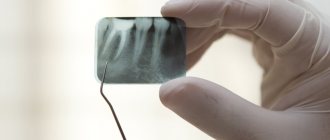
Before and after panoramic dental x-ray (optg)
A panoramic photograph allows you to study the structure of all tissues of the oral cavity with maximum accuracy; the roots, canals, joints and other details are clearly visible in the photograph. When visiting a professional medical institution, sheets with explanations are immediately issued for the images. In the text you can familiarize yourself with the details of the study; the ongoing pathological processes and deviations from normal indicators when they are detected will certainly be indicated.
Even an ordinary person can draw certain conclusions when viewing an image, because everything is quite simple. In the picture, wisdom teeth are clearly visible; they can grow in the right direction or at an angle in relation to adjacent units. Also, the number of previously filled canals and composite fillings will definitely catch your eye; quite often inflammation in the pulp chamber, changes in the root apex, etc. are clearly visible.
Without much experience, it will be more difficult to assess changes such as a decrease in bone volume or the degree of caries damage to the tooth. Bone deficiency can occur due to advanced periodontitis and other serious dental problems. Thus, the image provides comprehensive information about all the nuances of the location and structure of individual parts of the oral cavity, which, of course, is very helpful in drawing up a treatment plan.
Possibilities of orthopantomogram
A panoramic dental x-ray is absolutely painless and safe, takes only half a minute, and allows you to obtain a lot of information about the condition of the jaws. This research helps:
- assess the condition of all dental units, maxillary sinuses, temporomandibular joint;
- see the presence of carious lesions, neoplasms (cysts and granulomas);
- identify periodontal and bone tissue diseases at the initial stage;
- control the quality of any dental services, including root canal filling;
- monitor dental development pathologies in children.
The advantage of an OPTG image of teeth is the high accuracy of planning for orthodontic correction, prosthetics, dental implantation and others. The price of such a study will not affect the cost of treatment, but the specialist will accurately diagnose dental diseases.
Rehabilitation after panoramic dental imaging (optg)
Unfortunately, many patients are afraid to have x-rays and panoramic photographs because they simply do not have enough information about these procedures. Because of this misconception, fear and mistrust arise, but it's not that bad at all. Some sources claim that photographs are allowed to be taken only once a year, otherwise you can harm your body. This is also not true! Lack of timely treatment or doctor's work without prior examination can be more dangerous.
As with an X-ray, there are no special recommendations after an OPTG; you do not need to make changes to your schedule, go on a diet, or stop exercising. Just a couple of minutes after you leave the room with the device, you can forget about visiting it. It is extremely rare for people to experience pain and discomfort, but this is associated more with the fear of the dentists themselves than with the influence of the equipment.
There are no consequences from radiation either. Doctors give standard recommendations regarding recovery: you need to drink more warm liquids, eat soups, and it is best to avoid eating spicy foods, coffee, strong tea and alcohol. If mild stress occurs, you can take a prescribed sedative.