In the Ruddle on Rotating Files series, dentists will find a lot of useful information to help them shape root canals for three-dimensional obturation. Our articles will allow clinicians to learn more about the safe use of nickel titanium (NiTi) rotating files. In short, it will be interesting and educational for all clinicians performing root canal preparation procedures, regardless of the instruments and treatment methods they choose.
The first article discusses the factors influencing the success of rotating nickel-titanium files and the advantages and disadvantages of the most commonly used systems.1 The second covers the basic principles of proper access, strategies and sequences for preparing root canal shapes, and irrigants and their role in root canal cleaning.2 This article will introduce NiTi progressive taper rotating files (ProTaper) (Dentsply Tulsa Dental; Tulsa, Oklahoma), describe their geometry, properties, features, and basic principles of their use.
Advantages of working with universal propapers
Protapers are made of a special alloy, which is 56% nickel and 44% titanium. They have the same design as the machine versions, but are used in more complex clinical situations. Due to their ultra-flexibility, high safety and cutting efficiency, they are widely used by dentists in clinics.
Advantages of manual pro tapers:
- Easy to use.
Thanks to color coding, it is easy to select the sequence of instruments, regardless of the shape of the root canal. Drying and obturation products have the same color scheme.
- Speed.
The work requires only three tools that have high cutting efficiency.
- High performance.
Treatment of the root canal and removal of dentinal filings occurs efficiently due to the increased taper of the apical part.
- Safety.
The likelihood of deviating from the channel passage is minimal due to the rounded guide tip.
Most dentists prefer the manual option due to better tactile control when performing complex manipulations from an anatomical point of view.
Advantages and disadvantages
Modern prop tapers have many advantages: they are durable, easy to use, and highly effective. In addition to these qualities, they have additional advantages that make the doctor’s work easier:
- To completely clean the canal, 3 to 6 propapers are required - the operating time is reduced;
- due to its shape, dentine filings are instantly cleaned when removing the instrument;
- each item has its own color coding, so the doctor will not confuse the sequence of devices during treatment;
- the likelihood of tooth damage when the tool deviates from the trajectory is reduced;
- Suitable for processing rounded channels.
At the same time, propapers have a number of disadvantages that limit their use:
- the length of the processed channel should not exceed 31 mm, because there are no longer pro-tapers;
- channel width – up to 0.3 mm (up to 30 pro taper size);
- they do not have systems to prevent obstruction (blockage) of the canal.
Protapers require careful handling and compliance with the instructions prescribed by the manufacturer. Otherwise, you won't be able to reuse the tool. It can only be used by a dentist who has undergone preliminary training.
Working with manual tapepers: classification
Dental tapepers for endoscopic treatment are used strictly according to the instructions. Each set contains six tools, which can be divided into two groups: shaping and finishing files.
Shaping files are designed to give the root canal a specific shape.
Types of forming files:
- SX - used to work with short root canals or to give the required shape to the coronal part of long passes (the length of the protaper is 19 mm, the diameter at the tip of the working part is 0.19 mm, the base circumference is 1.20 mm).
- S1 – used for preparing the upper third of the root canal (the size of the manual taper can be 21 or 25 mm, the diameter at the tip is 0.17 mm, the taper increases along the entire length of the working part from 0.02 mm to 0.11 mm).
- S2 – equal in length to the previous version, but designed for preparation of the middle third of the root canal (circumference at the tip – 0.2 mm, taper increases gradually from 0.04 mm to 0.115 mm).
Finishing files are used at the final stage to shape the apical third, align and widen the middle third of the canals. They are divided into the following categories: F1, F2 and F3. All have a fixed taper, are quite flexible, but differ in length. Size F1 - 0.2 mm, F2 - 0.25 mm and F3 -0.3 mm.
It is possible to use auxiliary shaping files (shapers), which are used to give the optimal shape to short channels, to access long passages or to determine the directional channel.
Finishing tools for machining
Finishing instruments F1, F2, F3 are intended to complete the formation of the apical third of the root canal and expand and level it in the middle third. But usually one finishing file is enough to work on the apical third.
ProTaper Next Tool Set
A feature of their geometry is the different levels of taper of different blade segments. At D0:
- at 0.20 for F1;
- at 0.25 – for F2;
- in 0.30 mm – for F3.
The taper in D0-D3 is fixed at 7, 8 and 9%.
Over the course of D4-D14, each of the tools, with a steady increase in diameter, has a decreasing percentage of taper. This design feature allows for greater flexibility of individual sections of the file blade, reducing the risk of tool blocking.
Finish tapers are marked with rings on the handles of yellow, red and blue colors in accordance with the diameter of the blade and the sequence of use of the tool.
Marking of hand tools
The manual ProTaper is used for the same manipulations as the machine one, but in more complex cases. However, the markings on the products are the same. The standard set consists of six tools, which are distinguished by the color of the handle depending on the technical data.
Standard marking of the handles of assorted manual tapers:
- Sx – orange;
- S1 – purple;
- S2 – white;
- F1 – yellow;
- F2 – red;
- F3 – blue.
These symbols make it convenient to use hand tools in the required order. There are also F4 (with a black handle) and F5 (with a black and yellow), the length of the active area is 22 mm. They are intended for initial or final treatment of root canals.
Introduction
The new NiTi rotating ProTaper files revolutionize root canal preparation.3 They are specially designed to provide good flexibility, efficiency and significantly greater safety. The unique design features of ProTaper files allow clinicians to create uniformly tapered shapes over and over again in anatomically difficult or highly curved canals (Figures 1 and 2). The kit consists of just six (6) easy-to-use files, a series of three 'shaping' and three 'finishing' tools, currently available in two sizes: 21mm and 25mm (Figure 3). The following section describes the geometry of the ProTaper.
Picture 1
The anatomically challenging maxillary second premolar has an additional pulp shelf, and three canals are carefully processed with ProTaper files. (Property of Dr. Philip Lumley; Birmingham, England)
Figure 2
The significantly curved and split canals of the mandibular second molar were gracefully shaped using ProTaper files. (Property of Dr. Elio Berutti; Torino, Italy)
Figure 3
The ProTaper system revolutionizes endodontic preparation because it consists of only three forming files and three finishing files.
Design Features
Knowing the operating features and order of manual protapers, it is worth noting their design characteristics. It is thanks to them that the dentist’s work in hard-to-reach places is greatly facilitated.
The design feature that provides operational benefits is as follows:
- Multi-stage taper.
Improves flexibility and cutting efficiency, making it unnecessary to go through the canal repeatedly. For example, an Sx file between D1-D9 has a progressive taper from 3.5% to 9% and a fixed taper of 2% between D10-D14. Also, file S2 has nine values, and S1 has 12.
- The trihedral cross-section is convex.
Thanks to this, the main rod is strengthened, and the tool itself becomes super flexible. It also significantly increases safety, since the torsional load is reduced and the likelihood of contact between the walls of the channel and the tool blade is minimal.
- The spiral steps and angles change.
The helix and pitch angles are constantly changing, making it easier and more efficient to extract waste material.
- The tip diameter differs depending on the file.
Files, both finishing and forming, have different diameters for safe and efficient advancement deep into the canal.
- The guide tip is modified.
Due to the shape of the tip, the use of the instrument does not damage the canal walls, penetrating through soft tissue.
- Short handles.
The handles are up to 12.5 mm in size, which improves access to chewing teeth.
- Set of six tools.
Allows you to prepare canals of any complexity, making hand instruments universal.
Main characteristics
Protapers have unique features that allow treatment of complex and atypical canals.
- Convex triangular cross section. This design increases the strength of the profile and reduces the contact area with the canal cavity, thereby making treatment safe and reliable.
- Multiple tapers increase tool flexibility and efficiency.
- Varying angles and pitches of spirals. This feature allows you to clean the cavity as efficiently as possible, reducing the risk of clogging.
- Progressive taper. This design increases the flexibility and cutting ability of the file, thereby reducing the time for canal processing, as well as increasing the safety of the manipulations.
Indications and contraindications for use
The method used by manual tapers is so delicate and filigree that the devices are used only in a medical facility by specialists who have undergone special training. The instruments are designed for shaping and cleaning root canals. Their super flexibility makes the channel easier to clean.
Their introduction is carried out with light pressure, which ensures patency of the area. But at the same time, you must observe the speed limit within 500-700 rpm. There are no contraindications to the use of hand tools.
Actuators and handpieces
ProTaper instruments must be used in handpieces with electric drives that have high torque at speeds of 250-300 rpm. For example, the new AT&R Teknika electric motor (Dentsply Milefer) has software that allows you to select both rotation mode and speed for each ProTaper file (Fig. 18). The new device will make clinical work easier and increase the safety of using NiTi instruments.
Figure 18
AT&R Teknika endodontic motors have many programmable functions, including torque control.
Disadvantages of working with propapers
Despite having many advantages, manual tapers also have disadvantages.
The disadvantages include:
- inability to process wide canals (more than size 30), since there is no instrument with a large apical diameter;
- the maximum length of the canal that can be processed reaches 31 mm;
- a mechanism to prevent obstruction is not provided; a layer of lubricant may remain on the canal walls, which will subsequently prevent medications from entering the canal.
Tools for retreatment
The standard consists of a set of 3 sequentially used devices (D1 – D2 – D3) with different lengths and blade tapers, created for ease of filling teeth.
The shortest tool D1 is used for penetration of the coronal, the middle one D2 - for the middle, the longest one D3 - for the apical zone of the canal.
In addition to the dark gray color of the handles with a length of no more than 11 mm, the simplicity of the application algorithm (from shortest to longest) is ensured by the presence of one, two or three white rings on the handles, respectively.
The active cone tip of the D1 initiation file facilitates initial penetration into the roots.
Precautionary measures
When using hand tools, you should adhere to certain rules.
Precautions are as follows:
- many instruments are marked for “single use” (reusable sterilization and disinfection increase the risk of file fracture);
- Protapers are not immersed in sodium hypochlorite solution, the concentration of which exceeds 5%;
- cleaning is carried out strictly according to the instructions;
- manual propapers are used at a constant speed within 150-350 rpm;
- the file is checked for possible deformation, the grooves should be cleaned as often as possible;
- to create straightforward access to the canal, it is worth using shaping files with separate sweeping movements;
- sweeping movements are not applied to finishing files;
- Using finishing files over the entire working length, you should immediately remove the tool.
How to choose the right one
Each dentist chooses a prop taper model, focusing on ease of use and the methodology of their work. This is an individual tool, so there is no universal selection algorithm.
But since this is a relatively expensive device (the cost of one set of 6 files costs approximately 2000 rubles), it is important not to buy a fake. Its signs will be:
- the working part is matte (high-quality files are polished to a mirror finish);
- the color marking is glossy, the paint is unevenly distributed over the colored part;
- the file is curved;
- the working part of the propaper is visually different from the original, has chips or other defects;
- the price is significantly lower than usual;
- no marking stripes;
- packaging differs in appearance or quality.
The appearance of the packaging does not always indicate a fake. You can call the manufacturer's hotline and find out if the design has changed.
The original packaging must have a holographic sticker.
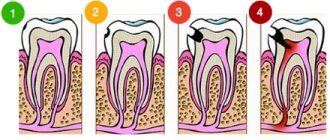
Method of mechanical treatment of root canals
Processing of canals using hand instruments is carried out in different ways, which directly determines the use of manual protapers in order.
Types of processing techniques:
- Standard.
Manual endotonic instruments are used starting from the smallest size. As you move along the channel, increasingly larger diameter propapers are used, reaching all the lengths of the passage. Processing occurs with one large K-file. Once fully inserted into the canal, the instrument is rotated 90 degrees clockwise in depth.
The reverse rotation occurs with slight pressure counterclockwise 270 degrees. Then follows another turn in the opposite direction by 180 degrees. After this, the instrument is removed from the mouth, treated with medications and reinserted into the canal for its entire length.
- Step-back.
First, the root canal must be expanded to the apical foramen, after which a K-file is inserted one size larger, but 1 mm less than the working length. Afterwards, the tool is changed, in which the working length gradually changes (by 2, 3 mm, and so on). With the help of H-files, the surface of the root is smoothed, and thus its taper is formed.
- Crown-down.
The formation of the ostial middle part and access to the apical third of the canal is carried out after expansion of the orifice. Next, the working length and tooling are determined. The canal walls are leveled in the final part.
ProTaper: User Guide
If the rules of use are followed, ProTapers provide excellent results and a high degree of safety. First you need to prepare a cavity for direct access to the mouths of the canals. The pulp chamber should be filled with sodium hypochlorite or a root canal enlargement agent. Rotary instruments should be inserted into the orifice of a canal that has previously been examined for patency using classic 2% flexible hand files or special C+ files designed for this procedure. Using the same instruments, you can further expand the mouth of the root canal and provide straight-line access, as well as determine the diameter of the canal and some of its anatomical features. When using the ProTaper tool, the clinician must follow specific guidelines for its use: select the recommended speed and torque for each file type:
Straight Line Access
Preparing the dental cavity for the shortest possible access is an integral element of successful endodontics.4 This requires removing the roof of the pulp chamber and the excess dentin covering it on top. The size of the cavity is determined by the location of the orifice(s). The walls expand so that the mouth(s) are inside. The inner walls are prepared and smoothed to provide easy, straight access to the root canal orifice. (Fig. 13). It is also possible to further expand the coronal part to eliminate interference during subsequent instrumentation. Our goal is achieved when all orifices can be seen without changing the position of the mirror. Ideally, the access cavity should comply with the standards of restorative dentistry. Creating good access greatly increases the chances of a perfect cleaning and shaping of the root canal. The instrument is easily inserted into the mouth of the canal and, when rotated, slides along the smooth walls of the cavity.5-7
Figure 13
The photograph shows the creation of straight-line access to the root canal orifices located within the access cavity.
Irrigation and Lubrication
No instrument should be inserted into the root canal until the pulp chamber is filled with irrigant. Once direct access has been achieved and all orifices have been identified, the pulp chamber must be filled with warm 5.25% NaOCl solution. For greater safety, NiTi rotary instruments are always used with a Glyde type lubricant (Dentsply Maillefer).
Creating a Guide
Before using any NiTi rotary instruments, it is necessary to examine the channels and determine the "guide" at the mouths. For this purpose, classic steel hand files of 2% taper in sizes 10 or 15 are used, as well as C+ files specially designed for this procedure (Dentsply Milefer, Tulsa, Oklahoma). With the advent of NiTi rotary tools, the role of hand-held instruments is reduced to nothing. Many clinicians use small hand instruments primarily to obtain initial information, confirm access, or, if necessary, to create sufficient space in the canal orifices before using more efficient NiTi rotary instruments. Among other things, they can provide additional information during tactile studies:
1) Transverse Diameter
Using manual files, you can immediately determine the estimated diameter of the canal, obtain information about whether it is open or closed and how calcified it is. Before inserting a rotating NiTi instrument into the canal, it is necessary to provide space for insertion and guidance of its tip. In other words, a depression must be created at the canal mouth and a smooth “guide track” must be created for the insertion of NiTi rotary instruments.
2) Straight Line Access
Straightness of access is confirmed by the position of the hand file handle. If a straight-line access is created in the coronal part of the root, then the handle of the file inserted into the canal will be located vertically, and the instrument itself will be parallel to the long axis of the tooth. In cases where the file handle is displaced from the long axis of the tooth, is located at an angle and is difficult to insert into the canal, it is necessary to make additional expansion and achieve a vertical position of the handle. (Fig. 14).10,11 This requires further expansion of access and sometimes even selective removal of a triangle of dentin from the coronal third of the canal. Creating a straight-line access simplifies all subsequent preparation procedures and practically eliminates many mistakes and failures when preparing canals for obturation.
Figure 14
The hand files are displaced relative to the central axis of the tooth due to the protruding dentinal ridge at the canal mouth.
Traditionally, a series of Gates Glidden drills have been used for such procedures. Today, just one instrument, the ProTaper Sx, can help remove the dentine triangle quickly, effectively and safely (Fig. 15). It provides excellent access to the depth of the root canal, serves to prepare the middle and apical third of the root canal, simplifies the use of any subsequent instrument and increases the safety of the procedure.
Figure 15A
The Sx file requires a return brushing motion to cut the dentin and safely align the direction into the depth of the canal as the dentin overhangs the orifice.
Figure 15B
Sx is primarily used to remove dentin and create lateral space, as well as to obtain a more progressive preparation into the depth of the canal.
Figure 16
Determination of the working length and diameter of the apical constriction before completing the preparation.
3) Anatomy of the Root System
Manual files can also provide very useful information about the anatomy of the root system. For example, a detailed examination can show the degree of curvature, the shape of the canal, and the presence of splitting or fusion of the canals. Please be aware that certain anatomical features of the root canal configuration make it impossible to safely use NiTi rotary files.
Apical Constriction Patency and Working Length
After finishing treatment of the apical third of the root canal, it is necessary to ensure its patency to the radiographic apex (RA). Typically, manual, flexible, small files are used for this. Working with a hand file, you can simultaneously remove the remains of spent dentin particles, moving towards the apical constriction. In this case, it is very important, while maintaining the patency of the apical constriction, not to push the waste masses into the periodontium. This will allow you to avoid apical blocks of dentin particles, as well as ledges and perforations. Most researchers and clinicians are of the opinion that passing the canal to the radiographic apex moves the tip of the instrument beyond the apical constriction. Since the apical third of the canal ends at the dentin-cement junction (DCJ), the so-called “apical constriction” or physiological apex, experienced clinicians suggest that this point be considered the starting point when measuring the working length. But this point varies significantly from tooth to tooth, from root to root, and it is not always possible to determine it tactilely. Previously, when determining the working length, clinicians relied on the radiographic apex. But with the invention of electronic apex locators, determination of the apical constriction has become more accurate than radiography. Apex locators, embodying all the best technological advances of recent years, provide greater accuracy in determining the working length, even for channels containing exudates and electrolytes (“Root ZX”). It should not be assumed that apex locators can completely replace X-ray film. In our opinion, only a combination of electronic apex locators and x-ray examination provides the most accurate diagnostic result when determining the working length of the canal.
Method of Use
Rotary instruments should be used passively within the canal and can continue to be used as long as they move freely toward the apex. When working with ProTaper, the “pencil lead analogy” is used to determine pressure. The pressure on the instrument should be equivalent to our efforts when writing with a finely sharpened pencil. The instrument should be lowered into the canal in the apical direction until slight resistance is felt. If any ProTaper tool stops advancing, remove it and determine which of the following factors is preventing the rotating file from moving:
1) Inadequate Channel Diameter. Determine if the diameter at the tip of the tool is too large to move any further; Is its progress hampered by calcification? In addition, NiTi rotating instruments will not be able to advance through a canal that is sharply bent, divided, or has walls that have resorption defects. In case of calcification, use lubricant in combination with 10 and 15 hand files and, if necessary, some larger hand instruments to create an extension that matches the tip of the NiTi instrument.
2) Presence of sawdust in the channel. To remove accumulated sawdust in the canal, rinse the root canal generously and file it with a #10 hand file to break up the sawdust and turn it into a solution. Then rinse again and remove the waste sawdust.
3) Sawdust between the cutting edges. Sawdust between the cutting edges interferes with the normal operation of the tool, since they push its active part away from the channel walls. In this case, remove the instrument, clean it, rinse the canal, then re-pass the hand file to ensure patency and rinse the canal again.
4) Anatomy of the Root Canal. Some root systems have difficult anatomical configurations that prevent the tip of the rotating instrument from moving easily, accurately, and safely through the canal (Figure 17). In this case, rinse the canal and re-pass it with small hand files to increase the diameter of the “guide track” to facilitate the use of rotary NiTi instruments. It should be recognized that in some cases it is best to shape using hand files. However, the ProTaper No. 1 shaping file, which has a D0 diameter of 0.17 mm and a modified guide tip, can follow the smooth guide path that was explored with hand files 10 and 15. Using the smooth, repeatable guide path, ProTaper rotary instruments will gradually form a channel with the same taper along its entire length.
Figure 17a
Using hand files to study the walls of the apical third of the canal.
Figure 17b
If small hand files cannot easily slide along the canal, then the use of rotating NiTi files should be avoided.
General recommendations
According to the methodology, manual protapers undergo disinfection, cleaning and sterilization before use (18 minutes using an autoclave and at a temperature of 134 degrees, pressure no more than 3 bar).
General provisions include the following:
- Disposable instruments cannot be reused.
- The doctor who uses the instrument is responsible for the sterility of the product.
- To avoid contamination, you should use personal protective equipment (goggles and gloves).
- Disinfectant solutions must be of high quality.
- Hydrogen peroxide destroys tungsten carbide burs, nickel titanium instruments and plastic stands.
- Do not use caustic soda solutions, alkali or mercury salts.
- The maximum number of hand tool processing is limited to five. After this they begin to collapse.
conclusions
This article described the geometry, properties and advantages of ProTapers. The basic principles of their use were also discussed. Our main goal is to help clinicians understand the benefits of this innovative yet easy-to-use toolkit.
The ProTaper rotating file system, introduced over the past few years, is the fruit of the joint efforts of Drs Ben Jonson, Pierre Mastu, Clifford Ruddle and John West, as well as engineers François Haebou and Gilbert Roth (Densplay Maillefer, Balague, Switzerland). Dr Ruddle would like to thank everyone involved in this project for their assistance.
REFERENCES 1. K.J. Ruddle: "Nickel-Titanium Rotary Systems: A Review of Existing Instruments and Geometries," Dentistry Today 19:10, pp. 86-95, 2000. 2. K.J. Ruddle: “Modern concepts in root canal preparation,” Dentistry Today 20:2, pp. 76-83, 2001. 3. J.D. West: "Introducing a New Rotating Endodontic System: Progressive Taper Files," Dentistry Today 20:5, pp. 50-57, 2001. 4. H. Levine: "Access Cavities," Dental Clinic of North America, pp. 701-710, November 1967. 5. H. Schilder: “Cleaning and shaping the root canal system,” Dental Clinic of North America, 18:2, pp. 269-296, 1974. 6. K.J. Ruddle: “Endodontic canal preparation: revolutionary strategies for cleaning and shaping,” Dentistry Today 13:2, pp. 44-49, 1994. 7. P. Mast: “Endodontics - a clinical guide.” Paris: SDP publication, 1993. 8. E. Berutti, R. Marini: “Evaluation using a scanning electron microscope of the cleaning ability of sodium hypochlorite at different temperatures,” J Endod 22:9, pp. 467-470, 1996. 9. E. Berutti, R. Marini, A. Angeretti: “Penetration ability of various irrigants into dentinal tubules,” J Endod 23:12, pp. 725-727, 1997. 10. K.J. Ruddle: “Chapter 8, Cleaning and Shaping Root Canal Systems. In Cohen S, Burns RS, editors: Pulp Paths, pp. 231-291, 8th ed., Mosby, St. Louis, 2001. 11. M.J. Scianamblo: Chapter 15, “Preparation of Endodontic Cavities.” "Endodontics", pp. 374-391, Castellucci, Edizione Odontoiatrice Tl Tridente, Prato, Italy, 1993. 12. K.J. Ruddle: “Chapter 25, “Nonsurgical Endodontic Treatment Revision.” In Cohen S, Burns RS, editors: Pathways of the Pulp, pp. 875-929, 8th ed., Mosby, St. Louis, 2001. 13. Pineda F, Cuttler W: Mesial-distal and buccal -lingual x-ray study of 7275 root canals,” Oral Surgery 33:101, 1972. 14. S. Shabagarg, V.V.U. Guun, A.H. Gluskin: “Laboratory evaluation of the Root ZX electronic apex locator,” J Endod 22:11, pp. 616-618, 1996. 15. Results of a study of leading endodontists from North America, South America, Asia and Eastern Europe. Regional meetings in 2000 and 200.
Brief reviews of the most popular models
The choice of tapeper for each dentist is based on personal preference. It cannot be said unequivocally that a less popular instrument is inferior in quality.
Let's look at the most popular models among doctors.
MTwo
Dentists have good reviews of the MTwo protector from the German manufacturer VDW GmbH. It has highly efficient cutting properties. Its tail is 5 mm shorter than similar instruments, which makes it easier to insert when access to the canal is difficult.
The protaper keeps the shape of the canal cavity unchanged and does not injure it. It is also durable and does not break. When properly processed and used, it lasts for several months. The cost of a set of 6 files is from 2000 rubles.
ProTaper Universal
Manual and automated ProTaper Universal instruments from the Swiss manufacturer Dentsply Maillefer have high cutting efficiency.
In most cases, the doctor only needs 3 tools to work. This significantly reduces the time spent on treating one patient.
They are effective and safe, and have a rounded tip. The set includes paper pins that match the size of the finishing tools. This allows you to quickly dry the canal if liquid gets into it. The approximate cost of the set is 3000 rubles.
Next
The latest development of Swiss dentists is the NEXT protaper from Dentsply Maillefer. Its innovation is that it can move in waves.