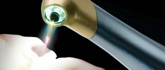
Diode laser
Modern technologies are increasingly used in medicine. Today, laser treatment of periodontitis is commonplace. The healing properties of a narrowly targeted flow of energy are manifested in dentistry, creating the possibility of quick and painless dental treatment. The wide range of services provided with its help, coupled with relative affordability, contributes to the growing popularity of this method.
Treatment principle
Laser treatment is based on the principle of local exposure of the beam to a specific type of tissue. The beam does not affect neighboring areas. Targeted action allows you to remove carious and inflamed areas without affecting healthy enamel, gum tissue and dentin.
Carious tissues contain more moisture. Under the influence of the laser, moisture evaporates from them intensively - tissues affected by caries are gradually destroyed. In 3-5 therapy sessions, the doctor removes all diseased tissue without affecting healthy areas.
When treating periodontitis, it is necessary to remove infected areas that have changed their structure. To do this, the dentist treats the gums with a special solution that reacts with the affected cells. The solution stains the pathological areas, after which the laser acts on them. It evaporates and destroys them without damaging healthy areas.
Principle of operation
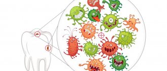
By heating the surface of the tooth, most of the liquid is removed from it. After this, the “protected” inflamed space is released. The laser beam burns out all harmful microorganisms and frees up space for further mechanical cleaning.
Treatment of dental cysts with a laser is carried out similarly to any other operations. A cyst is a formation with dense, hard walls, inside of which there is a large amount of bacteria or dead tissue. Outwardly it may not be noticeable, but in everyday life it causes great discomfort. In particular, previously a dental cyst was treated with great effort.
This purulent sac forms in the roots, so to remove it, in any case, it would be necessary to remove the tooth, clean out the abscess and install an implant in its place. There is another method - surgical. To carry it out, an incision is made in the desired place of the gum, corresponding to the cyst, the dentist-surgeon uses instruments to pull out the bag, and then sutures the tissue.
Laser treatment is also used to treat periodontitis.
What dental diseases can laser treat?
The use of laser is equally effective in pediatric and adult dentistry. It is shown:
- in the treatment of caries;
- in bone and soft tissue surgery using a laser scalpel;
- when installing implants;
- in the treatment of inflammation and infections of the oral mucosa;
- when removing soft tissue formations;
- in the treatment of periodontal disease and periodontitis;
- as a prevention of oral diseases.
All the best for children
Laser therapy is successfully used in the fight against caries even in baby teeth, although only at the initial stage of the lesion. It is advisable for the child to be at least seven years old, but diligent and calm children can take advantage of the latest technology at a younger age.
For the treatment of small patients, the following rules apply:
- it is necessary to use only equipment with the ability to adjust the radiation power;
- During the procedures, a slight tingling sensation may occur, which the child should be warned about.
All other advantages of laser dentistry are common to children and adult patients.
Benefits of laser therapy
Laser therapy has many advantages over traditional treatments. Let's list the main advantages.
- The laser beam acts locally and briefly, without irritating the dental nerves or causing thermal reactions, so patients do not feel pain.
- The painlessness of the treatment allows you to eliminate or reduce the amount of painkillers, which is important for patients with intolerance or hypersensitivity to anesthesia.
- The laser does not produce unpleasant sounds characteristic of a drill - patients in the dental chair do not experience stress.
- In combined treatment regimens, laser radiation enhances the effects of medications and increases the effectiveness of treatment in general.
- Laser radiation is detrimental to pathological microflora, so there are no side effects, including inflammation and infection of healthy areas.
- The rehabilitation period is reduced by 2-3 times.
Diseases of hard dental tissues of non-carious origin
Hypoplasia, erosion and pathological abrasion of enamel are pathologies caused by neuroendocrine disorders or heredity. Doctors developed a universal rehabilitation program (URP) based on laser exposure and conducted a study in which 185 patients took part.
Each of them received 5-7 sessions of laser therapy with a pulse frequency of 50 Hz. The duration of the scanning effect varied from 1 to 5 minutes and covered the following areas:
- apex beat zone;
- epigastric zone;
- sternum;
- right and left hypochondrium;
- subclavian fossa;
- kidneys and adrenal glands;
- suboccipital fossae.
After treatment, 52 people experienced significant improvement in their overall health. They noted improved sleep, high resistance to stress, absence of pain, irritability, and fatigue.
As for the results of treatment of hard dental tissues, it occurred 10 days faster than in patients receiving drug treatment. 150 out of 185 patients got rid of not only the underlying disease, but also sinusitis, tonsillitis, and radiculitis.
Treatment of caries
Caries is the destruction of hard tooth tissue due to chemical exposure and the activity of pathological microflora.
Another example from practice. 270 patients underwent laser treatment, and Moscow dentists diagnosed multiple caries in 150 of them. The treatment was carried out using a RIKTA laser therapy device using dental attachments. Doctors acted on the affected teeth for 1-2 minutes with a pulse frequency of 50 Hz.
In case of multiple caries, radiation was applied to the submandibular and parotid glands. This effect stimulates the production of immunoglobulin and reduces the viscosity of saliva. The duration of the procedures is 2 minutes, with a pulse of 50 Hz.
Each patient received a course of 8-10 laser therapy sessions. Over the next year, 86% of patients did not develop caries. For comparison, in patients undergoing traditional treatment, this figure was 61%.
How laser therapy affects tissue: biopsy results
Research on the effectiveness of laser therapy is carried out not only by measuring the effectiveness in patients, but also in laboratories, at the cellular level. One of these experiments made it possible to track changes in the cellular structure of the periodontium over 5 sessions of laser exposure and, based on its results, determine the effectiveness of therapy.
Histological and cytological studies of tissues before the start of treatment showed that the mucous membrane was affected by the inflammatory process, wound healing was sluggish, and there were practically no full-fledged cells.
After two laser sessions, the healing process intensified. After 4 sessions, the studied samples already showed signs of bright coagulations, a gradual restoration of erythrocytes and leukocyte cells.
After the 5th session of laser therapy, a biopsy showed the presence of full-fledged cells - red blood cells, leukocytes, and fibroblasts. The results indicate the patient's complete recovery.
Among odontogenic acquired cysts, the most common is the radicular (perihilar) cyst. It accounts for about 86% of all odontogenic cysts. This is a cyst of inflammatory origin, developing from cystogranuloma. The growth of the cyst occurs not so much as a result of the proliferation of the epithelium, but as an increase in intracavitary pressure. Due to this, the cyst increases in volume with resorption and restructuring of the surrounding bone tissue. The cause of the development of a radicular cyst in 40% of cases is a tooth affected by a carious process. In 60% of cases, a cyst occurs as a complication of endodontic treatment: pushing necrotic pulp to the apex of the tooth with the subsequent development of periodontitis, trauma to the pulp during odontopreparation, especially under anesthesia [2, 4].
X-rays reveal such complications of endodontic treatment as root perforation, missed canals, inhomogeneous filling, excessive removal of material into the periodontium, lack of sealing of the mouth or coronal part of the canals [4-6].
The main surgical methods for treating radicular cysts of the jaws according to their size are cystotomy and cystectomy. In some cases, surgical treatment is complicated by secondary infection, relapses, and the formation of fistulas due to the persistence of bacterial colonies in the complex branching system of the root canal of the tooth, which has additional lateral canals, deltas, and anastomoses. The likelihood of these undesirable consequences of treatment can be reduced by using modern laser technologies for surgical treatment.
Dental Er:YAG and Nd:YAG lasers, which are capable of emitting short powerful pulses, are distinguished by the fact that their operating parameters (energy, duration, frequency and pulse shape) can be changed automatically depending on the surgical treatment being performed by setting the desired mode on the display, simply by selecting the name of the intervention. The energy of the laser light wave is concentrated in space in the form of a very narrow beam, which is highly directional, monochromatic, and transmits energy continuously or in the form of short pulses. The focused laser light beam serves as an extremely sharp and sterile cutting instrument, with which it is possible to perform operations without direct contact with the tissue, with pronounced hemostasis, and with a decrease in pain intensity. The laser beam of an erbium laser makes it possible to cut both soft and hard tissues. The process of laser exposure to tissue occurs without pressure and friction, and is not accompanied by vibration.
As a result of the thermomechanical process in tissues under the influence of laser radiation, the process of ablation (evaporation) occurs. The chromophore, a substance capable of absorbing optical laser energy and transforming it into thermal energy, for an erbium laser is water. It is water that evaporates from tissues during laser exposure. The ablation process occurs only when a certain energy density (ablation threshold) is exceeded. This threshold is approximately 3.3 J/cm2 for enamel, 2.8 J/cm2 for dentin, 1.3 J/cm2 for bone and 0.8 J/cm2 for skin. When working in the subablative range (below the ablation threshold), only heating and drying of the tissue occurs [7, 8, 10, 11].
The optical energy of the Nd:YAG laser is significantly absorbed by protein structures and pigment deposits (pigment deposits on the root surface). The result of this interaction is homogeneous photothermolysis. The essence of the phenomenon comes down to the absorption of laser energy by the target tissue and its distribution in the surrounding tissues in the form of heat. The depth of the critical temperature effect when using a pulse duration from 90 to 180 μs does not exceed 0.3 mm.
When treating canals during endodontic treatment, Nd:YAG laser radiation (pulsed) due to the lack of absorption by hydroskilpatite and water penetrates as deeply as possible, laterally from the canal to a depth of 1000 µm (1 mm). Thanks to the high peak energy (up to 5 kW), it provides a sterilization effect of more than 99%. In this case, the phenomenon of inorganic melting and sealing (soldering) of lateral practically sterile channels is observed - the effect of glazing of the channel wall (described only for a high-power Nd:YAG laser). Due to the spread of heat over a radius of 1 mm, the effect of tissue sterilization is achieved without opening the top of the canal, sterilization of periodontal tissues in the lower third of the root (root wall thickness less than 1 mm). Measurements on the surface of the tooth root after a 45-second sterilization procedure at a frequency of 15 Hz, a power of 1.5 W and a pulse duration of 100 μs showed an increase in temperature to 38 °C, which does not exceed the physiological norm [7, 8, 10].
This phenomenon is very relevant when performing resection of the apex of the tooth root in patients with radicular cysts of the jaws. In this regard, it is of interest to study the effectiveness of laser cystectomy with sequential use of Er:YAG and Nd:YAG lasers. The resection itself is carried out with an erbium laser, and then the resected root surface is treated with laser radiation from a neodymium laser. After surgical treatment, it is advisable to fill the resulting bone tissue defects with large cysts with biocomposite materials [1]. The use of the domestic drug Osteoplast K in patients with radicular cysts of the jaws helps to accelerate the regeneration of postoperative bone defects and stimulate the activity of cellular factors of local immunity [9].
The purpose of the work is to study the effectiveness of surgical treatment of patients with radicular cysts of the jaws using Er:YAG and Nd:YAG lasers.
Material and methods
We examined and surgically treated 35 patients (23 women and 12 men, patient ages ranged from 21 to 62 years) with radicular cysts of the jaws of various locations: on the lower jaw in 9 patients, on the upper jaw in 26. In 12 patients, for replacement The biocomposite material Osteoplast K was used in the resulting bone cavity. In 19 patients, the traditional method of cystectomy was used, in 6 of them - in combination with the use of Osteoplast K. In 16 patients, laser cystectomy was performed with the combined use of Er:YAG and Nd:YAG lasers, in 6 of them They were also used by Osteoplast K.
Er:YAG laser (DECA “Smart 2940D plus”, Italy) has the following characteristics: wavelength 2940 nm, energy 50-500 mJ, pulse frequency 10-30 Hz. The pulse duration varies from 230 to 700 μs (Fig. 1).
Figure 1. Er:YAG laser DECA “Smart 2940D plus”, Italy.
Neodymium Nd:YAG laser (DECA “Smart A10”, Italy) has a wavelength of 1064 nm, radiation energy of 40-200 mJ, pulse frequency of 5-200 Hz, exposure mode continuous and pulsed. In pulse mode, the exposure duration can be set from 0.1 to 60 s (Fig. 2).
Figure 2. Nd:YAG laser DECA “Smart A10”, Italy.
Laser cystectomy is based on the traditional method of cystectomy. Using an erbium laser, a trapezoidal or arcuate incision was made in the area of the transitional fold according to the localization of the cyst at an energy of 100 mJ, a pulse frequency of 10 Hz in the “very short” mode (short pulses - 230 μs). Then the bone was expanded or the bone was perforated in the projection of the cyst, followed by expansion to the diameter of the cyst at an energy of 150 mJ, a pulse frequency of 10 Hz. Tooth root resection was performed at an energy of 200 mJ and a pulse frequency of 20 Hz. The resulting bone cavity was treated with a defocused laser beam (at a distance of 1.5 cm from the tissue) at an energy of 100 mJ and a pulse frequency of 10 Hz. After that, the resected root surface was exposed to neodymium laser radiation at an energy of 100 mJ, a power of 1.5 W, a pulse frequency of 15 Hz, in the Enh3 mode (very short pulses) for 15 s, and then the open lateral tubules and additional channels were melted at increasing power to 3 W for 5 s.
To justify the sequential use of erbium and neodymium lasers, we used scanning electron microscopy (SEM) to study the structure of the surfaces of transverse and longitudinal sections of extracted teeth, which served as experimental models of tooth roots after resection of their apices by mechanical and laser action during cystectomy.
Results and discussion
According to SEM data, during a survey study of the sample surface under the influence of Er:YAG and Nd:YAG lasers, the surface was flat, the channel branching system had a configuration characteristic of type VIII according to the classification of channels in one root according to Vertucci (1979) [6] (Fig. 3).
Figure 3. Electronic photography. Overview image of the sample (cross section of the tooth).
When examining the cut surface under the influence of an Er:YAG laser, it was revealed that it was finely lumpy, firmly connected to the underlying layer, and the dentinal tubules were open (Fig. 4).
Figure 4. Electronic photography. The sample surface after exposure to an Er:YAG laser in the mode: 200 J, 20 Hz, 4 V, “very short” pulse type.
The cut surface after sequential exposure to Er:YAG and Nd:YAG lasers has a finely lumpy structure, the dentinal tubules are sealed as a result of inorganic melting, additional lateral canals are open, and filling material is not detected in the lumen of the canals. In the depths of the channels, residual collagen structures are visualized, which appear after laser exposure in ablation mode (Fig. 5).
Figure 5. Electronic photography. The sample surface after exposure to an Er:YAG laser in the mode: 200 J, 20 Hz, 4 V, pulse type “very short” and Nd:YAG laser in the mode: 100 mJ, 20 Hz, 2 V, pulse type Enh3.
When studying the cut surface after sequential use of Er:YAG and Nd:YAG lasers with an increase in the neodymium laser power to 3 W, a finely lumpy structure of the dentin surface is determined; the dentinal tubules are sealed as a result of inorganic melting. There is an additional lateral channel in the viewing area. The canal lumen is empty, the filling material is not visualized, melted structures are determined, characteristic of the glazing of the canal wall as a result of partial melting of dentin, collagen structures are not determined (Fig. 6).
Figure 6. Electronic photography. An additional lateral channel after exposure to an Er:YAG laser in the mode 200 J, 20 Hz, 4 V, pulse type “very short”, Nd:YAG laser with parameters 100 mJ, 20 Hz, 2 V, pulse type Enh3 for 15 s and for 5 s in mode: 200 mJ, 15 Hz, 3 V, pulse type Enh3.
The surface of the sample subjected to the mechanical action of a carbide bur is flat with a tuberous-scaly structure of dentin, streaked with stripes left by the cutting part of the bur (Fig. 7).
Figure 7. Electronic photography. The surface of the sample after mechanical impact with a carbide bur, the torque of which is 20:1, 50 Ncm, 800 rpm, cooled with saline solution. On the surface there is a sintered weakly attached (lubricated) layer, consisting of fragments and agglomerates of tissue sawdust, caused by temperature and mechanical factors of influence; along the grooves there are straight cracks left by a mechanical bur, signs of melting and cracking of the tissue are determined, the dentinal tubules are open. In the viewing area there is a longitudinal crack going deep into the dentin tissue, formed as a result of the mechanical and thermal action of a rotating instrument.
On the section obtained by breaking the sample under the influence of Er:YAG and Nd:YAG lasers, the surface is finely lumpy, demonstrating a strong connection with the underlying layer; no signs of delamination and cracking of dentin are detected (Fig. 8).
Figure 8. Electronic photography. The fracture surface of a sample after exposure to an Er:YAG laser in the mode: 200 J, 20 Hz, 4 V, pulse type “very short” and Nd:YAG laser in the mode: 100 mJ, 20 Hz, 2 V, pulse type Enh3 for 15 s and for 5 s in 200 mJ mode, 15 Hz, 3 V, pulse type Enh3.
On the longitudinal section, after exposure to carbide bur, a relatively flat surface is determined, loosely connected to the underlying layers. Free-lying upper dentin layers are visualized; the underlying dentin layers are in places connected to the underlying ones. There are signs of tissue cracking and melting (Fig. 9).
Figure 9. Electronic photography. The fracture surface of the sample after exposure to carbide boron, the torque of which is 20:1, 100 Ncm, 800 rpm, cooled with a NaCl solution.
Analysis of SEM data showed that sequential exposure to Er:YAG and Nd:YAG lasers when cutting tooth roots is less traumatic compared to the mechanical action of a physiodespensor, and no cracks or smear layer are formed. As a result of Nd:YAG laser radiation, the process of recrystallization of dentin occurs, in all likelihood, the bacterial flora is destroyed, the melting of dentin leads to the sealing of dentinal tubules and small lateral canals, as a result of which their permeability decreases. The result of the study indicates that the combined effect of Er:YAG and Nd:YAG lasers during resection of the root apex should promote sterilization and sealing of dentin tissue, and this in turn should reduce the risk of infection of the periapical zone from the dental canal system in the postoperative period in patients with radicular cysts of the jaws.
According to clinical research methods, the advantages of using laser technologies have also been identified. Thus, severe postoperative pain syndrome, requiring the use of painkillers, was determined in patients after traditional cystectomy. Moreover, pain was recorded for a longer time after surgery - up to 4-5 days, while when using laser technologies, less intense pain was noted that did not require taking painkillers for 1-1.5 days. This may be due to the fact that when exposed to a laser, there is no stress effect on nerve cells, since laser energy is absorbed by cellular fluid and not by nerve endings (Fig. 10).
Figure 10. The severity of pain in patients in the postoperative period depending on the treatment method.
When using the traditional cystectomy method, collateral soft tissue swelling was observed within 3-5 days. When using Er:YAG and Nd:YAG lasers, collateral edema was mild and was detected within 2-3 days. This may be facilitated by the absence of pressure, friction and vibration of tissue during surgery, which results in minimal trauma to surrounding tissue. The healing process of a “laser” wound is accompanied by the absence of neutrophilic tissue infiltration, which is so characteristic of incised wounds using the traditional method [10].
Epithelization with the traditional method of cystectomy was observed on the 7-8th day, while when using surgical lasers, epithelization occurred on the 5-6th day, which made it possible to remove the sutures at an earlier date (Fig. 11).
Figure 11. Dynamics of the main indicators of the postoperative course depending on the method of treatment.
It should be noted that in 6 patients, treatment was carried out for a single or double recurrence of a radicular cyst. After laser cystectomy, no relapses or fistula formation were observed for 1-2 years.
According to the X-ray examination conducted 1, 3 and 6 months after surgery, the activation of bone tissue regeneration processes in the area of the postoperative defect in patients after laser cystectomy was determined. Bone beams filled the entire cavity of the bone defect. At the same time, the X-ray picture in patients after traditional cystectomy is characterized by a small number of bone beams. 12 months after laser cystectomy, the process of bone formation was completely completed; with the traditional method of treatment, the process of osteogenesis proceeded more slowly, and a pattern of alternation of young and mature bone was observed, with less mature bone.
X-ray revealed a feature of bone restoration in the area of the postoperative defect - early formation of the cortical layer along the periphery of the defect was noted, and then restoration was observed in the center. In the case of restoration of a bone defect after traditional cystectomy, a slower, uniform restoration of bone along the entire length of the defect was observed. The data obtained correspond to the results of an experimental study of bone tissue regeneration in rabbits depending on the method of exposure. Thus, according to microfocus radiography data, the authors of [3] identified the same feature of healing of a laser defect caused in the area of the angle of the lower jaw of a rabbit with an erbium laser.
So, analysis of data from clinical research methods showed that the combined use of Er:YAG and Nd:YAG lasers helps to reduce the pain response, reduce postoperative swelling, and shorten the time of epithelization (see Fig. 11). SEM data indicate a more gentle effect of Er:YAG and Nd:YAG lasers on tooth tissue during resection of root apices compared to mechanical lasers. As a result of Nd:YAG laser radiation, inorganic melting of dentin occurs, which leads to sealing of the dentinal tubules and small lateral canals.
As a result, their permeability decreases, which does not happen after exposure of dentin tissue to a rotating instrument (carbide bur).
According to radiological research methods, the formation of bone beams in the postoperative area was determined at an earlier time than with traditional treatment.
Here is a clinical example.
Patient K.
33 years old, was referred to the Center for Dentistry and Maxillofacial Surgery (CS and Maxillofacial Surgery) by a dental surgeon with a diagnosis of recurrent radicular cyst of the upper jaw on the right in the area of 2.1 teeth (Fig. 12, 13).
Figure 12. Orthopantomogram. Radicular cyst of the upper jaw in the area of tooth 2.1.
Figure 13. Scarred mucous membrane, after a previously performed cystectomy in the area of the causative tooth 2.1, in the projection of the root apex there is a fistulous tract. In a community clinic in 2008, with an interval of 6 months between surgical interventions, cystectomy and cystotomy were performed. In the CS and maxillofacial surgery, after endodontic treatment of tooth 2.1, surgical treatment of patient K. for recurrent radicular cyst was carried out using the combined use of Er:YAG and Nd:YAG lasers. Using an Er:YAG laser in ablation mode, an incision was made in the area of the alveolar process of the upper jaw (Fig. 14)
Figure 14. Arc-shaped incision of the mucous membrane using an erbium laser. the mucoperiosteal flap was peeled off (Fig. 15).
Figure 15. Peeling of the mucoperiosteal flap. Then, using an Er:YAG laser, the bone structure was expanded to the diameter of the cyst (Fig. 16, 17),
Figure 16. Bone structure in the area of tooth 2.1.
Figure 17. Expansion of bone structure in the area of tooth 2.1 using an erbium laser. resection of the tooth root at the level of the lower wall of the cyst (Fig. 18-20).
Figure 18. Resection of the root of tooth 2.1 at a lower level.
Figure 19. Resected part of the tooth root 2.1.
Figure 20. Removal of the resected part of the tooth root 2.1. The cyst shell was removed with a curettage spoon (Fig. 21). Figure 21. Removal of the radicular cyst shell. The resulting bone cavity was treated with a defocused laser beam (Fig. 22),
Figure 22. Sterilization of the bone cavity with a defocused erbium laser beam. after which, using Nd:YAG laser radiation, antibacterial treatment was carried out on the resected surface of the root of tooth 2.1, then melting of the open lateral tubules and additional canals, the lumens of which were determined on the resected surface of the tooth root (Fig. 23-26).
Figure 23. Treatment of the resected surface of the tooth root 2.1 with a neodymium laser.
Figure 24. Five additional lateral channels. Figure 25. Nd:YAG laser refusion of the exposed lateral tubules of the resected root surface.
Figure 26. View of the melted surface of the root of tooth 2.1 after treatment with a neodymium laser. The wound was sutured tightly (Fig. 27).
Figure 27. Suturing. On the 6th day, the postoperative wound was epithalized (Fig. 28).
Figure 28. Condition of the mucous membrane on the 6th day after removal of the sutures. A series of control radiographs can be used to trace the process of bone regeneration in the postoperative area. 1 month after the operation, the contours of the bone defect are still determined (Fig. 29, a).
Figure 29. Radiographs. a — 1 month after surgery; b - after 3 months; c - after 6 months; d — 12 months after surgery. After 3 months, formed bone beams are already visible (Fig. 29, b). After 6 months, the bone defect was completely restored (Fig. 29, c). After 12 months, mature bone tissue is determined without signs of relapse (Fig. 29, d).
Conclusion
Analysis of the results of SEM clinical and radiological studies and the characteristics of the combined use of Er:YAG and Nd:YAG lasers indicates that the sequential use of erbium and neodymium lasers during cystectomy can be effectively used for the surgical treatment of radicular cysts of the jaws, as this helps to sterilize the surgical area from microflora, less tissue trauma during surgery, shorter treatment times, reduced risk of infection of the periapical zone from the tooth canal system, prevention of disease relapses.