A fistula always comes suddenly, when the inflammation has already been cured or the surgical wound has long healed. Its appearance cannot be predicted, although its formation does not occur without symptoms, but it is impossible to guess that a fistula will occur in this place. But it is very difficult to confuse an emerging fistula with something else.
- What is a fistula?
- Types of fistulas
- Causes of fistulas
- Symptoms of appearance
- Diagnostic methods
- Methods for treating fistulas
- Prevention of fistulas
What is a fistula?
A fistula is an anastomosis formed as a result of a pathological process between the hollow organs of the gastrointestinal tract and/or genitourinary system. A fistula is also a convoluted tubular passage from an organ, passing through soft tissues with access to the skin.
A more euphonious name for a fistula, borrowed from Latin, is “fistula,” but this medical concept is broader, since it also includes artificially formed anastomosis, such as a surgically created fistula between a vein and an artery in dialysis patients. In one form, a fistula is a fistula; in another, it is an artificially created anastomosis; a fistula is always a miraculous pathology.
A fistula always has a beginning - an internal opening, localized in the primary focus of inflammation with suppuration. The beginning of a fistula can be a non-healing wound from injury or surgery, which is especially typical for organs of the gastrointestinal tract that produce secretions: gastric, intestinal or pancreatic juice, bile.
The external opening of the fistula can open into another organ or onto the skin, as in the case of a rectal fistula, but this opening is not necessary - some fistula tracts blindly end in soft tissues, muscles or fiber, forming purulent cyst-like cavities there - leaks.
Anesthesia and pain relief after surgery
The vast majority of surgical interventions for rectal fistulas are performed under subarachnoid or, as it is also called, spinal anesthesia. It refers to regional anesthesia methods that block the transmission of nerve impulses in a specific part of the body. A distinctive feature of any method of regional anesthesia is the minimum number of complications, in particular from the cardiovascular system, respiratory system, and brain. At the same time, you are conscious or, at your request, are in a superficial medicated sleep (sedation).
In the case of subarachnoid anesthesia during perineal surgery, the blockade of the nerve impulse occurs at the lumbosacral level. In our clinic, anesthesiologists and resuscitators prefer to perform a “saddle” block, which provides ideal anesthesia of the perineum with virtually no motor block, i.e. with preservation of motor function. If necessary, anesthesia can be supplemented with intravenous sedation. All this ensures maximum comfort for the patient and ideal working conditions for the surgeon.
If you are constantly taking a number of medications, for example, antihypertensives, those that affect blood clotting, or others, be sure to inform your anesthesiologist-resuscitator about this a few days before surgery.
Surgical interventions on the perineum can also be performed under general anesthesia.
After the operation, you will be prescribed pain relief as planned for several days, which can be easily done at home using tablets. These include local anesthetic drugs, intravenous analgesics and, in case of severe pain, potent drugs.
Types of fistulas
The classification of fistulas is diverse; each organ has its own gradation of fistulas by location, sometimes the degree of involvement of surrounding tissues, and even by the volume of secretion released through the fistula.
Complete fistulas have an external and internal opening, incomplete fistulas have only an internal opening.
Those opening in the skin are called external fistulas, while fistulas connecting organs are called internal .
Depending on the number of organs involved in the process, internal fistulas can be combined or isolated . Isolated fistulas are named after the organ that gave rise to them: pancreatic, biliary, intestinal, vaginal, ureteric, and so on.
When there is an anastomosis of two or more organs - a combined type of fistula - a “combined” name is used, so with a fistula between the rectum and the vagina - rectovaginal fistula, with a fistula from the gallbladder to the stomach - cholecystogastric fistula, between the pancreas and the stomach wall - pancreatogastric or pancreas-gastric fistula.
According to the number of tracts, fistulas are divided into single-channel or simple and multi-channel or complex , as well as branched or indirect and unbranched or direct .
According to the condition of the tissues and course - infected or complicated fistulas, as a rule, purulent and “clean” uninfected or uncomplicated fistulas with the release, for example, of bile or pancreatic juice.
| Depending on the number of holes | Complete fistulas | They have two openings - they connect two organs to each other or one organ to the surface of the skin. |
| Incomplete fistulas | They only have one hole. | |
| Depending on the location of the second hole (for complete fistulas) | Internal fistulas | Connects internal organs. |
| External fistulas | Connect internal organs to the surface of the skin. | |
| Depending on the number of organs involved | Isolated fistulas | They are named after only one organ that gave rise to the fistula. |
| Combined fistulas | Two or more organs are involved. | |
| Depending on the course of the fistula | Single channel (simple) | One fistula tract. |
| Multichannel (complex) | Several fistula tracts. | |
| Branched (indirect) | There is a branching of the fistula. | |
| Unbranched (straight) | No ramifications. | |
| Depending on the presence of complications | Uncomplicated (uninfected) | There are no signs of infection. |
| Complicated (infected) | There are signs of an infectious process in the fistula. | |
| Depending on the current | Primary | The fistula was discovered for the first time. |
| Recurrent | The process either subsides or reactivates: inflammation develops, new fistula tracts appear or old ones open. |
Fistulas are divided into primary and chronically occurring - recurrent , when the process either subsides or becomes inflamed again with the formation of new passages and, sometimes, the closure of old ones.
A rectal fistula is classified in relation to the anal sphincter, and fistulas that are localized above the anus and go around it like a horseshoe, with an opening into the rectum, are also graded according to 4 degrees of complexity.
Complete pancreatic fistulas can be small with the release of up to half a glass of juice, medium - up to 700 milliliters and large.
There are as many classification options as there are types of fistulas in human nature.
Anatomy of the rectum and perianal region, functions of the obturator apparatus
The rectum and anal canal are the final sections of the digestive tract. The immediate tasks of the rectum are the accumulation, formation and excretion of intestinal contents and gases. The functions of the anal canal are unique: a system of nervous reflexes and a complex muscle complex provide one of the most important functions of the body - control of bowel movements, as well as differentiation of the composition of intestinal contents without the need for conscious assessment and subsequent “smart” control of evacuation. In other words, a healthy person most of the time does not think about what kind of contents are in the rectum, whether it is necessary and whether it is possible to retain it in a given situation. All this is controlled unconsciously, thanks to multi-level neuromuscular self-regulation of the holding process.
The approximate boundary between the rectum and the anal canal on the side of the mucous membrane (lumen) of the intestine is formed by the so-called dentate line. There are vertical folds on it, alternating with depressions - crypts. The ducts of the anal glands open into the crypts. The mucus produced by these glands facilitates the sliding of intestinal contents as they pass through the anal canal. Circumstances such as trauma, swelling of the mucous membrane due to defecation disorders, chronic intestinal diseases can lead to inflammatory changes in the crypts and anal glands.
Outside the mucous membrane there is a complex of muscles - these are the sphincter muscles mentioned above, which provide the function of retention and excretion. There are internal and external sphincters. The internal sphincter forms the so-called resting tone, uninterruptedly ensuring the tightness of the anal canal. Its regulation and reduction occur unconsciously, that is, without our will. The external sphincter surrounds the internal one and consists of several layers (portions). Its reduction occurs due to our volitional effort (Fig. 1).
Figure 1. Schematic representation of the rectal sphincters
Causes of fistulas
Most fistulas are formed as a result of a complicated course of the inflammatory process : in acute pancreatitis, inflammation of the tissue surrounding the rectum - paraproctitis, ulcerative necrotizing colitis - Crohn's disease.
Destructive processes in internal organs can also give rise to the formation of a fistula tract, as occurs when pressure sores of the gallbladder wall are caused by a large stone, when compression of the tissues leads to their thinning and subsequent rupture. In three out of four women with cholelithiasis, bedsores are the leading cause of the formation of a biliary fistula.
A similar mechanism of local tissue destruction is activated when a gastric ulcer perforates or penetrates into the pancreas, resulting in the opening of the gastro-pancreatic fistulous tract.
Some researchers believe that the cause of fistula formation is ischemic changes in the rectal wall with frequent use of rectal suppositories with NSAIDs.
Surgical , birth and spontaneous are one of the leading causes of fistula formation. Surgical injuries can be accidental - not noticed during inspection and almost microscopic dissections of tissue in the operation area and delayed, when the body rejects the materials used in surgery: suture silk or stapler staples.
In addition to accidental surgical trauma, fistulas can be caused by surgical assistance during the opening of purulent paraproctitis. We are not talking about inadequate surgery, it’s just such an unfavorable place: poor blood supply to fiber - the main site of action, with the impossibility of stopping the transit through the intestine of feces, which are replete with intestinal microflora. There is no sterility and pathogenic microorganisms are always present - there are no conditions for the surgical wound to heal.
Injuries during childbirth , especially ruptures due to inadequate obstetric care, as well as injuries undetected during postpartum examination, are the most common cause of vaginal fistulas.
Changes in soft tissues after long-term radiation therapy , especially with combined irradiation of cervical or uterine cancer, and rectal cancer, can also lead to the formation of fistulas. Several factors predispose to the formation of a tract: impaired tissue nutrition as a result of post-radiation fibrosis in the presence of abundant intestinal and vaginal microflora. Post-radiation fistulas are quite difficult to differentiate from true malignant fistulas that form due to necrosis of a progressive or recurrent malignant tumor.
Pancreatic cysts, filled with caustic pancreatic secretions, are able to spontaneously find a way out for their contents, melting the tissue with enzymes and forming fistulous tracts from the abdominal cavity to the chest - between the pancreas and the pleura or bronchi.
A similar result, only with access to the skin, is possible with puncture drainage of cystic cavities of the pancreas, performed for therapeutic purposes in a weakened patient, or with drainage of a common bile duct blocked by a stone or tumor.
Each fistula has its own specific cause and combination of unfavorable conditions for healing.
Symptoms of appearance
The process of fistula formation is difficult to track; it can take several days, as happens with acute pancreatitis, or several months, as with post-radiation tissue changes.
Manifestations at the initial stage of fistula formation are due to its root cause, as a rule, a local inflammatory process resulting in purulent melting of tissue with pain and infiltration, often intoxication and fever.
Without an exacerbation of the inflammatory reaction, the fistula can be felt as a cord. The size of the compaction around the fistula tract is due to inflammatory infiltration and branching of the fistula tracts themselves, cicatricial changes in the surrounding tissues, previously involved in the inflammatory conglomerate.
A formed fistula has an entrance and, sometimes, an exit, the tissues around it are compacted, and discharge can be squeezed out of the hole: pus, bile, pancreatic juice, and so on. With a fistula, feces may leak from the intestine into the vagina from the genital organs; when there is an anastomosis of the intestine with the bladder, urine leaks from the anus. The discharge from the intestinal fistula has a fecal smell, and the purulent secretion from the vagina also has a specific smell. The smell of a discharged fistula, leading from the zone of disintegration of a malignant tumor, seems especially heavy to others.
Inflammation causes pain ranging from slight discomfort to unbearable pain. Tumor fistulas do not hurt because they form inside a disintegrating tumor.
When the infection activates with the formation of streaks of purulent contents, a general reaction occurs: intoxication, high temperature, sweating and pallor, palpitations and rapid breathing.
Postoperative care
In the postoperative period, all patients must undergo local and general anesthesia. Local anesthesia includes ointments and suppositories, which can locally reduce the severity of pain. Tablets, capsules, intravenous drugs are classified as general anesthesia. If the pain is significant, potent drugs (narcotic analgesics) are also used in a hospital setting. Your doctor will tell you the regimen for using painkillers, methods of administration, and specific names of drugs.
The average hospital stay is 1-5 days. It depends on the extent of the operation, the presence of complications and some other individual characteristics. During the postoperative period, you will undergo daily dressing changes and monitoring of wound healing. Antibacterial therapy is not indicated for all patients, but only if there are indications determined by the doctor. Mandatory wound care includes washing it with a stream of water at room temperature 3-4 times a day. This facilitates mechanical cleaning and will reduce the risk of infectious complications. The doctor will also show you how to properly perform dressings so that you can do them yourself at home.
Diagnostic methods
Diagnosis of a simple unbranched single-channel fistula is not difficult - it is enough to feel a cord in the local seal, from which contents can leak when pressed.
All external exits of the fistula are examined with a button probe, this is how the localization of the passages is determined. The probe is inserted from the side of the skin, carefully pushing it all the way; if a rectal fistula is being examined, then the passage of the probe inside the rectum is determined with the index finger.
Next, a test is carried out with a dye - methylene blue, which is injected into the outer hole with a syringe. In case of rectal fistula, a cotton swab is inserted into the intestine before the test, and the exact location of the internal opening is determined by the dye marks on it.
For any fistula, the paint output can be recorded during endoscopy: anoscopy, rectoscopy, colonoscopy, cystoscopy, colposcopy, and so on. Endoscopic examination is one of the leading ones and is performed repeatedly in the process of diagnosis and treatment.
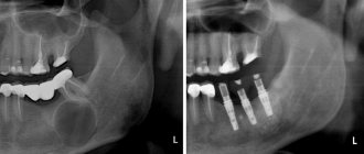
In some cases, fistulography is performed - an x-ray of the anatomical area before and after the injection of a contrast agent into the fistula. The procedure is not required only for simple and short rectal fistulas without exacerbation of inflammation.
Imaging methods - CT and MRI also make it possible to clarify the localization of tracts and leaks, branching and the root cause of the disease.
When the rectum is involved, ultrasonography (ultrasound) with a special rectal sensor is informative, when a computer program allows you to see the pathology in a three-dimensional image. When planning the operation, the function of the anal sphincter is additionally determined.
| Examination stage | Techniques | What are they used for? |
| Techniques used in the doctor's office during the initial examination |
|
|
| Lab tests |
|
|
| Instrumental studies |
|
|
What complications can there be after surgery?
Currently, with the availability of standardized technology and an integrated approach to the diagnosis and treatment of rectal fistulas, unpleasant consequences are minimized. However, it should be noted that the risk of complications always exists.
The most serious complication after surgery for rectal fistulas is the development of anal incontinence. The risk of its occurrence is especially high during repeated interventions, when the anatomy is significantly changed and the holding function may be initially compromised. It is worth noting that when operations are performed by an experienced specialist who performs this type of intervention on an ongoing basis, the risk of incontinence is practically absent.
In addition, bleeding may develop both in the early postoperative period and several days after surgery. The nature and severity of the complication are determined only after examination. Stopping bleeding is usually possible in a dressing room. In some cases, repeat surgery may be required.
In addition, when treating complex and recurrent fistulas, due to pronounced scar changes, the sutures fixing the mucomuscular flap may diverge, which leads to inflammation in the wound and requires repeated interventions.
Methods for treating fistulas
Fistulas rarely close on their own; this can only be hoped for by creating favorable conditions, for example, limiting and partially controlling the movement of feces through the rectum using cleansing enemas. In the vast majority of cases, conservative therapy is ineffective; the only radical treatment is surgical, that is, excision of the pathological area, including reconstruction of the missing tissue.
Technically simple surgical intervention, including endoscopic, and a hundred surgical modifications cannot cure about half of the patients who suffer from relapses. It is especially difficult to achieve success with intestinal and urinary fistulas, since they are always contaminated with microflora. In some cases, it is necessary to resort to the formation of an intestinal stoma, temporarily stopping the movement of feces through the pathologically changed area of the intestine for several months.
In isolated cases, they resort to “old-fashioned methods” of treatment with scraping the mucous membrane of the tract, burning it with chemical reagents and enzymes, achieving sticking of the walls. Better results - in approximately 50% - are achieved by introducing fibrin glue into the fistula tract, which glues the walls together.
Tampons made of biomaterials act similarly to glue, sealing the internal opening; emptying of the passage can cause the walls to stick together and close the fistula.
Until now, the role of antibiotics in the treatment of fistulas caused by inflammation has not been determined, since drugs are not able to penetrate into the infiltrate due to massive scar changes. However, with fistula tracts due to Crohn's disease, specific drug therapy is mandatory and not unsuccessful.
Surgery for fistulas using the LIFT method
Prevention of fistulas
Not all diseases can be prevented, especially fistulas, which complicate the course of purulent paraproctitis. However, it is possible to adequately treat the diseases that lead to paraproctitis - hemorrhoids and fissures, and this is precisely what will prevent the formation of fistulas.
Complicated childbirth cannot be prevented, but high-quality and timely obstetric care, attentive attention to the woman and a thorough postpartum examination are available preventive measures.
The high incidence of post-radiation tissue damage and fibrosis progressing over time forced oncologists to abandon high doses of radiation therapy and even change approaches to the treatment of malignant tumors of the genital area.
Particular importance is attached to the correct choice of method of surgical treatment of diseases of the hollow organs and adequate management of the postoperative period.
In our clinic, a complicated course of the disease is very rare, because we not only know about prevention methods, but also actively use them.
| Price : | |
| Primary consultation with a surgeon | 5100 rub. |
| Repeated consultation with a surgeon | 4600 rub. |
| Consultation with a surgeon Ph.D. | 6900 rub. |
| Primary consultation with an oncologist | 5100 rub. |
| Repeated consultation with an oncologist | 4600 rub. |
| Consultation with oncologist Ph.D. | 6900 rub. |
| Consultation with an oncologist, MD. | 10500 rub. |
| Fistulography | 41800 rub. |
Book a consultation 24 hours a day
+7+7+78
Bibliography:
- Shelygin Yu.A., Blagodarny L.A. /Handbook of coloproctology// -M.: Litterra; 2012.
- Becker A., Koltun L., Sayfan J. /Simple clinical examination predicts complexity of perianal fistula// Colorectal Dis.; 2006; 8.
- Gaertner WB, Hagerman GF, Finne CO, et al. /Fistula-associated anal adenocarcinoma: good results with aggressive therapy// Dis Colon Rectum; 2008; 51.
- Genadry RR, Creanga AA, Roenneburg ML, Wheeless CR /Complex obstetric fistulas // Int. J. Gynaecol. Obstet.; 2007; Vol. 99; Suppl. 1.
- Ommer A., Herold A., Berg E. / S3-Leitlinie: Rektovaginale Fisteln (ohne M. Crohn) // Coloproctology; 2012; Vol. 34.
- Sahni VA, Ahmad R., Burling D. / Which method is best for imaging of perianal fistula?// Abdom Imaging.; 2008; 33.
- Zoulek E., Karp DR, Davila GW/ Rectovaginal fistula as a complication to a Bartholin gland excision // Obstet. Gynecol.; 2011; Vol. 118; N 2.
Why do you choose us?
Part of the Perm State Medical University named after. I.M. Sechenova Clinic of Coloproctology and Minimally Invasive Surgery is an example of a new generation of medicine, harmoniously combining the deepest fundamental knowledge, honed skills, a modern multidisciplinary approach and attentive attitude to any category of patients.
KKMH is a guarantee that the surgical interventions performed within our walls correspond to the most current ideas about colorectal surgery. This includes our own department of anesthesiology and intensive care, whose employees ensure a smooth course of the operation and the early postoperative period and treat seriously ill patients. This is an “open intensive care unit”, where you can not only find out the most complete information about the condition of a loved one, but also be with him during a difficult time for him.
The doors of our Clinic are open to patients who were denied treatment in other hospitals due to the complexity of the surgical intervention or the neglect of the process. Elderly patients, patients with a “bouquet” of concomitant diseases (so-called comorbid) are an area of our special interest. The presence in the hospital of such highly professional specialists as a cardiologist, pulmonologist, urologist, etc. allows us to treat patients of any age and with any concomitant diseases.
KKMH is a dynamically developing team of specialists who sincerely and deeply love their work, who are continuously learning and teaching others, who are interested in keeping you healthy.