Dental diseases are pathologies that can greatly worsen the quality of human life and lead to serious consequences, such as disruption of the functioning of the gastrointestinal tract, cardiovascular system, and others. An open source of infection in the mouth in the form of caries will constantly provoke relapses of nasopharyngeal diseases, since the infectious agent penetrates the blood and tissues of the mucous membrane.
Having learned more about dental diseases, everyone will be able to prevent them through timely prevention, as well as recognize and seek medical help. What dental pathologies are, what symptoms indicate them, and how to prevent them - the attending dentist can also tell you.
Caries
This is the name given to a common pathology of hard dental tissues, which is characterized by demineralization and subsequent destruction of the molar. The disease begins with the formation of a small spot on the enamel, which is not even noticeable at first. Then it acquires a yellow-brown color, the affected teeth hurt, and react sharply to hot, cold and sweet foods.
If the development of the disease is not stopped at the initial and middle stages, it will be accompanied by pulpitis (nerve inflammation) and periodontitis, which can cause tooth loss. Also, chronic foci of infection in the form of carious cavities increase the risk of recurrent diseases of the body.
Reasons for development:
- insufficient oral hygiene (irregular brushing of teeth, ignoring rinses and dental floss after meals);
- poor nutrition, in which carbohydrate foods predominate, with a lack of vegetables, proteins and vitamins;
- hypovitaminosis – a lack of essential microelements in the body;
- living in unfavorable environmental conditions, with insufficiently fluoridated water or where food that does not contain phosphorus and calcium predominates;
- improper formation of teeth in children due to rickets, tuberculosis and other diseases;
- gastrointestinal pathologies;
- a sharp decrease in immunity.
The listed reasons cause changes in the structure of the enamel layer, as a result of which plaque cannot be completely cleared from the surface of the teeth, and the process of bacterial growth accelerates. Caries develops in several stages. As already mentioned, in the initial stage it has the form of a barely noticeable spot on the enamel; it becomes rough, with elements of demineralization.
Further, if the patient does not notice the first sign of pathology, the second stage begins, with a carious cavity forming under the enamel layer with dentin damage. Next, deep caries occurs, with the transition of the inflammatory process beyond the dentin, which is already fraught with various complications.
Starting from the second stage, the destruction process is accompanied by tedious pain that intensifies at night; analgesics help relieve them, but only temporarily
To avoid tooth loss as a result of its complete destruction, it is necessary to consult a dentist in a timely manner. The doctor will determine the degree of development of the problem and select the appropriate treatment method. At the stain stage, remineralization of the enamel is sufficient (it is restored using a special solution); at the superficial, medium and deep stages, this measure will no longer be enough; treatment followed by filling is necessary.
If the pathology is advanced, medicated pads impregnated with calcium hydroxide are used, which promotes the formation of secondary dentin, to strengthen and prevent tissue destruction.
Important for patients with cardiovascular pathology -
Many patients with gum inflammation try to self-treat, which involves regular use of antiseptics. Antiseptic solutions are not able to completely “disinfect” dental deposits (consisting of pathogenic bacteria) - to their entire depth, because antiseptics affect only the most superficial layer of microbial plaque or tartar. Therefore, firstly, the pathogenic effect of bacteria will continue even against the background of antiseptic rinses. But this is not the most dangerous thing.
Scientific research has shown that antiseptics interfere with the ability of oral bacteria to convert nitrates (found in foods) into nitrites. This bacterial activity is called “nitrate-reducing” and is one of the mechanisms that regulates our blood pressure. The fact is that nitrites formed by bacteria are then absorbed into the blood, and they help lower blood pressure. Therefore, if you regularly use antiseptic products (even toothpastes), this will reduce the production of nitrites by oral bacteria and may cause an increase in blood pressure.
This may not be noticeable in young healthy people, but will be significant in patients with chronic cardiovascular pathology. All these data are confirmed by scientific research - “The increase in plasma nitrite after a dietary nitrate load is markedly attenuated by an antibacterial mouthwash. Nitric Oxide" (2008), authors – Mirco Govoni, Emmelie Å. Jansson, Eddie Weitzberg, Jon O. Lundberg. If you wish, you can read this study using the link above using a browser translator. We hope that our article was useful to you!
Sources:
1. Dental education of the author of the article, 2. Based on personal experience as a periodontist, 3. American Academy of Periodontology (USA), 4. “Therapeutic dentistry. Textbook" (Borovsky E.V.). 5. “Non-surgical periodontal treatment” (Roncati M.).
Pulpitis
The most common dental diseases, in addition to caries, include pulpitis - this is the last stage of its development, when the lesion reaches the nerve area. The pulp is a connective tissue with a fibrous structure, it contains a large number of capillaries, lymphatic vessels and nerves, therefore the main symptom of damage to these tissues is considered to be acute, excruciating pain.
The acute form of the pathology is manifested by attacks of spontaneous acute pain, which can be provoked by hot or cold drinks, sweet food, or frosty air. If caries pain goes away immediately after eliminating the irritant, pulpitis is distinguished by the persistence of severe discomfort for another 15–20 minutes. When the disease becomes chronic, acute pain syndrome ceases to bother the person.
Anomalies of the dentition
During exacerbations, attacks can recur, especially when biting an inflamed molar. Reasons for development:
- advanced caries - a huge number of pathogenic bacteria located in the tooth cavity provoke rapid tissue destruction, penetrating into the deepest layers, reaching the pulp;
- poor-quality treatment of a carious lesion - some infected tissue may remain under the installed filling, causing re-inflammation and further destruction;
- thermal burn of deep tissues - while drilling the affected dentin, the dentist may be in a hurry and perform poor cooling, as a result the pulp is burned.
Also, pulpitis can develop not through caries, but, for example, with periodontitis, through infected gum tissue, to the roots of the tooth and above. When a molar is injured, sometimes the pulp tissues die due to the cessation of their blood supply.
Diagnosis of teeth affected by pulpitis is based on radiographic examination and data from the collected anamnesis (on the frequency of pain attacks and their intensity). Treatment involves removal of the inflamed nerve, root canal treatment and subsequent filling. In this case, pulp removal is carried out using vital (to remove any form of pathology) and deviant (for chronic form) methods.
Tartar
Like other dental diseases, tartar is formed due to the increased activity of pathogenic microflora in the mouth. During the life of microbes, especially with irregular oral hygiene, a large amount of soft bacterial plaque is formed. Under some circumstances, it is this plaque that becomes the basis for the formation of tartar.
Provoking factors include, in addition to the lack of proper hygiene, improper placement of molars, their crowding, which makes normal brushing difficult, features of the composition of saliva, and systemic pathologies that affect the pH level in the mouth. Symptoms of tartar:
- accumulation of a large amount of bacterial plaque on the tongue, at the roots of teeth, in periodontal pockets;
- gradual hardening of plaque and the formation of gray or dark brown stone, mainly on the posterior surfaces of the lower molars;
- inability to remove hardening and darkening with a toothbrush and abrasive pastes.
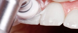
Dentists begin to treat many dental diseases with the removal of hard stone, since this measure is the prevention of diseases of the teeth and gums, such as periodontitis, gingivitis and periodontal disease. There are different types of removal procedures, one of which is ultrasonic cleaning. The tip of a special device vibrates at ultra-high frequencies, providing chipping of hardenings, polishing and grinding the enamel of molars.
At the same time, the doctor gives the patient advice on choosing a toothbrush and paste, using floss and mouthwash after meals, explaining how to prevent the re-accumulation of plaque.
This is what tartar looks like
Periodontitis
The inflammatory process that occurs within the connective tissue located around the tooth root is called periodontitis. The disease can be caused by many reasons, including:
- untreated caries;
- pulpitis;
- dental injuries (mechanical and those that develop gradually, when chewing hard objects, opening bottles with teeth, cracking seeds);
- improperly performed dental procedures - installation of fillings, dental reconstruction;
- penetration of the infectious agent through the general bloodstream, from infected maxillary sinuses, jaw bones;
- negative impact on periodontal tissue of strong toxins.
The most pronounced symptom of pathology is an “overgrown” tooth; to a person it seems too long and interferes with eating, talking and sleeping. If at the initial stage of the disease the pain is aching and of low intensity, then as it progresses it becomes more and more pronounced. In advanced stages, when a person suffers for a long time and does not see a doctor, the pain syndrome prevents normal life and radiates to the temple, eyeball, ear and neck on the affected side.
The lymph nodes under the lower jaw noticeably enlarge, and at the same time the temperature rises. If you touch a sore tooth, press on it, or drink a hot or cold drink, it responds with a flash of acute pain. Pus accumulates inside the connective tissue, which can lead to blood poisoning (sepsis).
Treatment involves removing the inflamed tissue, opening the canals of the affected tooth, placing medications in the cavities and subsequent filling. After which the doctor prescribes antibacterial and antiseptic drugs in order to eliminate inflammation and prevent its further development.
Tooth cyst and granuloma
A cyst is a periodontal disease in which a new growth forms in the area of the root apex. It is a round cavity located inside the bone tissue and filled with purulent contents. Depending on the reasons that provoked the problem, there are several types of cysts:
- eruption cyst – occurs in children during the period of replacement of milk teeth with permanent ones;
- paradental – formed due to problematic eruption of the “wisdom tooth” or a chronic inflammatory process during the period of its growth;
- follicular – formed when the molar bud in the gum becomes infected, or when an extra, supernumerary incisor grows;
- primary – its occurrence is influenced by a violation of tooth development from the remains of tooth-forming tissue;
- radicular – formed in the root area due to chronic periodontitis;
- residual - located in the bone after the molar has already been removed.
Granuloma is a type of cyst and also belongs to the category of neoplasms. This is a small round formation, localized in the area of the apex of the molar root, it takes a long time to form, is asymptomatic, and can be detected during an examination. The pathology can worsen under the influence of the same factors that lead to the formation of a cyst - these are caries, pulpitis, chronic infections of the nasopharynx, trauma and inflammation of molars, periodontitis, chronic pathologies developing under the crown, weak immunity.
However, there is still a difference between a cyst and a granuloma - the latter does not have a capsule, its boundaries are not so distinct, and it itself is a focus of inflammation of the connective tissue. Formations can be detected by characteristic symptoms - aching pain that becomes more intense as the disease progresses, swelling of the gums, headaches, fever, general malaise, suppuration and gumboil.
Therapeutic treatment of a cyst involves cleaning the tooth, treating it with antiseptics and filling; a copper-calcium suspension is also injected into the root canal and the tissue is exposed to small electrical impulses. To get to the cyst, the crown part of the molar is released, then the pus is pumped out, and instead of exudate, a paste is introduced into the cavity to promote the growth of new tissue. Only after these stages is filling carried out.
In the later stages, surgical treatment is suggested using cystectomy (when the formation is removed along with the membrane and the affected root tip) and cystotomy (in which the anterior wall is removed, and the oral cavity communicates with the cyst). The simplest surgical method is hemisection - here the cyst, tooth root and part of the damaged crown are removed.
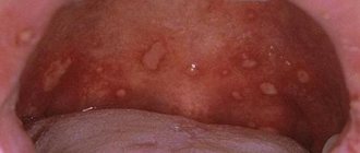
Pericoronitis
So, the inflammation that develops during the eruption of a wisdom tooth is called; in advanced cases, it manifests itself as purulent periostitis (suppuration of the gums). During the growth of the problematic eighth molar, this problem occurs frequently - in 60–80% of patients. The main cause is considered to be microbial flora (streptococci, staphylococci, anaerobes) that live in the oral cavity and accumulate in the gingival pocket overhanging the tooth.
Here are the main factors that can influence the development of pericoronitis:
- injury to the mucous tissue of the gums and the formed hood over the molar;
- the appearance of erosion and lesions on the surface of the epithelium;
- timing of tooth eruption.
The disease begins with the appearance of acute pain in the area of the erupting tooth, while it becomes difficult for a person to chew, and touching the inflamed gum causes a lot of discomfort. After 2–3 days, the general condition worsens, the pain syndrome becomes intense, the temperature rises, the pain radiates to the temple, ear and orbit. In the advanced stage, pus begins to secrete, the edge of the mucous tissue is damaged and then scarred, which causes secondary infection.
To make a diagnosis of pericoronitis, a general examination and radiography are performed.
Therapy involves excision of the mucous membrane overhanging the crown and anti-inflammatory treatment (if the tooth grows in the right direction). If it grows towards the tongue, cheek or horizontally, its removal is indicated. After the operation, the patient is prescribed antibiotics, followed by rinsing and treatment with antiseptic ointments on days 2–3.
Gum disease: treatment
It is unfortunate, but the most adequate patients who follow absolutely all the recommendations of a periodontist are those who have self-medicated for a long time with various rinses, ointments, and also used different toothpastes “for the gums.” When such patients come with loose teeth and suppuration from periodontal pockets, they no longer need to be convinced: 1) of the ineffectiveness of such self-medication, 2) of the need to regularly spend money on removing dental plaque, 3) of the need to brush their teeth after every meal, including flossing rather than twice a day.
Yes, such patients have a difficult situation in the oral cavity, but they are easy to work with. And when, after just a few days of adequate therapy, they see the first positive results, and you further recommend splinting their mobile teeth with fiberglass or curettage of periodontal pockets - they sign up without any questions and do it. But there are other patients for whom “not everything is so bad,” and who almost always listen with distrust to your recommendations - remove dental plaque, do curettage, splinting, prosthetics. They always think that they are being deceived and cheated out of money, and finally they always ask “how much does it cost” and then disappear for 1-2-3 years.
When they come again, they are also ready to do anything to avoid having their teeth removed and wearing removable dentures, but in many cases this is all that remains to be done. Therefore, when reading this article, you have 2 choices. The first option is that you can take only the anti-inflammatory therapy regimen for yourself, and then treat yourself at home. The second option is to also undergo a course of home anti-inflammatory therapy, but before that, visit a periodontist at least to remove the causative factor of gum inflammation, i.e. supra- and subgingival dental plaque.
Important: remember that gingivitis is reversible, and its proper treatment leads to a complete cessation of inflammation in the gums, and if after treatment, oral hygiene is good and regular, then you will never have gingivitis again. The development of periodontitis begins with the destruction of the dentogingival attachment (i.e., the attachment of the gingival margin to the necks of the teeth). As soon as the fibers of this attachment are destroyed, a path is opened for pathogenic bacteria under the gum, and there they do what they do best - they begin to destroy bone tissue, as well as the periodontal attachment (due to which the roots of the teeth are attached to the bone).
It is no longer possible to restore the destroyed dentogingival attachment and periodontal attachment. The resulting periodontitis can only be stabilized at the stage at which you consulted a doctor. Below we will talk about the general principles of treating gum disease, and you will also find useful links where some treatment issues are discussed in much more detail (than in this review article).
How to treat gums with gingivitis -
In general, you need to start with a consultation with a periodontist, and if periodontal pockets are found during the examination, the doctor will refer the patient to a panoramic photograph of the teeth - with a preliminary diagnosis of “chronic generalized periodontitis.”
If a diagnosis of “catarrhal gingivitis” is made, then this is a reason for joy, because in this case, treatment will consist only of removing dental plaque, a short course of anti-inflammatory therapy, as well as training in oral hygiene. Removal of microbial plaque and tartar is most often carried out using ultrasonic cleaning. This is a completely painless procedure, provided there is no increased sensitivity of the necks of the teeth (otherwise, anesthesia may be required). Usually, with gingivitis, only 1-2 visits to the periodontist are enough, who will remove dental plaque, polish your teeth, and also prescribe antiseptic rinses and gel applications to the gums. Well, he will definitely teach you how to use dental floss and brush your teeth correctly.
Video of dental plaque removal with ultrasound –
Anti-inflammatory therapy –
Anti-inflammatory therapy is prescribed by the dentist immediately after ultrasonic teeth cleaning, and its duration will be 7-8 days for the treatment of catarrhal gingivitis, and 10 days for periodontitis. In most cases (with the exception of patients with purulent discharge from periodontal pockets), there is no need to carry out antiseptic rinses and treat the gums with gel at a dentist’s appointment; you can do this just fine at home.
The procedures are carried out 2 times a day - in the morning and in the evening (after breakfast/dinner and subsequent oral hygiene). Please note that the sequence should be “breakfast → brushing teeth”, and not vice versa. First, you should perform an antiseptic mouth rinse with chlorhexidine solution. Depending on the severity of gingivitis, the effective concentration of chlorhexidine should be from 0.12 to 0.25%, but if we are talking about periodontitis, then rinsing your mouth with only 0.2-0.25% chlorhexidine.
Why exactly these concentrations - you can read at the link above, and in the same article you will find information on specific mouthwashes that contain chlorhexidine in this concentration. To rinse, you need to put 10-12 ml of solution in your mouth (this is about 1 sip) and, without spitting, rinse your mouth for 1 minute. After spitting the solution, it is important not to rinse your mouth with water! And then you will need to apply an anti-inflammatory gel to the gums.
Gel application regimen - I have treated thousands of patients with gingivitis and periodontitis, and in my opinion, the most effective anti-inflammatory gel is the drug Cholisal. If its price is high for you, then an alternative may be Parodontocid gel (however, it will work effectively only if used with antiseptic rinses, where the concentration of chlorhexidine will be at least 0.2%).
Before applying this gel to the gums, it is advisable to dry the wet mucous membrane of the gums using a dry gauze pad, because in this case the gel will stick better. Apply the gel only in front of a mirror, with your teeth bared properly (so that you can clearly see the gingival margin around the necks of the teeth). The gel should be applied only to the marginal part of the gum around the teeth, i.e. on the gingival margin - using a clean finger. To do this, you will squeeze portions of the gel onto the tip of your index finger and transfer the gel from it to the gum.
It is optimal to first treat the gums around the lower teeth, and then around the upper ones. On the gingival margin from the front surface of the teeth, the gel is best applied in 2 stages. First, rub small portions of the gel with light massaging movements, after which the second stage is to apply the gel without massage and leave it on the gum. On the palate/tongue side, it is also advisable to apply the gel, but here it is enough to only rub in small portions of the gel.
Important: we draw your attention to the fact that saliva will always be released during the application of the gel, but it does not need to be accumulated in the mouth or specially spit (swallow it as usual). In addition, after treating the gums with gel, it is undesirable to drink for 30-40 minutes, and it is also undesirable to eat or not rinse your mouth for 2-3 hours. The second treatment should be carried out in the evening according to a similar scheme (dinner → oral hygiene → antiseptic rinse → application of gel to the gums). And so on for 7-8 or 10 days, depending on the type of gum inflammation.
Useful links -
→ The best mouthwashes, → Rating of the best gels for gums.
Oral hygiene training –
Remember that the effect of treatment will not last long if you continue to not floss and brush your teeth after every meal. You can see how to brush your teeth correctly in the following video, and you will find comprehensive recommendations on oral hygiene in the article, the link to which is under the video.
→ Detailed recommendations for oral hygiene
How to treat gums with periodontitis -
You need to start with a consultation with a periodontist and analysis of a panoramic X-ray. Basic therapy will continue to consist of removing dental plaque and anti-inflammatory therapy, which may additionally include systemic antibiotic therapy. Periodontitis differs from gingivitis by the presence of not only supragingival, but also subgingival dental deposits, which are localized in periodontal pockets.
The problem is that not only ordinary dentists, but even many periodontists remove subgingival deposits quite poorly. The fact is that searching for subgingival dental plaque and removing it requires a large amount of doctor’s time (much more than for removing supragingival dental plaque), and time in modern dentistry is money. Therefore, with moderate/severe periodontitis, even 3-4 visits to the dentist may be required for high-quality removal of deposits. Removal of plaque is usually carried out using ultrasound (ultrasonic scaler), but we do not recommend using the Vector system apparatus for primary removal of dental plaque.
Depending on the degree of destruction of bone tissue and the depth of periodontal pockets, splinting of mobile teeth, depulpation of teeth, removal of highly mobile teeth, curettage of gum pockets, prosthetics of missing teeth, etc. may be required. Thus, further treatment for each patient will be individual, depending on the specific situation in the oral cavity. Read more about special additional treatment methods in the article:
→ Treatment of moderate and severe forms of periodontitis
How to treat gums with periodontal disease -
Treatment of this form of the disease is fundamentally different from the treatment of gingivitis or periodontitis. because The cause of periodontal disease is tissue degeneration and sclerosis, and not microbial factors and inflammation. The main treatment will be aimed at stimulating blood circulation in the gums (primarily physiotherapeutic methods are used for this), as well as at normalizing occlusal contacts between the upper and lower teeth.
Effective methods of physiotherapy for periodontal disease -
- vacuum massage of gums (including creation of vacuum hematomas),
- electrophoresis of drugs,
- the use of diadynamic currents,
- use of helium-neon laser.
All of the above methods are carried out only in a physiotherapy office; your dentist can give you a referral for the procedure. In addition to physiotherapeutic methods, regular massage of the gums, which can be done with a finger, can have an effect. An additional effect can be provided by regular courses of using Asepta gel containing beekeeping products, which will improve blood circulation in the gums.
Enamel lesions
A common problem among dental patients is abrasion of the enamel layer of molars, which has reversible consequences. It occurs due to a lack of calcium and fluorine in the body, food and water consumed, and development is also influenced by hereditary factors, bad habits, mechanical injuries and other factors.
Irreversible abrasion of enamel is observed after dental restoration, more precisely, during the installation of crowns, veneers and dentures, the top layer is removed by the orthodontist. But there are also certain pathologies that require long and difficult treatment; they also belong to dental diseases.
Enamel hypoplasia
This is a specific pathology in which the development of the enamel layer is disrupted due to an abnormal metabolic process. The body does not receive the necessary microelements, as a result the enamel becomes thin and vulnerable. Hypoplasia indicates protein metabolism disorders and serious health problems. Local hypoplasia affects individual teeth, and the enamel becomes yellow and is partially or completely erased.
The disease occurs in children and adolescents, but can also affect adult molars.
The systemic disease affects all molars at once, and is formed due to disturbances in intrauterine development (due to maternal illness with rubella, toxoplasmosis, use of tetracycline antibiotics), as well as in early childhood, due to unbalanced nutrition and metabolic problems. Symptoms:
- the appearance of yellow spots on the enamel layer, white stripes, dots and depressions;
- the presence of areas on the crown where the enamel is completely absent;
- a change in the shape of the teeth, when the enamel is erased on their entire surface, depending on the location of the modified molars, Fournier’s teeth (butterfly-shaped), Hutchinson’s (a notch on the incisors in the form of a semicircle), Pfluger’s teeth (the front teeth have underdeveloped cusps) and “tetracycline” (the enamel is underdeveloped and painted bright yellow).
Treatment can only be symptomatic, since the changes are irreversible. Therapy consists of enamel reconstruction; for large lesions, filling with a composite material is performed. If the top layer is completely missing, a crown is installed on the tooth.
Fluorosis
The disease fluorosis develops as a result of an excess of fluoride entering the human body and is endemic or occupational in nature. Most often, the disease is diagnosed in children living in unfavorable regions with water enriched with fluoride during the eruption of molars. In this case, baby teeth are not affected, since the placenta reliably protects the fetus from excess fluoride.
In mild stages of the disease, spotted and streaked forms appear; in moderate and severe stages, chalky-speckled, erosive and destructive forms appear. If the patient has a pronounced degree of pathology, the molars often suffer from its various forms. Symptoms depend on the type of disease:
- streaked form - chalk streaks and stripes appear on the surface of the molars, clearly visible when the teeth dry, the stripes can merge into spots;
- spotted form - spots of different diameters are noticeable on the enamel, merging with each other, their surface is smooth and shiny;
- chalky-mottled form - the affected teeth acquire a matte tint, pigmented dots and spots are visible on them, while the enamel is quickly erased, and brown dentin tissue is visible underneath;
- erosive form - areas of destruction on the surface are large, the enamel on them is completely erased;
- destructive form - erosions appear not only on the enamel, but also on the underlying hard tissues of the incisors, the tooth becomes fragile, its shape is disrupted, but the cavity does not open due to the formation of replacement dentin.
Treatment consists of changing drinking water if analysis shows that it is oversaturated with fluoride
The patient is advised to take calcium and phosphorus to prevent further tooth decay. Laser or chemical whitening is carried out, followed by remineralization with a course of 15-20 procedures, however, in severe forms these methods are not effective. For such patients, it is recommended to install veneers, as well as metal-ceramic crowns.
Gingivitis –
Gingivitis is an inflammation of the gingival margin, including the interdental gingival papillae (i.e., that part of the gum that is directly adjacent to the necks of the teeth). The main symptoms of this disease are periodic bleeding and soreness of the gums when brushing teeth, redness or cyanosis of the gingival margin, swelling of the gingival papillae, and often there is bad breath. Gingivitis with a predominance of such symptoms is called “catarrhal” by dentists.
Gum disease: photo of teeth with gingivitis
The cause of the development of catarrhal gingivitis is exclusively insufficient oral hygiene. As a result, soft microbial plaque accumulates at the necks of teeth, and hard dental deposits can also form (we are talking about tartar). In this case, the development of inflammation is due to the fact that pathogenic bacteria that are part of microbial plaque and tartar secrete various toxins and inflammatory mediators, which trigger a chain of inflammatory reactions in the gums.
Therefore, the main treatment for inflammatory gum diseases is always aimed at eliminating the causative factor, i.e. for the removal of dental plaque (+ normalization of oral hygiene). It should be noted that gingivitis is the simplest and most quickly curable form of gum inflammation, if, of course, the treatment is carried out correctly - with regular removal of dental plaque by ultrasound (the frequency of this procedure will depend on the quality of the patient’s oral hygiene).
Important: inflammation in gingivitis affects only the mucous membrane of the gums - without destroying the dentogingival attachment or bone tissue around the teeth (which is typical for a more severe form of gum inflammation - periodontitis). But the transformation of gingivitis into periodontitis is only a matter of time if the patient focuses his treatment on antiseptic mouth rinses and gel applications on the gums, rather than on the removal of dental plaque and high-quality hygiene.
Hypertrophic form of gingivitis –
Catarrhal gingivitis is the most common form of gingivitis, but not the only form. Sometimes patients can experience a hypertrophic form of gingivitis, which is manifested by an increase in the volume of the gingival papillae or the gingival margin as a whole (usually only in the area of the front teeth). Most often, this form occurs in pregnant women and adolescents, and is associated with hormonal changes in the body. There are edematous and fibrous forms of hypertrophic gingivitis.
In the edematous form, the enlargement of the gingival papillae occurs due to their chronic persistent swelling, which is practically untreatable. This form can occur not only in pregnant women, but also in patients with long-term catarrhal gingivitis (in the absence of adequate treatment). In the fibrous form, gum enlargement occurs due to the proliferation of fibrous tissue (Fig. 4). You can read more about the forms of catarrhal and hypertrophic gingivitis and their treatment regimens at the link below.
→ Treatment of gingivitis in adults
Prevention of dental diseases
- Experts recommend the following methods to prevent dental diseases:
- quitting smoking - when nicotine is used, the capillaries that feed the gum tissue narrow, resulting in a nutritional deficiency that leads to periodontal disease;
- proper nutrition - to prevent pathologies of teeth and gums, it is necessary to limit the amount of flour, carbohydrate foods, sweet and carbonated drinks;
- regular oral hygiene - includes brushing your teeth twice a day, using mouthwash and dental floss;
- timely treatment of caries will prevent complications in the form of pulpitis, periostitis and other dangerous consequences.
What other methods of disease prevention are there? The dentist will tell you, and you should visit him at least once every six months. After all, an ordinary patient may simply not notice developing problems with teeth and gums.
Today we talked about the main dental diseases, so that everyone could suspect problems in themselves in time, consult a doctor for diagnosis and choose the right treatment method. Ignoring strange stains on the enamel, pain, swelling and redness of the gums can lead to dangerous consequences in the form of suppuration of connective tissues and blood poisoning.
Treatment of dental diseases
Treatment of dental diseases always takes place within the walls of a dental clinic; at home, only auxiliary procedures can be carried out - rinsing, applications, taking tablets - prescribed by the doctor. Different pathologies require an individual approach to treatment, but the following procedures are most often performed:
- Filling with preliminary cleansing of destroyed tissue.
- Depulping – removal of the pulp when it is inflamed.
- Installation of prostheses and microprostheses to eliminate visible defects and strengthen crowns.
- Installation of structures for correcting dentition: braces, orthodontic caps.
- Surgical treatment – removal of the damaged part of the crown, root, tumor or the entire diseased tooth. Usually, removal is started if the dentist is unable to cure the disease, or when it is too dangerous for the patient’s health.
Dental problems are not only unpleasant, but also dangerous to health. The result of prolonged development of the disease may be a deterioration in the patient’s general condition, loss of teeth or the spread of infection to internal organs. It is impossible to prevent dangerous consequences with available home remedies; you need to seek help from qualified dentists.