Cleaning the tooth canals is a procedure for removing microparticles of the inflamed pulp from the roots, i.e. affected tissues. This is a particularly labor-intensive process, since the final effect of caries treatment depends on the quality of the work.
Clinical case: a patient has caries that has developed into pulpitis. When opening the tooth, the dentist diagnosed an infection and a severe inflammatory process; it was recommended to remove dead pulp tissue from the roots. If the work is not performed perfectly, an infection gets into the roots, a filling is placed on top, and the pathological process already develops under a thick layer of filling material. The toothache does not subside, the patient goes to the doctor again or endures the pain, quenching it with medications, and eventually loses the tooth. That is why, in case of caries, pulpitis and other problems, it is important to contact trusted doctors who perform high-quality root cleaning. It is advisable for the dentist to work with a microscope - multiple magnification allows you to monitor the process of cleaning the canals from remnants of pulp tissue.
One tooth may have two or more tubules, which are localized in the layers of the periodontium. The main task of the canals is to be a connecting link between the crown pulp and the nerves.
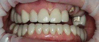
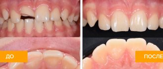
More than 10 years ago, deep caries and advanced pulpitis were the reasons for removing a unit, but now, thanks to effective cleaning and treatment of the canals, it is possible not only to preserve the crown, but also to restore its integrity to an ideal result.
Features of dental canal treatment
Differences in the structure and functions of the “representatives” of the dentition largely determine the nature of the treatment approach. Treatment of canals for different groups of teeth has its own nuances.
Front tooth canal treatment
The front teeth most often have one canal. They are often curved and difficult to pass through for instruments. In order to preserve aesthetics, the opening of the cavity of these teeth is carried out from the vestibule of the oral cavity. Due to their frontal location, it is important to prevent tooth enamel from darkening, so filling materials containing dyes are not used.
Treatment of wisdom tooth canals
Wisdom teeth can have more than 5 canals; they often have a branched structure, which is not always revealed by x-ray examination. These factors, along with the marginal location of the “eights”, significantly complicate the high-quality treatment of root canals. At the final stage of treatment, various filling pastes are traditionally used.
Root canal treatment of temporary teeth
Endodontic treatment of tooth root canals is usually used only at the stage of root stabilization. When choosing the optimal treatment tactics, it is necessary to take into account the specific structure of temporary teeth. The small thickness of the canal walls, the insignificant degree of dentin mineralization and the relatively large size of the apical foramen are the main reasons for special caution during instrumentation. Zinc oxide eugenol and iodoform pastes, as well as materials based on calcium hydroxide, are usually used as filling agents. They are not toxic to the permanent tooth germ and are able to dissolve along with the temporary root.
Possible problems during and after cleaning
Due to the carelessness of the dentist, poor technical equipment of the office, or when working with complex canals, an unpleasant effect is possible - breakage of the instrument, leading to a delay in its part in the canal. If an element remains in the farthest location, then sometimes it is impossible to extract it. Incorrect tool stop can also lead to artificial deformations in the canal. To prevent the formation of perforations of the canal walls and barrier steps, reamers equipped with a non-aggressive (non-sharp) tip are more often used in modern dental practice. It is also considered undesirable for the material used to extend beyond the root apex (in the absence of a cyst), which can lead to periapical pathology and a significant decrease in the effectiveness of endodontic therapy.
Fact! The formation of a crack in the root canal is fraught with the occurrence of a repeated inflammatory process due to interaction with the periodontal ligament.
Stages of tooth canal treatment
Endodontic treatment usually lasts several hours and includes a number of stages.
- Removal of the pulp (pulpectomy).
The inflamed soft tissue of the tooth is eliminated. - Root canal sanitation.
The procedure is a “cleaning” of bacteria and dead tissue elements. Pulpectomy and canal sanitation pursue one of the most important goals - eliminating existing inflammation. - Channel formation.
The root canal, freed from pathological contents, undergoes appropriate treatment. In addition to ensuring good passage of the canal, it is imperative to ensure that its apex reaches the apical part of the tooth. - Canal filling.
The last stage of the intervention is filling the root canal with filling material, followed by grinding.
Manual cleaning of canals with removed pulp
After poor-quality cleaning of the canals or removal of the nerve using the formalin-resorcinol method, infection and even pus may form in the canals if the inflammation is localized in the tissue area next to the tooth. The process of sclerotization of the canals becomes a barrier to high-quality cleaning; therefore, in this case, the procedure of widening the passages with the help of reamers is also carried out. After restoring the patency of the canals, chemical antiseptic cleaning is immediately carried out to remove any inflammation. In dental practice, irrigation is often performed using a cannula and irrigation needles.
Features of the traditional antiseptic rinsing method:
- the solution is fed through a syringe into the root canal;
- Pressure is applied to the syringe with the index finger;
- the released liquid is collected with a saliva ejector;
- the solution is held in the channel for 1 minute;
- The antiseptic is washed off with distilled water.
To enhance the impact, feeding can be done together with reciprocating movements, and not just by passive infusion. After the cleansing, treatment begins, which can last up to 2.5-3 months. During this period, the dentist places and periodically changes the disinfectant solution.
Treatment of teeth with problematic root canals
Filling dental canals
A relatively simple technology for treating tooth canals is filling with a special paste with or without a pin. According to the “gold standard” of endodontics, the canals are also filled with a latex-like material - gutta-percha. Several methods of its use have been developed, including the Termafil system, lateral condensation, injection or liquid thermogutta-percha (vertical condensation). In some cases, in particular when treating a tooth canal cyst, filling is carried out with a substance based on calcium hydroxide (copper-calcium hydroxide “depopheresis” method). However, special nanocomposite materials are increasingly used in dentistry.
Treatment under a microscope
The age-old “tough nut to crack” for the dentist is curved or branched root canals of the teeth. A dental microscope, often in combination with a laser, allows you to completely pass through them and adequately process them along their entire length to reduce tissue trauma. Sometimes it becomes necessary to treat a sealed tooth canal with the evacuation of remaining material of various nature, for example, fragments of fillings, tissue fragments and even instruments. Then a microscope also comes to the rescue. Read more about the technology in a separate article.
Cleaning canals in the presence of pulp
Carrying out a disinfection procedure is impossible without removing the fibrous nerve tissue - the pulp. The reason is that the channels are filled with this mass; an additional reason is the inflammatory process of the connective tissue, which in this case must be eliminated. To open the cavity, different places of influence are used with a drill: on the chewing ones - from the top, on the front - from the side.
Pulp removal algorithm:
- Introduction of anesthesia.
- Tooth preparation.
- Nerve killing.
- Placement of a temporary filling.
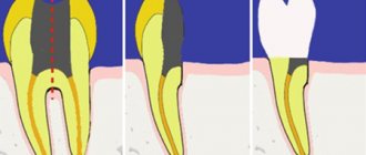
The pulp is killed by applying a special arsenic paste, or other options - Devit, Pulpex-S, paraformaldehyde variety. The use of these products allows us to call this part of the cleaning chemical. After a day or two, the tissues die, then the filling is repeated and a pulp extractor is used. By inserting it into the tissue and turning it in a circular motion, the dentist wraps the pulp around the device and removes it from the canals in parts.
After this procedure, reamers are introduced to help expand the mouth and the canals themselves. The thinnest instruments are used first, and then those with larger diameters. Periodically - usually after each manipulation - a disinfectant liquid (chlorhexidine, sodium hypochlorite, EDTA or hydrogen peroxide) is injected, which also helps to wash away detached areas of the pulp. If you do not carry out the procedure for expanding the canals, the quality of the filling will deteriorate significantly (voids may occur). The final part is a permanent filling.
Fact! The tooth being treated is protected with an insulating latex cloth to prevent saliva and bacteria from entering the canals.
If your tooth hurts after root canal treatment
If after root canal treatment your tooth hurts when you press it, this is normal. This phenomenon is associated with insufficient anesthesia in the area of endodontic intervention. Another reason why a tooth hurts after canal treatment is excessive treatment with the instrument moving beyond the apical foramen.
How long does a tooth hurt after root canal treatment? Sometimes pain persists for several days due to intensive intervention in the tissue structure in such a limited area. A similar situation occurs when an excess amount of filling material is placed into the canal, which causes discomfort when pressure is applied to the walls. As a result, the tooth “aches” after canal treatment. In any case, the presence of post-filling pain signals the need for a second visit to the dentist.
Endodontic instruments during cleaning
To effectively clean the canal, it is necessary to use several types of instruments, usually made of strong wire. Some of them are used in particularly difficult cases when the passage of channels is difficult. The specialist, taking into account the condition of the patient’s dental passages, can select rigid devices with a square cross-section, which are durable, but are less efficient at evacuating dentine filings. Another option is triangular-section reamers, which have increased flexibility but break more easily.
Groups of tools by purpose of use:
- channel expansion. The most commonly used options: Pro File 04, ProTaper, Rasp, K-flexofile, Rotary Instrument;
- increase in the diameter of the canal mouth. : Varieties: Largo and Gates Glidden;
- facilitating passage of the canal. Types: K-reamer, K-reamer farside;
- removal of tool remains. Instrument Removal System, Masseran Extractor Kit.
An auxiliary device is a microscope, which helps to visually zoom in on the channels and ensure high precision of the manipulations performed.
Advice! The safest instruments are considered to be nickel-titanium reamers, which bend easily and “remember” their shape after penetration into the canal.
Possible complications
Not all cases go smoothly. Let's consider the main problems that arise after the procedure.
- Perforation.
The phenomenon is the formation of holes between the dental canals and surrounding tissues. Treatment of perforation consists of medicinal treatment and filling. - Cheek swelling.
The reason why the cheek is swollen after root canal treatment is believed to be the impregnation of the periodontal tissues and mucous membranes with an anesthetic drug, which themselves are quite loose and easily absorb liquid. - Instrument fracture.
The probes for passing through the channels are very thin. If they break during medical procedures, the fragments are removed with special devices. Modern dental instruments made of nickel-titanium alloys are less susceptible to wear and break less often. - Adverse reactions to medications.
The range and severity of side effects of modern anesthetics are minimal. Before treatment, the doctor must collect an allergic history - information about drug intolerance - and the likelihood of a full-blown allergic reaction is practically reduced to zero. Adverse reactions of moderate and minor degrees are mostly short-term, can be easily corrected or can be overcome by changing the drug. - Other complications.
Situations such as swallowing particles of fillings, tooth dust, and small instruments now practically do not occur thanks to the use of a rubber dam - a latex plate that separates the tooth or teeth being treated from the oral cavity.
Preparatory procedures
In order for the dentist to choose the right treatment tactics, he should obtain maximum information about the condition of the tooth. Additionally, it is necessary to find out if the patient has an allergic reaction to medications, since they are required to be used when cleaning canals.
Collection of information:
- interviewing the patient (nature of pain, sensations);
- conducting an initial examination;
- sending a patient to get an image.
If the patient is a pregnant woman, then it is more rational to choose a study performed using a visiograph. The most informative is a 3D image created by a tomograph. Based on visual data, the dentist determines the length of the canal, allowing you to accurately select the instrument. There is a more accurate way to determine the distance using an apex locator, which displays a picture on the screen that allows you to determine the distance of the inserted instrument from the apex of the root. This method prevents breakage of endodontic devices during cleaning.
Fact! The most difficult cleaning is carried out when the canals are obstructed and are strongly curved.
When is tooth extraction necessary after root canal treatment?
It happens that you have to part with a treated tooth. Typically, such a sad outcome is due to the following factors:
- initially unsatisfactory root canal treatment with the development of poorly controlled inflammation;
- some cases of wisdom teeth treatment;
- the complexity of the anatomical structure of roots and canals in a particular patient;
- late request of the patient for specialized help, in which adequate therapeutic measures do not lead to the desired result.
The effect of treatment is short-lived
Wrong. Although nothing can completely replace a healthy tooth, quality root canal treatment followed by a suitable filling or crown can be very successful. In approximately 85% of cases, the effect of treatment is lifelong.
If a tooth becomes affected again several years after root canal treatment, it often needs to be re-treated. However, in some situations, such as a crack in the tooth, very severe tooth root decay, or severe loss of bone around the tooth, the dentist has no choice but to remove the affected tooth.
What is the cost of treatment?
How much does root canal treatment cost? The cost largely depends on the method used and the quality of the materials. When using laser and microscopic technology, nanocomposites and other advanced developments, the cost of tooth treatment with canal filling increases significantly. Approximate prices for root canal treatment in Moscow with gutta-percha filling and composite filling are presented in the table below.
| View | Price |
| Single channel tooth | 9,500 – 12,5000 rubles |
| Double channel tooth | 11,000 – 14,500 rubles |
| Three-channel tooth | 13,500 – 17,000 rubles |
The cost of root canal treatment should not be the determining factor in choosing a clinic. Contact only dentistry with a good reputation and experienced specialists who use modern equipment and the latest techniques in their work. Remember - the future fate of your teeth and the aesthetics of your smile depend on the quality of canal treatment!
What does the canal consist of and why should you clean it?
The inside of the dental canal is filled with pulp. It is a soft tissue penetrated by nerve fibers. In addition, it contains blood and lymphatic vessels and connective tissue. When an inflammatory process occurs in the pulp, the soft tissues inside the canal are also affected. Sooner or later it comes to such an extremely unpleasant thing as their necrotization.
The cause of inflammation can be caries or pulpitis. Therefore, in order to relieve the patient of acute pain and prevent tooth extraction, it is necessary to clean the dental canals of dead tissue.
The capabilities of modern dentistry in the vast majority of cases make it possible to completely cure and save the affected tooth, while just 10 years ago such inflammation invariably led to surgical removal of teeth.
Important! A disease that has become chronic may well be asymptomatic.
Signs of canal infection
In some cases, patients can independently determine the presence of problems with a previously treated tooth. The main signs of canal infection are:
- pain that did not go away after treatment or occurred some time after it;
- a feeling of fullness in the gums, discomfort when pressing on the tooth;
- the presence of swelling, swelling on the gums, as well as the formation of a fistulous tract (cyst breakthrough);
- changing the shade of the dental crown from light to gray. This may indicate dentin destruction;
- persistent unpleasant odor.
If you have the above symptoms, you should not waste time by self-medicating. At the IMEZA clinic, the patient can count on a quick, accurate diagnosis and adequate treatment.
Microscope or X-ray
Currently, dentistry uses two types of control over the treatment process: x-ray and using a microscope. X-ray is inferior both in terms of diagnostic capabilities and from a safety point of view:
- Some types of pins and composite materials are not visible in the photographs, which blurs the objective picture of the condition of the dental canals;
- optical distortions and channel overlap are possible;
- if there is liquid in the cavity, then the x-ray will not show it;
- To diagnose and monitor the treatment of the canals, a series of images will be required, and this, although insignificant, is still radiation.
A dental microscope is capable of magnifying the image 30 times - the doctor will notice the smallest tissue defects. You can easily detect the mouths of additional canals, examine their most tortuous configurations, assess root damage and monitor the quality of cleaning.
The microscope allows for unsealing with ultrasound, thereby preserving a large volume of valuable healthy tissue. Optics has significantly expanded the possibilities of dentistry in terms of restoring teeth that were previously considered hopeless. Treatment under microscope control almost completely eliminates the development of recurrent inflammatory processes and ensures long-lasting results.
Patients of the IMEZA clinic can count on high-quality canal retreatment using high-precision optical equipment, technological instruments and modern materials. It is enough to make an appointment, during which experienced doctors will identify all potentially dangerous elements and determine the optimal therapeutic tactics.
Expert opinion
Lyubov Ivanovna Kopylova
dentist-therapist
Experience: more than 10 years
If you have the opportunity to go to a dental clinic equipped with an electron microscope, take advantage of it! Even the most experienced dentist works with canals without a microscope by touch. This means that there is a risk of underfilling or, on the contrary, excessive deepening into the canal, leading to perforation. The use of a microscope, when the working field is clearly visualized and the doctor efficiently cleans and seals even the thinnest and most curved canals, will allow this situation to develop.
