Oral mucosa: what to pay attention to
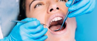
Regular dental examinations are an important part of the prevention of not only dental and periodontal diseases, but also pathologies of the oral mucosa. They make it possible to detect cancer, leukoplakia, stomatitis and other dangerous diseases in the early stages.
Inspection algorithm
The American Dental Association recommends that your oral examination include the following steps:
- a thorough visual examination of the lips, buccal mucosa, gums, anterior part of the tongue, floor of the mouth and palate;
- examination of the throat (pharynx), including tonsils, root of the tongue;
- palpation of the jaw and neck to detect nodes, tumors, any abnormalities or abnormalities.
This procedure allows you to timely identify foci of pathological processes in the oral mucosa (OM), including erosion, ulcers, compactions, and also assess the condition of the lymph nodes.
Russian specialists adhere to a similar inspection algorithm. He suggests that the dentist should pay attention to any visible abnormalities in the structure of the face, nose, cheeks or chin. Pathological changes, taking into account their localization, are recorded using special codes. Thus, ulcerations of the mucous membranes, wounds, tissue erosions, cracks, enlarged lymph nodes and any other lesions are necessarily noted.
Oral diseases
The most common symptoms of stomatitis encountered in dental practice. Acute herpetic stomatitis accounts for 85% of all diseases of the oral mucosa; chronic recurrent aphthous stomatitis is also common [1].
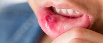
Aphthous stomatitis is considered a diagnostic feature of aphthous stomatitis - oval ulcerations on the oral mucosa with clear edges, with a diameter of 3-12 mm or more, surrounded by a halo of hyperemia. Herpetic stomatitis is characterized by blistering rashes not only on the surface of the mucous membrane, but also on the lips and skin around the mouth.
Data from multicenter studies in different countries of the world also indicate a high level of prevalence of other oral diseases: leukoplakia, candidiasis, lichen planus.
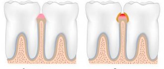
Leukoplakia is characterized by the presence of foci of keratosis in the form of whitish thickenings on the oral mucosa and the red border of the lips. With candidiasis, spots of white plaque with a cheesy consistency form on the mucous membrane, which are easily scraped off with a spatula. With secondary bacterial infection and erosion, the plaque may acquire a brownish tint.
The most common sign of oral lichen planus is also gray-white spots. They appear on the inner surfaces of the cheeks, tongue and gums. Due to the similarity of these manifestations with the symptoms of other diseases, in particular leukoplakia, visual diagnosis can be difficult.
Unfortunately, in recent years the number of cases of oral cancer has been increasing. The World Dental Federation attributes this to widespread smoking and alcohol consumption. Therefore, early diagnosis of cancer of the oral mucosa is becoming increasingly important. It is precisely this that makes it possible to identify precancerous and cancerous states of cells at stages when treatment can be anatomically gentle.
Experts advise paying attention to the following signs, which may be symptoms of oral cancer:
- long-term non-healing wound or irritation;
- red or white spots;
- sensation of pain, numbness, sensitive areas in the mouth or on the surface of the lips;
- tumors, thickening, roughness, erosion and other changes;
- difficulty chewing, swallowing, speaking, or moving the tongue or jaw.
The number of visits to dentists by Russians is approximately 15 million per year [2]. This figure is higher than any other medical specialty. Therefore, the doctor’s compliance with the external examination algorithm, thoroughness and accuracy during its implementation are the key to the success of early detection of many dangerous diseases of the oral cavity.
List of sources
[1] Zarkumova A.E. Structure of morbidity of the oral mucosa // Bulletin of KazNMU. 2021. No. 3. URL: https://cyberleninka.ru/article/n/struktura-zabolevaemosti-slizistoy-obolochki-polosti-rta (date of access: 05/22/2020).
[2] Astakhov P. Chief dentist of the Ministry of Health: mortality in the dentist’s chair is below 0.001% // RT, June 7, 2021. URL: https://russian.rt.com/russia/news/397438-glavnyi-stomatolog-minzdrav-smertnost (access date: 05/22/2020).
Medical Internet conferences
Oral mucosa: barrier and immune functions.
Odekov D.M., Shcherenko S.P.
Scientific supervisor: assistant Kapishnikov M. S.
Federal State Budgetary Educational Institution of Higher Education Saratov State Medical University named after. V. I. Razumovsky Ministry of Health of Russia
Department of Clinical Immunology and Allergology
Relevance of the problem: The oral cavity has a close anatomical and physiological relationship with different body systems. Pathological processes in the oral mucosa are often the initial symptom of systemic pathology. Many childhood infectious diseases have manifestations in the oral cavity. Therefore, dentists, pediatricians and doctors of other specialties must know the changes in the oral mucosa in various diseases and be able to evaluate them from the perspective of the whole organism. On the other hand, damage in this area of the human body can lead to a fairly wide range of diseases. Up to 90% of the adult population of Russia periodically suffer from various inflammatory diseases of the oral mucosa. The oral mucosa protects us from many harmful environmental factors, and therefore knowledge and study of all its functional responsibilities is one of the most important factors for preventing disease.
The purpose and objectives of the study: to analyze the most important functions of the human oral mucosa and study the mechanisms of its protective reactions.
Materials and methods: we analyzed numerous scientific medical literature (more than 250 sources) in several databases. The key words during the search were: “oral mucosa”, “innate and acquired immunity”, “allergy”, “barrier function of the body”, “cellular composition of the oral cavity”
Results obtained: Performing numerous functions, the oral cavity is constantly exposed to influences that can lead to violations of the integrity of its integument, which in the future can lead to the development of more serious diseases.
The oral cavity is a unique complex of analyzers, thanks to which the body receives comprehensive information about the food it eats. Moreover, it has a complex immune defense system. The oral mucosa also performs barrier, sensory, absorption and plastic functions.
Conclusions: against the background of improper functioning or violation of the integrity of the oral mucosa, various diseases can develop. Therefore, knowledge of its normal state plays a vital role in diagnosing these diseases, as well as in maintaining human health in general.
Leukoplakia of the oral cavity: causes of the disease
The reasons causing the development of the disease include:
- Smoking. When using tobacco, the oral cavity is exposed to various irritants, including thermal (incoming smoke has a temperature of about 60 degrees Celsius) and chemical (nicotine, tar and combustion products). No less dangerous is chewing tobacco, which is also a provoking factor.
- Eating either very hot or very cold food on a regular basis for a long time.
- Mechanical trauma (bad bite, sharp edges of teeth, orthopedic structures installed with violations).
- Metal seals that cause galvanic currents.
- Inhalation of vapors of gasoline, benzene, varnishes and paints, as well as other resins.
- Hormonal imbalance, constant stress and lack of retinol.
Standards for a healthy oral cavity
Home / Articles / Standards for a healthy oral cavity
How is oral health determined? A healthy oral cavity is the absence of diseases, inflammations and pathologies. Orthodontist Farida Yusifovna Gasimova talks about the health standards of the oral cavity and dental system.
— Farida Yusifovna, how is dental health determined?
— Disorders in the dental system provoke malfunctions in the joints and muscles. Fixation of the lower jaw is carried out with the help of the muscles of the head and neck; the chewing process occurs when the temporal and masticatory muscles work. If the chewing apparatus is functioning correctly and the joints and muscles are anatomically normal, there is a distance (2 mm) between the teeth of the upper and lower jaws. Disturbances in the bite occur with excessive muscle tone in a calm state, which affects not only the teeth, but also general well-being: the patient complains of frequent headaches, pain in the neck muscles, in the back, there is pinching of the facial muscles, teeth are subject to wear, chips appear at the ends, and a wedge-shaped defect is often diagnosed. Disturbances in the dental system can be of a different nature; based on external symptoms, one can more or less understand that it is necessary to consult an orthodontist. If the patient notices stiffness when moving the lower jaw, jamming of the jaw, characteristic clicks when it moves (crunching, clicking) - this indicates problems with the temporomandibular joint. The action of the TMJ (temporomandibular joint) is aimed at ensuring normal movement of the lower jaw. The temporomandibular joint looks like a dimple with a head, with an articular disc placed between them. The absence of disturbances is manifested in the form of the location of the head in the fossa (resting state). When the jaw moves, the head is displaced by the articular disc. If there are disorders, the articular disc is displaced, the bite is disturbed and the jaw moves incorrectly. Dysfunction of the TMJ (temporomandibular joint) requires complex treatment from doctors of various specialties, including a gnathologist, an orthodontist, and a neuromuscular dentist. The condition of the dental system is significantly influenced by the patient’s spine and posture. If posture is impaired, the muscles of the face, neck, and back cannot work correctly, and this fact, in turn, affects the normal functioning of the lower jaw. The displacement of the lower jaw causes disruptions not only in the functioning of the dental system, but also in the body as a whole. An anatomically correct bite is not an indicator of a healthy dental system, as many patients think. Just like white, straight teeth. The dentofacial system generally cannot be characterized by any one of its organs, since, as I have already said, a violation in any of its components affects the entire system. The condition of the dental system is directly related to the work of the ear organs, temporomandibular joints, neck and head muscles. The psycho-emotional state of the patient also plays an important role. If pain in the neck and back periodically appears, this already indicates an existing disorder, including in the dental system. Therefore, only a dentist orthodontist in Anapa can determine its condition.
— What is the dental system?
— The dentofacial system is a complex apparatus in which the organs are anatomically united. The organs included in the dental system are bones (palatine, zygomatic, nasal, jaw), teeth, temporomandibular joints (temporomandibular joints, lips, cheeks, tongue, soft and hard palate, salivary glands, masticatory and facial muscles. Functioning separately and together, all these organs give us the opportunity to breathe, talk and eat food. I want to emphasize that in the dental system there are no “important” and “minor” organs, each individual component of the system plays its role. The growth and formation of the dental system begins in changes in the womb and throughout a person’s life. From childhood to final adulthood, modifications of the dental system are clearly noticeable, since during this period the bite changes, individual features of the cheekbones, tongue, and lips are formed. Then the changes are little noticeable, but they continue to occur. When there is a process of active formation (childhood), the Anapa orthodontist dentist can quickly correct the incorrect growth trend to an anatomically correct one, direct the growth of teeth and bones, so to speak, in the right direction. You should not wait until the jaw is fully formed (adulthood) for orthodontic treatment; in children, therapy is faster, easier, and cheaper. Dentistry Millennium Anapa Mayakovsky offers patients orthodontic services. It is also important to note the fact that a disorder in one organ inevitably affects the functioning of the entire system, therefore, if the pathology is not corrected during the period of formation and growth of the jaws, it can develop into a severe disorder in adulthood. It is necessary to undergo an examination by an orthodontist during the period of 5-7 years, which will exclude or identify a tendency to disrupt the development of the dentofacial system. A pediatric orthodontist in Anapa conducts a visual examination and diagnosis using special equipment. I repeat, it is much easier to carry out correction in a child than long-term therapy in an adult patient.
— Farida Yusifovna, how are teeth treated today?
— Treatment of the dental system must be carried out comprehensively, with mandatory visits to specialized specialists. Anapa Mayakovsky Dentistry Millennium dental treatment, in the clinic you can get a consultation with one of the best dental specialists in the city and undergo a full diagnosis of the dental system. Depending on the patient’s clinical picture, the orthodontist in Anapa prescribes consultations with doctors and computed tomography. Therapy of the dental system is always carried out according to an individual program; in case of violations, it is impossible to treat everyone according to the same scheme. Electromyography (myography) is used to identify dysfunction of the masticatory and temporal muscles and determine the affected nerve areas. Using electromyography, it is possible to determine the causes of paralysis of an individual muscle. The principle of operation of the myograph is that it records changes in the bioelectrical activity of muscles using special sensors attached to the facial muscles and connected to the myograph. Therapy of the dental system includes treatment with an occlusal guard and a splint (a removable device for correcting the bite). With the help of a mouthguard, pain in the TMJ, pinching of the lower jaw, headaches and other disorders are eliminated, the action of the occlusal splint is aimed at restoring the natural position of the muscles during the functioning of the jaw, with the help of the splint the symmetry of the face and anatomically correct muscle tone are restored. Another technique that deserves attention is electrical neurostimulation (TENS), the effect of which is aimed at relaxing the masticatory muscles that are in hypertonicity. This method effectively eliminates muscle spasms, saturates tissue cells with oxygen, ensures the correct position of the lower jaw, removes lactic acid, and restores proper muscle function. TENS therapy is carried out using a current that is passed through the patient's skin; the entire procedure takes about one hour. The facebow is used to accurately analyze the individual position of the jaws in three dimensions in order to then produce a prosthetic structure (veneers, dentures or crowns). The use of this mechanical device in dentistry can significantly reduce errors in the design of orthopedic devices. Making a structure for the dental system is a rather complex task that requires maximum attention, experience and professionalism; not only the individual characteristics of the position of the bones are taken into account, but also the dynamics of movement. When making orthopedic structures, it is very important to match the central occlusion and jaw relationship. When conducting therapy, dentists set themselves the priority tasks of restoring proper functionality, and only then the aesthetic factor. Of course, most patients are primarily interested in the beauty of a smile, but if beautiful teeth are not capable of fully chewing food, then, of course, it is much more important to recreate functionality.
Comprehensive hygiene for 2500 rubles! Air Flow 1000p for patients with braces!
A unique promotion for our regular patients! A set of hygienic procedures (removal of tartar and plaque using Air Flow and ultrasound , scaling, polishing, fluoridation) for regular patients once every 6 months for RUB 3,000!
Air Flow and polishing for only RUR 1,500 for patients with braces!
Kinds
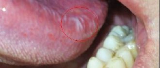
- The most common is simple or, as it is also called, flat leukoplakia . It is usually discovered by chance during an examination by a dentist, since the patient does not experience any subjective sensations. A burning sensation occurs extremely rarely, and the appearance of the mucous membrane may change. If the disease affects the tongue, loss of taste may occur.
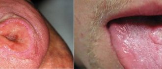
- Hairy leukoplakia of the tongue resembles stomatitis. The shape of the spot that appears, as well as its size, can be different, the color - from pale gray to white. The surface of the mucous membrane at the site of the lesion becomes slightly rough, which can be felt to the touch. On the cheeks it appears as solid or broken lines. It can also be found on the lips, where it looks like thin paper pasted on.
- Verrucous leukoplakia is the second stage of the development of the disease. The keratinization thickens, the affected area seems to rise above the nearby tissues. When you touch it with your fingers, you feel a compaction.
- Erosive form . Untimely diagnosis of the two previous stages of the disease leads to a worsening of the situation - the person feels pain when exposed to any irritants, erosions or ulcers are visible in the mouth.
- Soft leukoplakia is a type of cancer. Its distinctive feature is peeling of tissue in the area of the lesion. To clarify the diagnosis, a histological method of studying cells is required.
- Tappeiner's leukoplakia . This form of the disease affects people who abuse smoking. According to studies, daily smoking 10 cigarettes a day increases the chance of developing the disease by 50 times (as the number of cigarettes increases, the risk also increases)
The disease begins with the formation of lesions on the roof of the mouth (sometimes they appear on the gums). The mucous membrane changes its color to a pronounced gray or bluish, which is noticeable to the naked eye, folds appear on it. Reddish nodules may begin to appear, which is accompanied by infectious inflammation of the oral cavity (caused by the accumulation of salivary gland secretions in the tissues).
Oral leukoplakia: how to treat it at home
In addition to drug treatment, oral leukoplakia can be treated with traditional medicine.
There are many recipes, here are just the main ones:
- rinsing with herbs (infusions of oregano, chamomile, ginseng and other adaptogens that reduce the inflammatory process and increase the body’s resistance to harmful factors are suitable);
- regular consumption of nuts and tinctures based on them;
- rinsing with decoctions of calendula, St. John's wort, eucalyptus. Alternation works well - once the oral cavity is rinsed with a soda solution, after a couple of hours - with an infusion of herbs. This procedure should be repeated at least 5 times a day;
- lubricating the lesions with sea buckthorn and olives (the fruits must first be mashed in your hands so that the juice appears).
Timely detection of the disease and compliance with all doctor’s recommendations is the key to recovery in the shortest possible time and reducing the risk of complications. If you start treatment at the initial stage, you can reduce the likelihood of complications to almost zero.
Symptoms
The first signs of the disease often go unnoticed because they do not cause any pain or discomfort in the patient. Nevertheless, a specialist will be able to determine the onset of leukoplakia by the appearance of the mucous membrane, lips and the area where the teeth meet.
The first sign of the disease is the appearance of a keratinized gray area, which can appear on the palate (in smokers), in the corners of the mouth, on the inside of the cheek, etc. An easily removable white plaque forms in this area, but after a few days the formation makes itself felt again . The patient may feel tightness in the mouth, but, as practice shows, most people simply do not pay attention to this.
Plaques with a diameter of no more than 4 centimeters are formed. They may appear:
- on the inner surface of the cheeks;
- on the tongue (on the back or sides);
- in the sky;
- on the gums;
- in the corners of the mouth.
The process of plaque formation takes up to one month. At the first stage, the area of the future formation seems slightly swollen; when you feel it with your fingers, the compaction is not felt. However, over time, another symptom of oral leukoplakia appears - the mucous membrane at the site of the swelling loses its original shine and becomes rough, which is noticeable when touched.
There is no pain in this case: only sometimes a feeling of dryness at the site of the outbreak is possible.
Gradually, the color of the spots changes from gray to bright white. The spots in most cases have clear boundaries. Their increase is possible when the disease enters its second stage, called verrucous.
The disease often causes candidiasis and malignant cancers. In an advanced state, leukoplakia is very difficult to treat: the affected areas become even more keratinized, ulcers can form, and the infection gradually spreads to other areas of the mouth.