Why can odontogenic sinusitis occur?
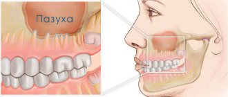
The maxillary (maxillary) sinus is a cavity with bony walls that is located inside the upper jaw. The cavity communicates with the nasal cavity through the anastomosis, which is located on the side wall of the sinus facing the nasal cavity. The lower wall has contact with the upper teeth. Quite often (in about 15% of cases) the apex of the tooth root lies directly under the mucous membrane of the bottom of the maxillary sinus, and there is no bone septum between them.
When an infection enters the maxillary sinus, inflammation of its mucous membrane or sinusitis occurs.
There are two ways of infection:
- rhinogenic - through the nasal cavity. In this case, the infection penetrates from the nasal cavity through natural or artificial (after surgery) communication;
- odontogenic - through the tooth or tissue around it.
Odontogenic sinusitis most often develops slowly against the background of chronic infection in the area of the tooth root. As a result of chronic inflammation, a cyst forms in the root area, which destroys the barrier between the sinus and the tooth. Pathogenic microbes gradually penetrate the sinus mucosa, causing inflammation.
Also, odontogenic sinusitis can occur as a result of the actions of the dentist. Often, after the removal of the upper tooth, the thin barrier between the sinus and the oral cavity may be damaged. As a result, a gateway for dental infection appears. In this case, the anatomical features of the patient with a thin bone septum between the sinus and the tooth root or its complete absence are of key importance.
Infection can occur when cleaning the canals and filling them. In some cases, the filling material gets inside the sinus, causing the formation of fungal sinusitis, and the zinc contained in the filling material promotes the growth of mold fungi (Aspergillus, Mucora). Also, odontogenic sinusitis can develop after the sinus lift procedure and the installation of dental implants in the upper jaw.
What are the maxillary sinuses
The maxillary sinuses (also called the maxillary sinuses) are special cavities on both sides of the nose that are filled with air. Each cavity is connected to the nasal passage by small openings called anastomoses. The cavities are covered with mucous membrane. The function of mucus is to trap bacteria and harmful particles in it, and then remove them from the body through those same anastomoses. When edema occurs, the excretory opening becomes very narrow, as a result of which mucus, along with harmful particles and bacteria, cannot come out and stagnates. At this time, the patient begins to experience bursting pain in the cheek area - this is how inflammation of the maxillary sinus begins. Treatment of the maxillary sinus should not be neglected, since inaction can provoke serious consequences, including sepsis and meningitis.
Classic sinusitis can be bilateral, when both sinuses are affected. In the odontogenic form, the inflammatory process starts in the sinus on which side the diseased tooth is located.
Complications
With odontogenic sinusitis, a chronic inflammatory process occurs. Dental microflora appears in the sinus, not typical for the upper respiratory tract, which can destroy bone tissue. Due to the fact that the paranasal sinuses have contact with the orbit and brain, odontogenic sinusitis can lead to severe complications:
- intraorbital (orbital phlegmon, ophthalmitis, optic nerve neuritis);
- intracranial (meningitis, encephalitis, brain abscess).
Therefore, at the slightest suspicion of this disease, you should consult a doctor.
Sinusitis or tooth – what to treat first?
In this case, many otolaryngologists prescribe tooth extraction.
In our clinic, we collaborate with specialists who treat odontogenic sinusitis or help the patient prepare the sinus mucosa for sinus lifting.
Of course, it is not always possible to prosthetize and treat a tooth with a source of infection in the periodontium in the upper jaw. Therefore, such teeth often still need to be removed.
Tooth extraction surgery is a major source of irritation for the body and the immune system. After it, you need to wait 4-6 months until the porous bone tissue of the alveoli is restored.
In this case, conservative anti-inflammatory therapy, antibiotics, and active copious rinsing of the nasal sinuses are prescribed. Treatment lasts about a month after tooth extraction.
If CT and diagnostics revealed a source of infection (cyst or granuloma) on the tooth, then treatment for odontogenic chronic sinusitis begins immediately after removal. Then the chance to reduce inflammatory changes in the mucosa is quite high.
Otherwise, the patient risks getting large growths, which sometimes occupy more than half the height of the sinus.
This is manifested by nasal congestion, because when we have ARVI, the nasal mucosa swells greatly. And our nasal mucosa is exactly the same as in the sinus: the same columnar epithelium. It swells very much when initially, as a result of constant irritation, the infection has already grown by more than half.
In this case, the half of the nose that is more blocked will be an indicator to check what is wrong with the teeth on the same half of the jaw.
Treatment
The treatment of odontogenic sinusitis requires an integrated approach. As a rule, treatment requires the simultaneous participation of an otolaryngologist and a dentist. Isolated antibacterial and conservative therapy lead only to temporary relief of the condition and removal of the severity of the process.
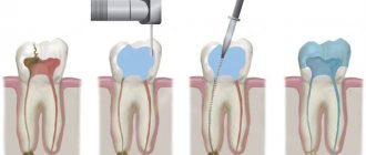
For a complete recovery, it is necessary to eliminate the source of infection - remove or treat the causative tooth while simultaneously sanitizing the inflamed sinus.
In case of foreign inclusions in the sinus (filling material, sinus lifting material, fungal bodies), their complete removal is necessary. For this, endoscopic techniques are used. They allow you to remove these formations through the nasal cavity. If there is a connection between the sinus and the oral cavity (oroantral fistula), it must be closed using special bioinert collagen-based membranes and mucosal flaps.
Methods for diagnosing perforation of the maxillary sinus floor
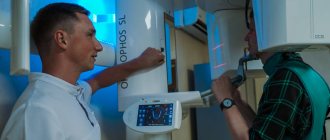
During diagnosis, a comprehensive study and analysis of the typical clinical picture observed with perforation of the maxillary sinus is used. The condition of the tooth socket, the presence of air bubbles, nosebleeds, and pain are taken into account. X-ray diagnostics are also performed, which show the area of perforation with high accuracy.
The dentist can perform a probing procedure for a perforated canal or an extracted tooth using a thin medical probe. This helps to reliably understand that there is no bone bottom in the wound. The instrument itself must pass freely through soft tissue without encountering obstacles along the way.
X-rays of the sinuses can detect characteristic darkening in the images, which are formed as a result of the accumulation of blood clots. Among other things, X-rays can show filling material, implants, and fragments of dental roots. X-rays with a contrast agent are often taken to help clearly visualize the condition of the bone tissue and surrounding tissues.
In such situations, a contrast agent is introduced into the cavity through a perforation fistula. An additional method of instrumental diagnostics is computed tomography, which determines the degree of perforation and the presence of foreign bodies, such as tooth fragments or residual filling material. If perforation is suspected, general clinical tests are required, which can show the presence of a focal infection in the patient’s body.
General information about sinus lift surgery
A sinus lift is a surgical procedure that uses the human maxillary sinuses (maxillary sinuses) to fill the bone deficiency in the area of the anterior chewing teeth. The essence of the operation is to separate the mucous membrane from the jaw bone and fill the resulting space with osteoplastic material.
Surgery requires preliminary anesthesia, which can be either local (with or without sedation) or general. Access to the maxillary sinus depends on the type of sinus lift:
- Open method. An incision is made on the front surface of the upper jaw. The technique is performed in cases of severe bone volume deficiency and is considered quite traumatic. The graft healing process lasts on average 3-6 months, after which dental implantation can be performed.
- Closed method. This sinus lift is often performed together with implantation, since the incision is made at the site of the future installation of the artificial root. This operation involves less trauma, but is used only for small deficits, up to 4-6 mm.
The duration of the operation depends on its type, but on average it lasts 1-2 hours. During the rehabilitation period, the patient must strictly follow the doctor’s recommendations in order to prevent serious complications from occurring. These include, first of all, proper oral hygiene, as well as taking the necessary medications.
Features of the maxillary sinuses
Sinus lift surgery is closely related to the anatomy and functioning of the maxillary sinuses. Each maxillary sinus has the same structure, but has certain differences that the dentist must pay attention to when planning the operation.
There are two main types of maxillary sinus:
- Hyperpneumatized. It is a large volume cavity with increased airiness.
- Hypopneumatized. A small space characterized by sclerotic areas.
When assessing the results of radiography or computed tomography, the dentist always pays attention to the structural features of the maxillary sinus in order to accurately calculate his actions during the operation. In addition, examination of the maxillary sinus is required in order to exclude any signs of an inflammatory process, which the patient may not even be aware of.
Carrying out a sinus lift after a maxillary sinusotomy
Sinus lifting for sinusitis is not performed under any circumstances, so the patient is first treated for the inflammatory process. Surgical intervention to replenish bone tissue deficiency after a manipulation such as maxillary sinusotomy is considered possible. The doctor must take into account that the patient has a history of a similar procedure and act much more carefully.
Sinus lifting after perforation of the maxillary sinus is dangerous because the risk of infection in the sinus increases. This occurs due to perforation of its wall, especially with an open operating technique. To avoid such complications, the doctor carefully plans each step of the future surgical intervention, focusing on the data of instrumental studies, in particular computed tomography.