An oral mucosal cyst (also called a mucocele or mucocele) is a small capsule filled with fluid. According to dentists, such cysts are generally harmless and painless, but they can cause discomfort because they feel like a foreign object.
Mucoceles usually occur on the inner surface of the lips, but can also appear on the tongue, palate, buccal mucosa, and floor of the mouth. These cysts also form around tongue or lip piercings. A cyst on the bottom of the mouth is called a ranula, and on the gum - an epulis. Mucous cysts are bluish in color and filled with clear fluid. The dentist usually recognizes them at first sight.
Content:
- Why does a cyst grow in the oral cavity?
- Types of oral cysts
- Signs of a cyst
- Examination of patients who have a cyst in their mouth
- How to treat
- Treatment of cystic formation at home
- How to reduce the likelihood of developing education
Sometimes, when visiting a dentist, a patient hears a strange diagnosis - mucocele.
More simply, it sounds like an oral cyst. Underneath this disease lies a cavity tumor neoplasm containing mucous contents. It is benign and develops due to obstructed outflow of secretions produced by parenchyma cells. The peculiarity of mucocele is that it does not have a strong epithelial membrane. Usually localized on the mucous membrane of the lower lip or under the tongue. It can also form in the chewing area. Neoplasms of the submandibular zone are very rarely encountered in dental practice.
Conducted studies demonstrate that most often young people under the age of thirty experience mucocele. The disease also occurs in adolescent children.
Prevention of recurrence of tonsil cysts
After removal of the cysts, relapse of the disease may occur. To avoid this, it is necessary to prevent the occurrence of situations that provoke inflammation of the mucous membrane of the oropharynx. Try to promptly treat ENT diseases, especially tonsillitis, and avoid injury to mucous tissues. If you smoke, if possible, give up the bad habit or reduce the number of cigarettes you smoke to 1-2 per day. Tars and other substances contained in tobacco have an irritating effect on the throat and can provoke an exacerbation of sore throats.
If the patient has a history of frequent ENT diseases and relapses of tonsillitis, then the doctor may recommend removing the entire tonsil. A tonsillectomy will relieve you of chronic infection and many other health problems. But tonsil removal is carried out according to indications. You can talk about the need for total resection with the doctors of the CONSTANTA Clinic at your appointment.
If you have any questions or make an appointment with a specialist, please call: (4852) 37-00-85 Daily from 8:00 to 20:00
Sign up for a consultation
Why does a cyst grow in the oral cavity?
Most often, doctors are unable to determine the exact cause of the disease. It is believed that its development can be caused by:
- repetitive trauma to the mouth;
- inflammatory pathologies of the oral mucosa;
- congenital obstruction of the excretory ducts of the salivary glands.
Some doctors are inclined to believe that frequently appearing cysts of the sublingual salivary gland indicate a non-standard structure of the latter. There is also a dysembryogenetic version of the origin of tumors. But dentists give the main importance to the traumatic factor. Thus, blisters on the inside of the lower lip often form due to the habit of constantly biting it.
The pathogenesis of the disease can be described as follows:
- The excretory duct of the salivary gland becomes blocked for a certain reason.
- Internal hydrostatic pressure increases. The mucus accumulates and cannot come out.
- Throughout the day, the mucous secretion permeates the surrounding tissues.
- Swelling forms and the blood vessels are compressed. Tissue permeability is impaired.
- A capsule consisting of connective tissue is formed. She is gradually growing.
Treatment methods
Conservative treatment:
- washing the tonsils;
- inhalation;
- rinsing;
- ultrasonic influence;
- laser therapy.
These procedures cleanse the tonsils, increase blood circulation, have an anti-inflammatory and antiseptic effect, which has a positive effect on the recovery process of the tonsils and their proper functioning.
If conservative methods do not produce results, then surgical treatment is resorted to.
Types of oral cysts
Based on their origin, mucoceles are:
- true;
- extravasal.
The first ones lack their own membrane and are covered with a gland capsule. They occur due to blockage of the duct and accumulation of mucus. The second are post-traumatic. They are formed when the tightness of certain structures is broken and the mucous secretion enters the surrounding tissues.
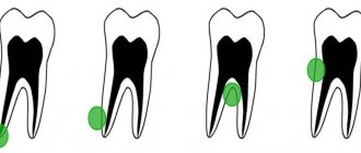
According to the localization criterion, the neoplasm is classified into:
- Sublingual. It is located in the hyoid-maxillary muscles or submandibular region. During rapid growth, it grows very quickly and then causes serious discomfort.
- Submandibular. Located in the lower submandibular region. It feels like a dense ball to the touch. Promotes disruption of the natural mechanism of salivary fluid secretion.
- Parotid. Rarely encountered in dental practice. It can be very painful when you open your mouth wide. It is formed due to impacts, injuries, after which the inflammatory process caused the closure of the salivary ducts.
- Extravasal. Most often found on the inner surface of the lip. Occurs due to mechanical damage. The inside is filled with granulation tissue.
Cyst on the gum: main causes of appearance
A cyst is a consequence of inflammation occurring in the tissues around the tooth.
Causes of inflammation:
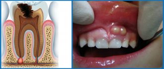
Advanced carious formation or pulpitis. If caries is ignored or delayed in going to the dentist, it can develop into periodontitis, characterized by the development of a purulent abscess in the upper area of the tooth root. The pus in the abscess will sooner or later find a way out: after it gets under the mucous membrane, a lump forms.
Pulpitis is another common disease when the inflammatory process occurs in the pulp (the connective tissue that fills the internal cavity of the tooth) without affecting other tissues. After the death of the pulp, the infection spreads to other tissues, resulting in the formation of pus in the upper part of the root.
Unsealed or poorly sealed root canals. Inaccuracies when filling canals are allowed by both beginners and experienced dentists. If you believe the statistics, then in more than half of the cases, canal filling is performed poorly, which is fraught with various complications.
Basically, dentists fill the canal without reaching the upper part of the root, as a result of which an infection begins to develop in the part of the canal not filled with filling material over time, gradually spreading beyond its boundaries and beginning to affect nearby tissues. An abscess occurs near the top of the roots. When there is too much pus, it will lead to the formation of a cyst.
Perforation. The second most popular mistake made by dentists is perforating the wall of the root canal when filling it. Perforations are small holes that occur when the canal processing technique is not followed and the pins are poorly fixed in them. Sometimes the pin is brought directly into the bone, which is why an abscess develops (if you look at the X-ray, it looks like a strong darkening).
Periodontitis. This disease causes deep pockets filled with pus. Quite often, the outflow of pus is disrupted, and then a purulent abscess occurs in the gum tissue.
Signs of a cyst
Among the main symptoms of the disease:
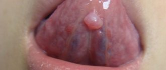
- The appearance of an unusual protrusion on the soft tissues of the mouth. It may resemble an abscess. It usually has a bluish color with a burgundy border, but it can also match the tone of healthy gums. The “older” the mucocele, the thinner its walls become. The “bubble” is movable; it is not fused to the surrounding tissues.
- Discomfortable sensations while chewing food. There is a feeling as if there is a foreign object in the mouth that is constantly in the way.
- An unpleasant feeling of constriction of the mucous membrane. In this case, pain does not occur.
The cyst may burst if there is a lot of pressure on it. Spontaneous opening sometimes occurs while eating. The difficulty is that afterwards it forms again - through the passage in the mucous membrane, the cleared cavity is refilled with liquid contents.
Symptoms of tonsil cyst
The clinical manifestations of this pathology depend on the size of this formation. For symptoms to appear, the retention cyst must exceed five millimeters. Before this, her discovery during examination was accidental. The location of the cyst also influences the clinic.
The patient is concerned about the following:
- sore throat; constant sensation of a foreign body; difficulty breathing; pain when swallowing saliva; there is a tingling sensation at the location of the cyst; have difficulty swallowing food and drinks; food entering the nasopharynx; change in voice timbre (hoarseness, hoarseness); sensation of unpleasant taste; constant coughing; bad breath.
If blood appears, it is necessary to suspect malignant degeneration of the tumor. When the cyst suppurates, the temperature rises, the throat turns red, and swelling of the tonsils is noted. The disease is differentiated from purulent tonsillitis.
Examination of patients who have a cyst in their mouth
Diagnosis of the described disease is simple.
The doctor examines and palpates the abnormal lesion and studies the symmetry of the face. If the mucocele has reached a diameter of more than one and a half centimeters, then its color is blue. When the lesion is opened, viscous yellow contents are released. If the resulting biological material is submitted for analysis, a large amount of salivary proteins and amylase will be found in it. If necessary, the Trommer reaction is performed to confirm the preliminary diagnosis.
During ultrasound diagnostics of the salivary gland, the doctor observes an anechoic formation of a round shape. Its borders are smooth. The fact that the patient has a mucocele is said:
- presence of granulation lining;
- absence of epithelial membrane;
- the presence of mucin and protective blood cells.
If there is doubt about the benignity of the tumor, the patient is referred to an oncologist.
Diagnosis of tooth root cyst
To make a diagnosis and carry out appropriate treatment, the dentist collects and analyzes the medical history. During the initial diagnosis, many patients report the fact of endodontic treatment performed to eliminate periodontitis or pulpitis. Some patients indicate an exacerbation of the disease after intraoral dissection.
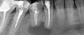
Radiography is used as the main diagnostic method. Below is a photo and x-ray of a dental cyst.
To obtain an x-ray, several methods are used, the first method is based on contact intraoral x-ray, the advantages of this technique:
- determining the degree of destruction of the jaw bones;
- assessment of the condition of the tooth root and tooth canal;
- assessment of the quality of canal filling;
- identifying the presence of perforations and fragments of instruments and materials in the tooth canal;
- determination of the relationship between the cyst and the roots located near the teeth.
The second method of performing radiography is an orthopantogram; the procedure is a panoramic photograph of both jaws and the maxillary sinuses of the upper jaw.
Another method of the procedure is a survey X-ray in the nasomental projection; the X-ray covers the bones of the skull from the nose to the chin; using the image, the doctor assesses the condition of the maxillary sinuses and detects cysts that have grown into the nasal cavity.
In addition to radiography, to detect a tumor, the patient may be prescribed an electroodontic diagnostic procedure. This technique helps to assess the degree of such an indicator as the electrical excitability of the teeth that are located next to the cystic tooth. If the value exceeds 60 microamps, the dentist prescribes endodontic treatment to the patient.
For diagnostic purposes, histological and cytological studies are used to determine whether the neoplasm is benign or malignant.
Diagnosing a dental cyst is not difficult, but only qualified dentists can carry it out in a hospital setting; under no circumstances try to independently determine the presence of a cyst and do not take therapeutic measures; strictly follow the doctor’s recommendations.
How to treat
It is very dangerous if a person tries to remove a cystic formation on his own and, to do this, puts pressure on it or bites it. Any mechanical influences have a negative effect on the course of the disease. Due to external pressure, the liquid inside the bubble begins to flow beyond its boundaries. Afterwards it accumulates again in the inflamed area. But at the same time, the risk of infection of damaged tissues increases significantly.
Doctors most often treat mucocele surgically. If the “bubble” is localized on the lower lip, two semilunar incisions are made in its projection, after which the internal neoplasm is isolated along with all its contents. Finally, stitches and a sterile pressure bandage are applied.
If the problem concerns the sublingual area, the following can be done:
- cystectomy;
- cystsialadenectomy.
In the first case, only the cyst itself is removed. It is cleaned and cut out. In the second type of surgery, the gland is also removed.
If there is a formation in the parotid zone, a parotidectomy is performed - complete or partial. It involves excision of the cyst and part of the parenchyma. Lesions located in the submandibular area are always removed along with the gland.
If we are talking about treating a child or a weakened elderly person, the dental surgeon may decide to excise only the dome (upper part) of the abnormal structure.
Signs, diagnosis and treatment
Manifestations of the disease depend on the location of the cyst. Most often, these are rounded formations that slowly increase in size, elastic and soft to the touch. They do not cause pain at first, rarely exceeding 1 cm in diameter. But in the absence of treatment, cysts begin to seriously interfere not only with eating, but also during conversation. The formation may disappear if, due to an accidental breakthrough, the contents come out. However, over time, the cyst forms again; if its size becomes very large, it acquires a characteristic bluish tint.
For diagnosis, methods such as visual inspection, palpation, and a number of hardware studies are used. Ultrasound, sialography to determine the problem with contrast, MRI and CT are more often performed. Additional examination methods may also be prescribed, for example, cystography of the bladder, probing of the salivary ducts, histology. If the diagnosis is questionable, a biopsy and puncture of samples are performed.
Conservative therapy does not bring any effect; when diagnosing a cyst of this type, surgical intervention is recommended. For this, local anesthesia is used, after which the formation is removed along with the affected gland and membrane to prevent relapses. If surgical intervention is refused, the formation often develops into phlegmon, and a tissue abscess develops.
The conservative method provides only temporary results. To do this, the membrane is punctured and the contents of the cyst are sucked out. But in all cases, the formation appears again, that is, this method of therapy is not recommended, as it is ineffective.
Surgery includes the following steps:
- inspection and surface preparation are carried out;
- local anesthesia methods are used for pain relief;
- the area with the cyst shrinks, which reduces blood flow and ensures stability of the tissue position;
- two oval-shaped incisions are made near the formation, after which the contents are husked;
- the affected lobes of the gland are removed, which helps to avoid complications or relapses in the future;
- the wound is sutured, thin sutures are applied, and self-absorbing sutures are often used.
The contents of the capsule are sent for additional research. This helps to eliminate serious risks, especially if the development of malignant processes is suspected. In case of serious intervention, plastic cystotomy is recommended. Typically, this situation occurs when removing a maxillary-hyoid formation.
Treatment of cystic formation at home
Home therapy for oral cysts only makes sense if for some reason you cannot get to a dental surgeon in the next few days. It consists of rinsing with herbal solutions and antiseptics. You can also make compresses with anti-inflammatory herbal medicines.
If the hearth breaks through, it’s too early to rejoice. Most likely, it will soon reappear in its original place. This is how cysts work - their contents expire, but the outer layer remains.
Under no circumstances should you treat a bulge on the gum or mucous membrane as a regular pimple. Any attempts to open it mechanically will not lead to anything good. But the resulting wound can become infected. Then the inflammation will spread to deeper layers in a fairly short time, and it will be much more difficult to cure.
conclusions
Having noticed inflammation, in no case should you engage in independent treatment or hope that everything will go away over time on its own. Even if the cyst opens, this will only slightly reduce the inflammatory process, but bone tissue destruction will begin in the upper part of the tooth, which will ultimately cause the tooth to be removed, since it will become impossible to cure it. Remember that at the site of the opened cyst there remains a hole through which pus will flow into the mouth: those around you will clearly smell this discharge, and the patient himself will also “enjoy” its taste. Treatment in case of cyst formation should begin with a visit to the dental surgeon, who will first take an image to make the correct diagnosis.
How to reduce the likelihood of developing education
To minimize the risk of mucocele formation, you need to follow the rules:
- lead a healthy lifestyle, quit smoking;
- carefully observe oral hygiene;
- rinse your mouth after every meal;
- eat a balanced diet;
- do not put foreign objects in your mouth;
- get rid of the habit of biting your lip;
- correct malocclusion;
- undergo oral hygiene every year;
- avoid trauma and chemical burns to the lips;
- promptly replace dentures;
- Follow the rules for wearing braces and caring for them.
People who follow preventive measures are much less likely to need treatment for oral cysts. If a lesion has appeared and is growing in size, there is no need to expect it to disappear on its own. This happens very rarely. It will either “deflate” or be filled again with contents and ultimately may grow to such a size that it will not be possible to do without emergency surgical intervention.
Rehabilitation, expected results
After surgery, there is slight swelling and pain at the operation site for two to three days. The doctor may prescribe medications to eliminate such unpleasant symptoms, for example, analgesics, antibiotics to avoid complications. Oral rinses using special antiseptic solutions are also prescribed.
If the healing process is normal, the sutures are removed after a week, after which a pressure bandage is applied. During recovery, it is recommended to avoid solid, spicy foods, too cold and hot dishes. Oral care also requires care to avoid damaging the operated area.
The prognosis for treatment is favorable; if therapy is carried out correctly and the patient follows all recommendations, there are no problems. But in some cases there are risks of complications. These include relapses, damage to the facial nerve or facial muscles when removing a cyst in the ear area. To reduce risks after completion of the recovery period, it is recommended to correct the shape of the tooth and install new orthodontic structures or dentures. In addition, prevention is needed, protection from injuries to the mucous membrane, and compliance with the rules of oral care.
Classification
Experts distinguish two types of tonsil cysts:
- Retention - formed after blockage of the ducts of the glands of lymphoid tissue, have a classic round shape, a thin capsule, filled with serous or purulent secretion. After emptying (evacuation of contents or spontaneous breakthrough), they are filled again.
- Dermoid or congenital - formed during intrauterine development, inclusions of embryonic tissue are always found in them. These true tumors, caused by genetic causes or harm suffered by the mother during pregnancy, are very rare. The capsule is usually dense and the contents are viscous.
Our services in otorhinolaryngology
The administration of CELT JSC regularly updates the price list posted on the clinic’s website. However, in order to avoid possible misunderstandings, we ask you to clarify the cost of services by phone: +7
| Service name | Price in rubles |
| Taking ENT smears | 600 |
| Videoendoscopy of the upper respiratory tract | 2 400 |
| Radio wave removal of skin (mucous) neoplasms of ENT organs | 4 000 |
All services
Make an appointment through the application or by calling +7 +7 We work every day:
- Monday—Friday: 8.00—20.00
- Saturday: 8.00–18.00
- Sunday is a day off
The nearest metro and MCC stations to the clinic:
- Highway of Enthusiasts or Perovo
- Partisan
- Enthusiast Highway
Driving directions
What do patients say about apex resection? Reviews are usually only positive!
“Resection of the top of the tooth took about an hour, the operation was performed under pain relief. It didn’t hurt very much near the tooth for about a month. A few months later I had to take an x-ray, the doctor showed that all the tissues had been restored."
“The cyst grew, they told me to remove it, it was a little unpleasant, but quite tolerable - the doctor quickly and accurately carried out the procedure. Everything was restored soon, now it’s completely unnoticeable that there was anything there.”
At Ritsa Dentistry, cyst removal operations are performed by experienced surgeons with extensive experience in performing such procedures. Therefore, you can rely on our professionalism and entrust the treatment of the cyst to our doctors.