Necrosis is a serious pathology accompanied by the death of gum tissue. It is quite rare in dental practice. The danger of this process lies in its irreversibility, asymptomatic course in the early stages of development and serious complications that it can lead to if the necessary measures are not taken in time. Find out what can trigger necrosis of gingival tissue, what the dead areas look like, what symptoms should alert you and where to go if they are identified.
Necrosis of gum tissue: how dangerous it is, what causes it and how to treat the pathological process
Necrosis is the process of gradual death of cells and tissues, leading to the cessation of their functionality and cessation of vital activity. Such a pathological phenomenon can begin in almost any part of the human body. In dental practice, gum necrosis usually occurs when the mucosal tissue begins to die, which is due to the cessation of blood circulation in them. There are many potential causes for this pathological process. Today we’ll talk about what this phenomenon is, what its characteristic symptoms are and how to treat the problem.
Possible reasons
Gum necrosis can be associated with various negative factors. The process of cell death is triggered by:
- chemical burns - doctor’s mistakes when killing the dental nerve with arsenic, treating gum fibromatosis using liquid nitrogen;
- prolonged exposure to low temperatures - applying ice to the gum after implantation and other dental procedures;
- chronic periodontitis is an inflammatory disease that affects all periodontal structures; in severe cases, soft tissues die;
- mechanical injuries of the gums - rubbing of the mucous membrane with a prosthesis, cuts with subsequent infection, jaw fractures, serious bruises;
- hormonal imbalances - pregnancy, age-related changes in the body, taking certain groups of medications;
- bad habits - smoking, use of narcotic and psychotropic substances.
To establish the cause of gum necrosis, it is necessary to undergo a comprehensive diagnosis. If the pathological process is not stopped, dangerous complications can arise. The most common include osteomyelitis of the jaw, sepsis, and loss of chewing function.
What is gum necrosis?
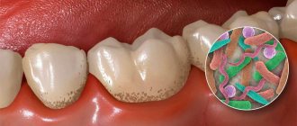
Gum necrosis is a serious pathological process leading to irreversible death of mucosal cells and its destruction. Symptoms of the initial stage are usually poorly distinguishable and rather vague, which makes it very difficult to diagnose the problem in the early stages of its development. Experts in the field of periodontics distinguish two forms: wet and dry necrosis. The affected areas noticeably darken and lose sensitivity. At the same time, the treatment is quite complex and lengthy. If we are talking about the wet form, it is important to urgently transform it into a dry form, in which surgical intervention is possible to stop the further spread of the process. You can see what the affected gum looks like from the photo below.
At the initial stage of the pathology, the symptoms are quite difficult to distinguish
There are many factors that significantly increase the risk of developing pathology. This includes aggressive chemical exposure, sudden temperature changes, disturbances in blood composition, penetration of harmful microorganisms, and much more. Symptoms that may indicate a problem include bleeding, a putrid odor, loose teeth, ulcers, and pain. However, similar symptoms are typical for many other dental diseases, so it is important to see a doctor as soon as possible and undergo a thorough diagnosis. In addition, gum necrosis often develops against the background of advanced diseases, so this process can be considered as a complication of many pathological conditions.
Necrosis is not reversible, which means that dead cells are not regenerated. Timely seeking specialized help will help stop its further spread, but the resulting defects can only be filled with the help of gum grafting.
Loss of dental tissue due to erosion
Tooth erosion
- progressive loss of dental tissue (dentine and enamel) of an insufficiently studied nature.
Some authors suggest that the cause of tooth erosion, like the wedge-shaped defect, arises solely from the mechanical action of the powder and the toothbrush. Others believe that the appearance of erosion is due to the large amount of consumption of citrus fruits and their juices. In any case, our dentists will be able to help in any clinical situation - they will provide high-quality treatment and save you from dental erosion.
A significant role in the mechanism of progression of erosion of hard dental tissues is assigned to increased function of the thyroid gland, and, in particular, endocrine disorders. According to experimentally obtained data, one of the main symptoms of this disease is considered to be a decrease in saliva viscosity and an increase in its secretion. It has been proven that tooth erosion occurs in patients with thyrotoxicosis twice as often as in people with normal thyroid function. At the Dantley Clinic, our dentists will perform remineralization as a treatment option for erosion. The number of patients with erosion of hard dental tissues increases by 20% with an increase in the duration of the disease by just one year.
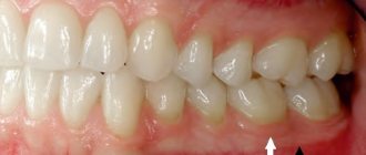
Erosion is the loss of dental tissue, which will continue to progress without medical intervention. Dental tissues are affected and thinned on one or more sides of the tooth.
Erosion of hard dental tissues mainly manifests itself on the symmetrical surfaces of the lateral and central incisors of the upper jaw, as well as on small molars and canines of both jaws. Erosion practically does not occur on large molars and incisors of the lower jaw. The lesion is most often observed in middle-aged people, and is characterized by a long course - up to 10-15 years. In St. Petersburg, the Dantley Clinic provides remineralization and restoration of teeth to patients of all ages. Over time, a large number of teeth become involved in erosion. Due to the significant impact of adverse factors, including environmental ones (Chernobyl disaster, etc.), the number of cases of dental erosion in fairly young people (18-25 years old) is significantly increasing.
Manifestation of tooth erosion
It is a round or oval enamel defect located in the transverse direction of the more convex part of the labial (vestibular) surface of the dental crown. The bottom of the erosion is shiny, hard and smooth. The gradual expansion and deepening of the boundaries of erosion leads to the loss of part of the dentin and all of the enamel.
There are two transitional stages of damage:
- initial (enamel erosion);
- pronounced (erosion of dentin and enamel).
According to the depth of the lesion, three degrees of the disease are distinguished:
first degree (initial)
- only the superficial layers of enamel are affected;
second degree (medium)
— the entire thickness of the enamel is affected, dentin is not involved in the pathological process;
third stage (deep)
— the entire thickness of the enamel, as well as the surface layer of dentin, is affected.
According to the course of the disease, two stages of erosion are distinguished - active and stabilized, but both of them are characterized by a chronic course. With any type of erosion, our dentists will carry out remineralization or filling, reducing the chronicity of the disease to zero.
The active stage is characterized by:
- rapid loss of hard dental tissues;
- increased sensitivity of the affected area to external irritants (sweet, sour, hot, cold, building up to erosion).
The stabilized stage is characterized by:
- slowly progressive loss of hard dental tissues;
- lack of increased sensitivity of the diseased area;
- possible transition from a stabilized stage to an active one.
Treatment of tooth erosion
Local treatment of the active stage:
- The goal is to stabilize the process. This result is achieved through additional remineralization of hard dental tissues using calcium electrophoresis or applications. Daily applications of a 10% potassium gluconate solution are prescribed for 15 minutes for 15-20 days. After which, for 3 days, a 2% sodium fluoride solution is applied to the area of erosion for 3 minutes. And the treatment is completed by coating the diseased surface with fluoride varnish. A 10% calcium gluconate solution is used for electrophoresis. The duration of the procedure is 5-10 minutes. After the electrophoresis session, a swab moistened with a 2% sodium fluoride solution is applied to the area of erosion. The entire course of treatment is 10-15 procedures.
- Fluoridation - restoration and strengthening of enamel with fluoride-containing and mineral substances.
- Filling erosion with composite materials fully restores the defect, the teeth look beautiful and natural.
- If there is a large area affected by erosion, it is worth making artificial crowns.
Local treatment of the stabilized stage:
- Phosphorus and calcium preparations, microelements and vitamins are prescribed internally.
- The task is tissue depigmentation. For this purpose, over the course of two or three visits, the affected surface is treated with an abrasive paste with a high fluorine content. Whitening, filling and restoration of the defect area are also used.
For what reasons can the process of mucosal cell death begin?
There are quite a lot of potential prerequisites for the development of such a process. The most common causes of destruction of mucosal structures are the following phenomena and conditions:
- low level of hygiene and, as a consequence, the appearance of abundant dental plaque, the development of gingivitis, periodontitis, advanced stages of these diseases,
- constant injury to mucosal tissue due to malocclusion, poorly installed filling, crown or prosthesis - everything that disrupts local blood circulation and leads to its stop in certain segments of the gums,
- hormonal disruptions during adolescence, during pregnancy, and in diseases of the endocrine system and hematopoietic organs.
Untimely treatment of gingivitis can provoke necrosis.
Pathology can appear against the background of severe hypothermia or temperature changes, after using arsenic to kill the pulp. If a temporary filling with this substance is not sealed tightly enough, it can come into contact with the mucous membrane and provoke an irreversible process of its gradual death. Much less often, a similar problem occurs after tooth extraction due to severe tissue trauma, as well as after anesthesia. So, for example, if a powerful anesthetic is administered too quickly, a spasm of blood vessels and nerve endings may occur, which will lead to poor circulation. This situation is especially dangerous for patients with diabetes and infectious diseases.
General overview
Necrosis is a dental disease resulting from mechanical damage or infection. Suppuration occurring in the facial hard tissues gradually destroys the cellular structure, which can ultimately lead to disability, and in particularly advanced cases, death.
- Prevention of necrosis, despite the severity of the pathology, is quite simple and includes the following recommendations:
- Abstinence from bad habits, including abuse of alcohol and tobacco products;
- Compliance with the correct approach to building a diet containing a sufficient amount of micro- and macroelements.
- Periodic dental diagnostics, including hygienic cleaning procedures - at least once every six months;
- Prompt treatment of identified local diseases, as well as compliance with hygiene procedures on a daily basis.
Risk factors that can trigger the development of necrosis include oncology treatment methods, which also necessitates a visit to the dentist after the next chemotherapy session.
How dangerous is this pathological phenomenon?
If you start the process, it will inevitably lead to irreversible consequences. The affected tissues do not have a chance to recover, and the further the necrosis goes, the higher the likelihood of losing teeth and losing chewing function. The addition of infections can provoke the development of osteomyelitis of the jaw - purulent-necrotic destruction of bone tissue structures. Less commonly, the pathological condition is complicated by general blood infection - sepsis.
You need to understand that necrotic areas in the oral cavity become a suitable environment for the proliferation of harmful microorganisms. The problem is that the initial stages of the pathology are quite difficult to diagnose. If the problem has already become pronounced, it will be much more difficult to treat.
Necrotic areas in the oral cavity become a suitable environment for the proliferation of harmful microorganisms
Classification
The course of the disease caused by infection can be acute, chronic or subacute.
According to location they are distinguished:
- osteonecrosis of the lower jaw;
- necrosis of the upper jaw.
By type of infection:
- endogenous - infection occurs as a result of dental diseases;
- hematogenous - pathogens enter the jaw through the bloodstream.
Also distinguished:
- medicinal (bisphosphonate);
- traumatic.
The American Dental Association defines the following stages of disease development:
Stage 0
– toothache that radiates to the temporomandibular joint or sinus. Mobility of teeth, with intact periodontium. The appearance of fistulas, cysts, fluxes.
Stage 1
– exposure of a section of bone without severe pain
Stage 2
– exposure of a section of bone, accompanied by pain and inflammation
Stage 3
- exposure of a section of bone that extends beyond the alveolar (the one in which the tooth is located) bone. With necrosis of the lower jaw, the jaw is affected down to the lower edge. Bone tissue atrophy occurs. This is the stage of pathological fractures. With necrosis of the upper jaw, the zygomatic arch and maxillary sinus are affected. Numerous fistulas are formed.
Types and symptoms of pathology
Necrosis develops gradually, moving from a mild stage to a more complex and advanced one. To choose the right treatment tactics, it is important to determine its form and degree of development:
- early stage – the symptoms are vague and weakly manifested. Dental tissues lose their shine, sensitivity to temperature changes in food occurs, and minor bleeding appears. The enamel becomes slightly rough, and the gums become pale, moving slightly away from the teeth,
- medium - the gingival papillae swell, destruction of their tips, bleeding, formation of a grayish coating, and pain when touched are observed. The affected area may become very pale or even darken, and an unpleasant odor may appear. Possible enlargement of the submandibular and other lymph nodes. At the same stage, ulcers may appear on the mucous membrane, the patient’s temperature rises to 38-39 ° C, headaches, appetite and sleep disturbances, nervous disorders occur,
- the latter – there is severe hyperemia, inflammation and swelling of the affected area. Tanya die off, decrease in volume and sometimes expose bone structures, and teeth begin to loosen. Abundant deposits of soft plaque appear, and the patient suffers from high fever, digestive disorders and general malaise.
At the initial stage, the gum turns white and moves away from the tooth
“I was diagnosed simply during a routine preventive examination. Can you imagine my shock? After the examination, the doctor said that I suspected necrosis of the mucous membrane near one upper tooth. After examination, the diagnosis was confirmed. I went through treatment and everything seems to be fine. Now I don’t miss a single scheduled examination. This is how nothing portends trouble, and therefore, like a bolt from the blue, your gums are dying...”
Natalya V., from correspondence on the forum www.32top.ru
As mentioned above, gum necrosis can be in one of two forms: wet or dry. In the first case, the dead areas dry out and significantly decrease in volume. In this case, signs of inflammation and intoxication are usually absent. The wet form is considered more complex and dangerous, accompanied by swelling and severe hyperemia. As part of treatment, they usually try to make it dry in order to surgically remove the affected areas and stop the spread of the pathology.
Acid necrosis of teeth
Acid necrosis of hard dental tissues is an occupational disease that occurs in people working in the production of inorganic acids. It is assumed that acid vapors reduce the pH of saliva, and the capacity of the buffer systems of the oral fluid and the remineralizing properties of saliva are also reduced. This contributes to rapid wear (abrasion) of the hard tissues of the tooth. Dental damage is widespread and the process develops slowly.
Clinical picture
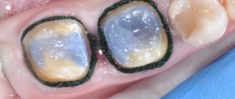
Acid necrosis of teeth develops slowly. With chemical necrosis, fangs and incisors are more often affected: in the area of the cutting edges, the enamel disappears, resulting in the formation of sharp edges. In the initial stage, patients have a feeling of teeth on edge, and there may also be a feeling of teeth sticking, which is associated with the “washing out” of minerals from the tooth enamel, resulting in a feeling of softness of the teeth when they are closed. In later stages of the disease, pain from temperature and chemical stimuli appears. However, it should be borne in mind that pain may not be observed, due to the fact that tertiary dentin is being produced. As necrosis progresses, the enamel loses its shine, becomes dull and rough. And after complete loss of enamel, dentin becomes pigmented and becomes dark. In advanced cases, complete dissolution and abrasion of the tooth crown may occur.
Differential diagnosis of acid necrosis
Acid necrosis of teeth must be differentiated from enamel erosion. Erosion is characterized by a hard, shiny surface, and with necrosis, softening of the enamel occurs.
Prevention of acid necrosis
Prevention
acid necrosis is to improve working conditions, use individual products, and conduct regular alkaline rinses.
Necrosis of the gums after the use of arsenic
Previously, such a pathological phenomenon was a fairly common complication from arsenic. Some time ago, this poison was quite widely used in dentistry to kill the pulp and subsequent depulpation - removal of the nerve.
Problem may arise when using arsenic for dental treatment
Until now, some budget clinics use arsenic-based substances, although today there are safer and less toxic devitalizing pastes. So, for example, necrosis from “Depulpin” (arsenic-free pulp killing agent) is an unlikely phenomenon. However, even when using it, it is important to act carefully, in no case exceeding the recommended dosage, otherwise an aggressive effect on surrounding tissues may provoke a similar problem.
Both arsenic pastes and more modern gentle agents have a strong toxic effect, directly related to the disruption of metabolic processes in the treated area - this is their direct purpose. But if excess composition gets on the mucous membrane and is in contact with it for too long, their effect will spread to healthy tissues, resulting in an irreversible process.
How is diagnostics carried out?
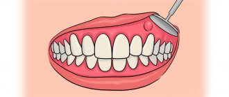
Often the problem is identified during a routine dental examination, without complaints from the patient. In other cases, symptoms appear: pain, hyperemia, bleeding. Other common signs of pathology are bad breath, inflammation and swelling of the mucous membrane, deterioration in general health, digestive problems, difficulty swallowing, etc. As part of the diagnostic examination, an X-ray is usually taken and an instrumental examination is performed. Radiography allows not only to identify the necrotic process, but also to assess its extent, as well as determine the stage and possible complications.
X-ray examination allows you to see hidden problems
On a note! Additionally, laboratory examination may be required, for example, microscopic examination of soft plaque. It allows you to study the microflora, identify the possible development of a fungal infection, determine the number of leukocytes, spindle-shaped rods and Vincent spirochetes1.
At the very initial stage, changes in gum tissue are still reversible, but making an accurate diagnosis requires the use of differential techniques to exclude other possible gum diseases. It is also important to identify associated pathological conditions and complications.
Symptoms of the disease
Signs of the disease differ depending on the stage.
Early.
The tissues are slightly affected, darkening of the enamel and pale gums may be observed.
Average
. Swelling of the interdental papillae, a gray coating forms on the mucous membrane, and bad breath appears.
Heavy.
Swelling of the gum tissue - it turns black and ulcers appear on it. Appetite disappears, body temperature rises to 39 C°.
Last
. The epithelium dies, severe pain is felt in the gums, the necks of the teeth are exposed, and the dental units become loose.
Features of treatment of the pathological process
How to treat necrosis directly depends on its form and stage, the presence or absence of concomitant diseases. This is an irreversible process, so the main treatment is aimed at stopping it and stopping further spread. It is important to stop cell death and restore blood circulation. Necrotic areas will have to be removed surgically. Depending on the form of the pathology, there are two treatment tactics:
- dry necrosis - the affected area is treated with antiseptics, after which dead tissue is removed, normal blood flow is established in areas that have not lost their functionality,
- wet – the causative area is treated with a solution of hydrogen peroxide, abscesses and ulcers are opened and drained, and a thorough antiseptic treatment is carried out. All this is necessary to transfer the condition to a dry form. It also happens that 2-3 days after the procedures the patient’s condition worsens, and in this case the affected tissue is removed urgently.
As part of treatment, antibiotics are usually prescribed to prevent intoxication of the body. After the necrotic process has completely stopped, a transplantation - gum grafting - can be performed to restore aesthetics.
After treatment you may need gum surgery
Therapy methods
Treatment of necrosis is long and quite complex. But with timely treatment, the prognosis for treatment of necrosis of both the lower and upper jaw is quite favorable (although damage to the upper jaw bone is considered more dangerous).
First, it is necessary to eliminate the primary purulent focus: remove the tooth, prescribe anti-infective drugs or surgically treat wounds after injury. Then the dental surgeon opens the purulent formations, carries out antiseptic treatment and prescribes antibiotics, painkillers, anti-inflammatory drugs, as well as agents for tissue regeneration.
Also, after removing dead tissue, the dentist can strengthen the teeth using splinting. As additional measures, doctors can prescribe hyperbaric oxygen therapy, ultraviolet irradiation of blood, plasmapheresis, local physiotherapy, including the use of ultrasound and UHF. If, due to chronic necrosis, it is necessary to remove areas of bone tissue, then these areas are filled with special materials with antibiotics.
Preventive measures - how to prevent the development of necrosis
Prevention is based on maintaining oral hygiene and preventing dental diseases. In this regard, experts suggest adhering to the basic rules:
- regularly brush your teeth with an individually selected brush and toothpaste - 2 times a day, rinse your mouth after each meal, use floss and irrigator,
An irrigator is an additional device for maintaining oral hygiene - correct the bite in a timely manner to avoid tissue injury and accumulation of bulky deposits,
- adhere to a healthy diet and a balanced diet, ensure a sufficient supply of vitamins, minerals and amino acids in the body,
- give up alcohol and smoking - these bad habits lead to severe irritation of the mucous membranes and dental tissues, provoke many pathological conditions,
- support the body's immune defense,
- promptly treat diseases of the teeth and gums, as well as systemic pathologies of the gastrointestinal tract and endocrine system, and control diabetes mellitus.
After treatment of necrosis, it is important to provide competent and regular oral care to prevent relapses. It is imperative to regularly visit the dentist’s office for routine preventive examinations - at least 2 times a year.
- Wolf G.F., Rateitskhak E.M. Periodontology, 2008.
Prevention
Prevention of this pathology, first of all, consists in eliminating aggressive factors that provoke tissue death, or limiting their impact.
In addition, it is necessary to adjust the diet, reducing the consumption of sour and sweet foods to a minimum.
It is also worth paying special attention to the quality of oral hygiene and regularly visiting the dentist.
If you find an error, please select a piece of text and press Ctrl+Enter.
Tags treatment necrosis tooth decay
Did you like the article? stay tuned
Previous article
Schutz implants: features of creation and installation
Next article
Chronic caries: characteristic signs, treatment methods