Difference between molars and premolars
The main difference between molar teeth and premolars is their location on the jaw.
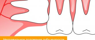
Premolars are located closer to the front of the dentition, and molars are slightly moved inward. Their quantity also varies. There are always two premolars on each side of the upper and lower jaws. There can be either two or three molars. Still others are also called “wisdom teeth.” They grow later than others and often appear only at 20-25 years of age. This process is often accompanied by malaise and fever. However, not all people on earth have third ones. A certain genetic predisposition can cause the absence of wisdom teeth in the oral cavity.
Timing of adult teeth eruption
The rudiments of the first teeth (on average, about 20 units) in infants are formed during the first two years of life. When the time comes to replace them with permanent teeth, the milk teeth become loose and fall out. There are no specific dates for the eruption of molars; many factors can affect the speed: environmental conditions, climate, water quality and diet. Genetic characteristics also play a certain role, some of which make themselves felt even during the formation of the fetus. The influence can be both positive and negative. If parents have healthy teeth from birth, then you don’t have to worry about the child’s teeth. If the first incisors, canines and premolars grow in 3 years, then the permanent ones take a long time to erupt. The first symptoms of dentition change can be seen at the age of 5, and it continues until the age of 21, when the third molars appear.
Features of the structure of molars
Molars have a characteristic structure. On the upper jaw they have three roots and four canals. The lower jaw has two roots and three canals. Moreover, the number of canals also differs depending on the location of the individual tooth. Thus, the first of them often have one more channel than the next two.
The main characteristic feature of this type of teeth is the area of their chewing surface. They bear the heaviest load when chewing food particles. The molars themselves also have differences among themselves, which are associated with the structure of the jaw.
Central lower incisor
Average age of eruption: 6-7 years
Average age of root formation: 9 years
Average length: 20.7 mm
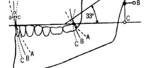
Narrow and flat in the labial-lingual direction, the lower central incisor is the smallest adult tooth. Radiologically it is visible in only one projection and thus appears more accessible than it actually is. The crown, narrow in lingual projection, has a limited area for access. For gentle access formation, a fissure bur and a spherical bur No. 2 are used. The access cavity should be oval and performed on the lingual side.
The lower central incisor often has two canals. One study reported that 41.4% of mandibular central incisors studied had two separate canals, of which only 1.3% had two separate apical foramina [1]. After completing the access formation, the doctor must examine the tooth cavity to identify an additional canal. Endodontic treatment failures in the lower incisor area are most often associated with an unidentified canal, usually in the lingual location. To create conditions for the straight entry of endodontic instruments into the additional canal, access can be expanded in the incisal direction.
There is a great danger of perforation of the vestibular wall, but it can be avoided if the doctor remembers that it is almost impossible to perforate in the lingual direction, since the bur axis is in contact with the incisal edge. The slit-like lumen of the canal is so common that it can be considered a normal variant, and this requires special attention when cleaning and shaping.
Lateral perforations and pulp anatomy will be discussed in an illustration of the lower second molar.
Molar Differences
Often the surface of each molar tooth is shaped like a triangle. There is a certain number of tubercles on it, which take an active part in chewing food. The number of such tubercles may vary. Usually there are three, but sometimes there are more.
Such mounds are connected to each other by special ridges. On the upper and lower jaws the structure of these elements is different. In the upper dentition, the apex of the surface triangle is directed towards the tongue. This form is called a trigon. On the lower jaw, the apex of such a triangle is directed towards the cheek, which is called the trigonid. The size of the first and second types of teeth is practically the same.
Possible damage to small molars
Carious lesions of primary and molar teeth in children are observed quite often. This is due to the bumpiness of their surface, which can trap food particles and bacteria. Since children at an early age do not always actively and correctly brush their teeth, these residues can lead to damage to the enamel and deeper spread of caries.
Depending on the degree of damage, caries can penetrate either exclusively into the enamel, or into dentin or even cement. In the latter case, we are talking about deep caries, which often requires serious treatment or even tooth extraction.
Another variant of damage is a change in tissue structure, which may be associated with metabolic disorders in the human body or other diseases. Such a disease can also be the result of poor nutrition or constant diets, in which the body does not receive enough nutrients.
At any age, a person can experience excessive tooth wear, which leads to the destruction of enamel and can cause other serious damage. Teeth can wear down in different situations, such as:
- if a person has bad habits of grinding his teeth, etc.;
- in case of improper or insufficient nutrition;
- in the presence of diseases of the endocrine system;
- due to a genetic predisposition to enamel depletion.
All of these lesions can be the result of both an incorrect lifestyle and various diseases or heredity. Regardless of the reasons, it is necessary to begin treatment of emerging injuries immediately so as not to aggravate the process.
Lateral lower incisor
Average age of teething: 7-8 years
Average age of root formation: 10 years
Average length: 21.1 mm
Very similar to the lower central incisor, so access cavity preparation follows the same principles.
Their similarity can cause rare but serious errors. Hasty installation of a rubber dam, identical fillings and carelessness can lead to preparation of the access cavity on the wrong tooth. This error can be prevented by marking the vestibular surface of the tooth with a felt-tip pen before applying the rubber dam.
Trauma, periodontal disease, carious lesions and malocclusion can lead to obliteration of the canal. When moving apically to identify the orifice, great care must be taken to prevent unnecessary destruction of the crown and root. Labial perforations were discussed in Illustration IX. If the bur is not oriented along the long axis of the tooth, there is a risk of lateral perforation. The situation becomes more complicated with the traumatic loss of an anatomical crown. Without anatomical landmarks, a lateral perforation can be easily made when moving in a coronal direction. To prevent this, access is carried out without a rubber dam so that the root can be palpated.
Lateral perforation with endodontic files and Gates-Glidden burs is facilitated by the presence of slit-shaped canals with a narrow, hourglass-shaped cross-section. To avoid vertical fracture along the approximal root wall, minimal expansion and preparation of the space for the abutment pin is indicated.
Apical curves and accessory canals are common in the lower incisors.
Replacement of primary molars with permanent permanent molars
Molars are the first permanent teeth to appear in a child's mouth. This begins around the age of five. Growth begins with the first molar tooth, which appears in a free space in the depths of the jaw, closer to the milk one that has not yet fallen out.
The second molar usually grows in at the age of 12-13 years. It also takes up free space and does not replace a baby tooth. The latter, or “wisdom teeth,” may take up to 25 years to grow or may not appear at all.
As for dairy products, they begin to fall out from the age of 9. In their place, permanent teeth grow, which occurs at approximately 10-12 years of age. Usually these are the last molars, the growth of which must wait to fill the adult dentition (not counting “wisdom teeth”).
Is it possible to loosen baby molars?
The process of tooth loss always begins with softening of its root. This occurs as the jaw grows, freeing up more space for a permanent tooth. Thus, while the baby tooth is still in the hole, the molar is already beginning to take its correct place.
In this regard, dentists categorically do not recommend deliberately loosening baby molars. If they fall out prematurely, jaw growth may be stunted. As a result, there will not be enough space for permanent teeth, and they will begin to grow crookedly, getting out of the general dentition.
Signs of imminent appearance of molars
The first signs that molars are about to appear are noticeable even before the baby teeth fall out. These include the following:
- widening of the jaw, which can be seen when gaps appear between other baby teeth;
- the appearance of sufficient free space behind the outer lateral milk teeth;
- swelling of the gums.
In this case, the temperature does not necessarily have to rise or the child’s well-being deteriorates, as was the case with the growth of baby teeth. This is why the appearance of the first molars often goes unnoticed.
Helping your child replace teeth
The process of replacing teeth often takes place without pain or discomfort. The roots of baby teeth dissolve on their own, and the dental crown falls out freely. However, there are a number of recommendations that should be followed during this period.
The main one is systematic rinsing of teeth for disinfection purposes. This is necessary in order to avoid bacteria from entering the hole that will form at the site of the falling tooth. In addition, the sharp edges of a crown that has separated from the gum may cause minor damage to soft tissue. To prevent them from becoming inflamed, it is important to exclude any infection.
If your child experiences any pain during the tooth replacement stage, you should immediately consult a doctor. Under no circumstances should pain be eliminated using improvised methods.
Molars and prevention of their loss
Molars are stronger than baby teeth. However, they need proper care to prevent their loss, because new ones will not grow in place of a lost tooth.
Prevention of molar tooth loss involves proper oral hygiene. It includes systematic brushing of teeth, use of dental floss and mouthwash. In addition, you should carefully monitor the condition of your teeth and consult a doctor if even minor damage is detected.
Proper balanced nutrition is also mandatory. Especially in childhood and adolescence, you need to ensure that your body gets enough calcium and vitamin D.
Caring for teething teeth
When changing teeth, caring for them must be especially thorough, because a lost tooth tears the tissue, and when infected, it quickly becomes inflamed. To prevent such problems it is necessary:
- teach children to regularly brush their teeth, use a scraper and floss, and rinse their mouths;
- to support the enamel, buy your baby a paste with added calcium and fluoride;
- Proper nutrition with limiting sweets and carbohydrates in favor of vegetables, fruits, and dairy products will help strengthen new teeth and protect them from caries;
- consult a doctor on the choice of vitamins (especially vitamin D) and gels to improve the mineralization of new teeth;
- In case of inflammation, before meeting the dentist, you should actively rinse the child’s mouth with antiseptics and herbal decoctions.
You can buy mouthwashes for children or prepare herbal teas for this purpose.
Bad habits interfere with the normal growth of adult teeth: sucking fingers or tongue, pacifiers and any objects. Despite the lost teeth, do not limit your baby to solid food. A piece of apple or carrot massages and strengthens the gums, freeing teeth from plaque.
Maxillary premolars
The crown of the premolars on the upper dentition has a prismatic shape. The buccal and palatal bones often have a convex surface. The first and second premolars in the row also have their differences.
First premolar
The difference between the first premolar of the upper jaw is the predominance of the vestibular surface over the palatal surface. Its contact surfaces are rectangular in shape. The buccal tubercle has two distinct slopes. The root of such a tooth often has a bifurcation.
Second premolar
In such a tooth, the buccal surface usually predominates over the palatal surface, which is more rounded. In most cases, this premolar tooth has a single cone-shaped root. However, there are also cases when it has a split.