Osteotomy is a surgical operation aimed at eliminating deformity or eliminating anatomical and functional disorders by artificially breaking the bone and correct fusion. This operation is performed to treat both congenital and acquired pathologies, for example, in case of improperly healed fractures.
Indications for osteotomy:
- improperly healed bone fractures;
- ankylosed joints;
- change, shortening of the limb;
- rachitic curvatures;
- formation of a false joint;
- osteomyelitis;
- osteoarthritis, spondioarthrosis;
Depending on the surgeon’s goal and the patient’s clinical picture, the following types of osteotomy can be performed:
- wedge-shaped;
- linear (oblique and transverse);
- hinged (can be arc-shaped, that is, made in one plane, or angular, spherical - in several planes);
- staircase;
- Z-shaped;
- derotational.
By purpose they are distinguished:
- Corrective osteotomy - to correct deformity after complications that led to malunion.
- Hinge osteotomy is an operation to lengthen the limbs.
Osteoplasty techniques are also divided into closed and open. The first is performed with minimal access, through a 2-3 cm incision. The open method involves wide access - an 8-12 cm incision is made exposing the bone. In some cases, when there is a risk of damage to nerves and large vessels, preference is given to open osteotomy, despite its low invasiveness, over closed one.
The choice of method depends on the type of pathology, volume and area for surgical intervention - pelvis, jaw, hip, etc. Most often, the operation is aimed at restoring the function of the musculoskeletal system and removing bone deformations.
To equalize the lengths of the lower extremities, osteotomy is performed according to the principle of oblique cutting of the bone in mandatory combination with extrafocal compression-distraction osteosynthesis. The design for osteosynthesis is selected individually for each patient.
Osteotomy: a scientific view
How does it work and why trim the bones? The entire human musculoskeletal system is penetrated by thin axes. They can only be seen in an anatomical atlas, but it is by them that the correct formation of the skeleton is determined. With pathologies, bones deviate from the anatomical axis. This is expressed by crooked legs, protruding joints, and other symptoms. In addition to aesthetic aspects, deviation from natural axes leads to diseases of the musculoskeletal system and disability. Bone tissue has an amazing ability: it is renewed throughout life. This happens quickly in childhood, but the ability decreases with age. However, bones can repair themselves by growing new tissue. This happens after injuries, fractures, operations with the help of nature. But sometimes the bones need help. Bone tissue is a material. It is the task of an orthopedic surgeon to mold it into beautiful shapes, adjust it and set the direction of correct growth. This is what corrective osteotomy does. An incision is made through the skin into the bone at the site where bone tissue needs to be built up. This position is fixed with a special device. There are many varieties of such devices. The only task is to hold the limb in this position long enough for new bone tissue to form. This usually takes 1-3 months. During this period, the patient can move independently with the device. Then it is removed. In leg surgeries, osteotomies of the femur and tibia are most often performed.
Possible complications
Some complications arise already during osteotomy, others during the rehabilitation period.
- Incorrect fusion of bones. Occurs due to improper fixation of bone tissue fragments during surgery. With such a complication, repeated intervention is necessary.
- Non-union of bones. It may occur due to severe concomitant pathologies, smoking, impaired blood supply to the operated area, or osteoporosis. For treatment, a repeat operation is performed and special rehabilitation is prescribed.
- Compartment syndrome. Occurs if, during surgical manipulation, the muscles were strongly compressed by a tourniquet. For treatment, certain drugs are prescribed; if the case is severe, a fasciotomy is performed.
- Improper functioning of the joints located next to the surgical field. This complication is typical for the absence or violation of rehabilitation rules. Exercise therapy is prescribed.
- Infections. They can be introduced during surgery or due to improper wound care. An antibiotic is prescribed for treatment; in severe cases, revision surgery will be required.
- Nerve damage is a surgeon’s mistake or a peculiarity in the location of the patient’s nerve endings. The function of a damaged nerve cannot be restored.
- Thromboembolism. Occurs when anticoagulants are prescribed incorrectly, getting out of bed late, or the inability to wear compression stockings. To eliminate this complication, high doses of antiplatelet agents and anticoagulants are needed.
Contraindications for osteotomy
Osteotomy is an operation. Like any intervention, it has contraindications. According to ISAKOS (International Society of Arthroscopy, Knee Surgery and Orthopedic Sports Medicine), bone correction cannot be performed with the following diagnoses:
- Rheumatoid arthritis;
- Osteoporosis;
- BMI more than 40;
- Impaired blood flow in the lower extremities;
- Extra-articular deformities;
- Previous infection;
- Reduced bone regeneration ability;
- Operations on the meniscus;
- Limitation of flexion more than 25 degrees;
- Some types of arthrosis.
In other situations, surgery can be performed, because for many, corrective osteotomy remains the last chance to return to motor activity, beautiful legs and a full life.
To eliminate lower prognathia, O. Neuer (1962) proposed.
The technique of decortication of the mandibular ramus in the form of a narrow cavity in the horizontal direction above the mandibular foramina, leaving only small bridges of the compact layer along the posterior edge of the ramus. The operation was performed using a submandibular approach. After 2 weeks, a blow to the chin caused a posterior displacement of the lower jaw (like a subperiosteal fracture). In the postoperative period, intermaxillary fixation was performed with a rubber rod. In our opinion, this method does not guarantee against relapse.
To eliminate prognathia of the mandible or its combination with an open bite, R-Ritter (1956) proposed performing an arcuate osteotomy in the area of the lower third of the mandibular ramus, i.e., above the opening of the mandibular canal (Fig. 29).
The lower jaw moves backwards, and in the presence of an open bite, upwards, the teeth are set in an orthognathic bite. Osteosynthesis of bone fragments is carried out using a wire suture. In the postoperative period, intermaxillary fixation is performed using dental splints. O. Neuer (1962), N. Kole (1963) performed the method of partial arcuate resection of the branches of the lower jaw in the same location and for the same indications as R. Ritter.
This technique, in our opinion, is technically complex and requires great precision. It is not without the same disadvantages as horizontal osteotomy of the branches (small contact area of the wound surfaces of bone fragments, the possibility of damage to the neurovascular bundle, etc.).
Among operations on the branches of the lower jaw, oblique “sliding” osteotomy, first performed in our country in 1928 by A. A. Limberg (1924) (Fig. 30), and abroad by G. Regtes (1924), occupies a significant place.
Its essence lies in osteotomy of the jaw ramus from the middle of the semilunar notch to the posterior edge of the ramus, then the large fragment is mixed downwards, sliding the osteotomized edge over the small fragment. The operation ends with osteosynthesis with a wire suture. This surgical method has found wide application in many clinics and specialized hospitals [Kabakov B. D., Vasiliev V. S., 1966; Rudko V.F., 1966 (Fig. 31); Vasiliev V. S., 1967, 1969, 1970 (Fig. 32) and others. ] .
Corrective osteotomy at the Ladisten clinic
The clinic specializes in minimally invasive orthopedic surgeries. More than 6,000 patients from all over the world have already undergone osteotomy and are satisfied. Each patient is offered a tour of the facility, a separate room during rehabilitation, and 24-hour medical supervision in the first days after the procedure. The operation itself is bloodless: a small puncture is made in the leg, through which doctors correct the bone. For fixation, a unique device from Dr. Veklich is used. This is an improved design of the Ilizarov apparatus. It is less bulky, weighs little and does not involve dangerous knitting needles. Doctors at the Ladisten Clinic have been performing corrective osteotomy procedures for more than 30 years; the price varies depending on the pathology and severity of the case. To find out the exact diagnosis, consult about contraindications and discuss the cost of osteotomy, just make an appointment by calling us at: +38 +38 or Write to WHATSAPP Write to VIBER We will choose a convenient time for you to visit the clinic or online consultation.
In the literature, this method is sometimes called “Swedish”.
In 1910, W. Babcock also used horizontal osteotomy of the branches at the level of their upper third. No bone fixation was performed. WW Babcock inserted a piece of ivory as a spacer between the fragments (Fig. 25).
In the postoperative period, the jaw was fixed using dental splints and rubber traction.
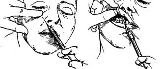
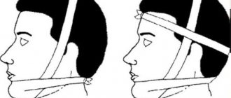
The Czech surgeon F. Kostecka in 1924 performed a horizontal osteotomy of the branches of the lower jaw without a soft tissue incision - blindly. The essence of the operation is as follows. Using a Kerger needle, an injection is made below the earlobe 5-6 mm from the posterior edge of the branch. Then, all the time feeling the medial surface of the jaw branch with the end of the needle, they move it anteriorly towards the lower edge of the zygomatic bone. At the front edge of the branch, the needle pokes out through the cheek. It is important that the needle passes above the entrance to the mandibular canal, between the medial surface of the ramus and the soft tissues containing the neurovascular bundle (inferior alveolar artery, nerve and vein). At the same time as the needle, a silk thread is passed through, held in a hole at the end of the needle. Then they do it in two ways: either the needle is removed, leaving a silk thread in the wound for further insertion of the Gigli saw, or the needle is inserted without the silk thread, and the Gigli saw is fixed to the end of the needle and brought to the level of the posterior edge of the branch. Using sawing movements in a horizontal direction (the skin at this moment must be protected from damage), the jaw branch is cut first on one side and then on the other. After this, the lower jaw is shifted posteriorly until the correct bite is established and fixed to the upper jaw using dental splints and rubber rings for a period of 2 - 3 months, and sometimes more (Fig. 26).
The main disadvantages of the Kostechka method are bleeding, the formation of salivary fistulas, damage to the facial nerve and frequent relapses (up to 50%, according to E. Reichenbach, 1955).
The operation using the Kostechka method in its initial version was used by many surgeons in our country and abroad [Kabakov B. D., Pasternak A. A., 1962; Mukhin M.V., 1963; Egiyan G. M., 1964; Klementov A.V., Vasiliev V.S., 1964; Mikhelson N. M. et al., 1965; Vasiliev V. S., Popov S. E., 1967; PopudrenkoP. I, 1970; Pichler N., 1928; Pearson W. N., 1943; Traunham V. N., 1944; Reichenbach E., 1955; Hinds E. S., 1957; Toman Y., 1957, 1968; Shira R.V., 1961; Urban F., 1961; Ullik R., 1962, etc. ] .
While maintaining the basic principle of the method, a number of surgeons made some modifications to Kostechka’s operation. Thus, A. Zindemann (1921) performed osteotomy of the branches not with a Gigli saw, but with a hacksaw. E. Skaloud (1954) performed this operation using intraoral access. S. M. Davydov (1960) proposed a protective plate to prevent injury to soft tissues and the neurovascular bundle. S. Moose (1945) performed osteotomy of the mandibular branches intraorally using a mechanical saw with a protective plate. To prevent anterior displacement of the small fragment, K. Mushka (1971) proposed fixing the fragments with a wire loop from the semilunar notch to the angle of the jaw; The author passed the wire subcutaneously using a special conductor.
Method of “intercortical” osteotomy of the branches of the lower jaw
Proposed in 1962 by G. G. Mitrofanov and V. V. Rudko. Its essence lies in the sagittal splitting of the lower part of the branches of the lower jaw when eliminating lower prognathia or its combination with an open bite. After exposing the bone using the submandibular approach, an osteotomy is performed with a fissure bur in the form of an oval (on the outer surface of the vegvi), as well as separation of the outer and inner compact plate in the retromolar region and along the posterior edge of the ramus. The internal compact plate is intersected in the transverse direction using a Gigli saw, moving upward from the mandibular foramen by 1 - 1.5 mm. The final splitting of the bone is carried out with a flat chisel. After the jaws are established in the correct bite, the fragments are fixed with one wire suture (Fig. 50).
Subsequently, one of the authors [Rudko V.V., 1975, 1976] called the osteotomy “planar.” The positive side of this method is a fairly large area of contact between bone fragments, a good postoperative effect, and the ability to form normal dimensions of the mandibular angle. The disadvantages of this method include the laboriousness of osteotomy with a Gigli saw, since damage to the neurovascular bundle cannot be ruled out; horizontal linear cutting along the inner surface of the branch makes it difficult to carry out rotational displacements of fragments.
Our experience in treating adult patients with malocclusion indicates the high effectiveness of sagittal retromolar osteotomy; 95.2% of patients had a good functional and cosmetic result. However, this surgical method was not without one significant drawback: the rectangular edges of the bone wound on the inner surface of the branch of the delicate jaw limited the mobility of the small fragment. With combined forms of malocclusion, it was necessary to constantly correct the bone edges. Otherwise, the fragments could not be in close contact with each other, and this in turn prevented favorable consolidation.
Our modification of the Dal Pont operation (1977) to form an oval rather than a rectangular incision on the inner surface of the jaw branches only partially eliminated this drawback (see Fig. 45). In this regard, we proposed to form an oval incision on the outer surface of the angle of the lower jaw [Sukachev V.A., Gunko V.I., 1977]. The experience of treating 14 patients showed that “oval planar retromolar osteotomy,” as we called it, without causing damage to the size of the area of contact of bone fragments, allows with a good outcome to eliminate all types of combined forms of malocclusion that cause deformation of the lower jaw (Fig. 51).

