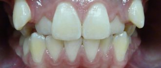
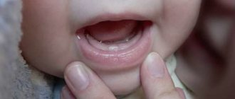
The development of malocclusion in children is a fairly common and common problem. Malocclusion can also develop in adults, but for children this problem is most relevant, since in childhood the processes of formation of the dentofacial apparatus are actively underway, and teeth during this period are easily amenable to movement.
For modern dentistry, malocclusion in children is not a big problem and can be easily corrected. The main factor for quickly and successfully correcting a malocclusion is a timely visit to a pediatric dentist, preferably before the child’s permanent teeth begin to erupt.
A malocclusion ruins your child's smile. For children, this can also result in psychological problems, since crooked teeth can become an object of ridicule among the child’s peers. In addition to the aesthetic problem, various other diseases of the teeth and gums can develop.
Correcting a malocclusion should not be delayed; at the first symptoms, you should contact a pediatric orthodontist.
Signs and symptoms of malocclusion in children
The development of malocclusion can be noticed in children as early as 3 years old. Parents can determine this themselves. Modern dentistry distinguishes several types of occlusion with symptoms and signs characteristic of each of them:
Deep bite . When a deep bite is formed, a pronounced protrusion of the upper jaw over the lower jaw is observed. With such a bite, the incisors interact poorly with each other. Symptoms may appear in the form of pain in the jaw joints. The development of a deep bite negatively affects chewing food.
Mesial bite . With a mesial bite, there is a significant movement of the lower jaw forward. Just like with a deep bite, this leads to poor functioning of the incisors, and, consequently, to insufficient chewing of food, which can ultimately cause digestive problems.
Crossbite . The peculiarity of this type of malocclusion is the overlap of the dentition when the jaws are closed. Pathology can manifest itself both from the front part of the dental rows and from the side. In addition to aesthetic troubles with crossbite, there are problems with the pronunciation of words, the risk of developing caries increases, and problems associated with diseases of the gastrointestinal tract may develop.
Distal occlusion is one of the types of malocclusion with a characteristic advancement of the upper dentition forward in relation to the lower one when the jaws are closed. With this type of bite, there is a slanting of the chin, shortening of the upper lip and retraction of the lower lip. With distal occlusion, various disorders associated with breathing, swallowing, chewing and speech can develop.
Open bite . With an open bite, there is incomplete closure of the lower and upper dentitions. Characterized by elongation of the face shape, disruption of the chewing and swallowing process, and poor pronunciation. An open bite is the least common form of malocclusion.
Errors in the treatment of mesial occlusion
All mistakes are made due to incorrect treatment planning.
Most often this is expressed in a discrepancy between the level of treatment and the level of the cause of mesial occlusion. For example, if the upper jaw is to blame, the lower jaw is operated on. Or the jaws are to blame (the problem is skeletal), and they are trying to solve the problem “on the teeth” (only with braces). Incorrect determination of the level of the problem occurs when diagnostics are insufficient in scope or incorrect interpretation of diagnostic data. Those. when they try to treat mesial occlusion only on the basis of external signs (essentially “by eye”). Without considering the real reason.
Attempting to control the growth of the lower jaw using a sling.
A very common mistake in the treatment of mesial occlusion in children is an attempt to restrain the growth of the lower jaw using a special sling.
Many people try, but no one has managed to “contain” anything yet. Because you can’t go against nature. She (nature) will still take her course. As much as is genetically determined, so much will grow. But you can easily get complications when using such a sling.
Treatment in installments
The Orto-Artel clinic offers installments for the entire process of treating any disease. Personal conditions are considered on an individual basis.
Find out more
or call 8 (495) 128-11-74
Causes of malocclusion in children
The formation of malocclusion in children can begin to develop from a very early age, in infancy. It often happens that pathology develops in children who receive artificial feeding. Due to the physiological structure of the jaws in infants, the lower jaw is slightly smaller than the upper jaw and alignment occurs during natural feeding. Also, during natural feeding, the baby actively works the facial muscles and moves the lower jaw. In the process of artificial feeding, these actions do not occur, and the lower jaw lags behind in development, which over time can lead to the formation of malocclusion in the child.
Breastfeeding a child for a long time can also cause the formation of malocclusion. It is important that children over 1.5 years of age have dense and solid foods in their diet that require thorough chewing. If this does not happen, then the lack of solid food, and, consequently, the lack of active work of the dentofacial apparatus, can lead to the development of malocclusion.
A child’s bad habits are also one of the main reasons for the development of malocclusion. Such bad habits include: frequent use of a pacifier, lip biting, the habit of biting nails or pencils, thumb sucking and many others. Long, monotonous position of the head, for example, during sleep or feeding, can have an unfavorable effect on the development of the dentofacial apparatus in children.
Other reasons can also contribute to the formation of malocclusion in children, such as:
— Hereditary predisposition;
— Loss of baby teeth prematurely;
— Violation of the timing of teething;
— Diseases of ENT organs;
- Jaw injuries.
How to get rid of prognathia?
Since distal occlusion is a complex dental pathology, its correction usually consists of several stages. In some cases, surgical intervention is not possible. However, this is not a prerequisite for the treatment of prognathia - modern dental technologies make it possible to solve the problem without surgery. The easiest way to treat distal occlusion is in children and adolescents, that is, during the period when the development of the dental system has not yet been completed or has just stopped.
Methods for correcting malocclusion in children
In modern dentistry, several methods are used to correct dental occlusion. These include:
Myotherapy . Myotherapy is a set of special exercises for the maxillofacial muscles, which are aimed at training and restoring chewing, facial and other muscles of the oral cavity. This training has a beneficial effect on the proper development of the jaws. Myotherapy is also widely used as a method of preventing the formation of malocclusion.
Orthodontic devices . A widely used method for correcting malocclusion is the use of special orthopedic devices. Such special devices help to force the teeth to move until they reach the correct position. As a rule, in young children under 6 years of age, plates , mouthguards or trainers .
Surgical intervention . In complex and advanced cases, if it is impossible to correct the malocclusion using conservative treatment methods, the surgical method is used.
Comprehensive bite correction . This approach combines two methods - the use of orthopedic devices and surgical intervention.
What is a distal bite?
A synonymous name for this type of dentoalveolar curvature is “prognathia.” Its peculiarity is an imbalance in the development of the jaws, in which the lower jaw lags behind the upper jaw. At the same time, the teeth of the upper jaw protrude above the lower ones, and the chin is underdeveloped and seems small in comparison with other parts of the face. The general facial expression changes. A person with prognathia appears surprised or unhappy.
The often used term “deep distal bite” seems to us not entirely correct, since deep occlusion is an independent disorder that can develop in a patient as a result of his existing prognathism.
The severity of the distal bite can be determined by the closure of the sixth upper tooth with its “neighbor” located in the lower dentition. In the severe stage, the first large molar of the upper jaw comes into contact simultaneously with the large and small molars of the lower jaw. If the tooth comes into contact mainly with its antagonist, then the degree of violation is regarded as mild.
Prevention of malocclusion in children
Like other pathologies, the formation of malocclusion in children is easier to prevent than to treat it later. When preventing malocclusion, all responsibility lies with the child’s parents, who must monitor him and the development of his teeth. Periodic visits to the pediatric dentist are necessary, especially when permanent teeth begin to emerge. Preventive measures also include the following:
— Application of a special set of exercises (myotherapy);
— Massage of the lips to increase their elasticity, massage of the frenulum of the tongue and the alveolar processes of some teeth;
— Preventing the child from developing bad habits (lying on one side, sucking fingers, grimacing, etc.);
— Timely treatment of ENT diseases;
- Teaching your child how to properly brush their teeth,
- Limiting sweets.
You can correct your child’s malocclusion by visiting our family dentistry “Orthodontics”. Our experienced pediatric dentists can help your child regain his beautiful smile. Our dentistry has everything to ensure that the treatment of dental diseases is effective, quick and does not cause any discomfort to your child. You can find prices for pediatric dentistry services directly on our website in the price list section.
Return to list of services
Correction of distal occlusion in adults
It is better to treat prognathism in childhood or adolescence, because later, to correct the distal bite, surgical intervention may be necessary - orthognathic surgery, which is also called genioplasty. It consists of correcting the shape of the chin. This is a rather complex operation, and many orthodontists prefer to mask the pathology by correcting the position of the teeth with braces or special mouth guards. In most cases, several teeth have to be removed. As a rule, these are premolars or molars. Thanks to masking treatment, doctors correct the patient's profile. However, in case of severe pathology, it is unlikely that it will be possible to achieve the desired results in this way.
After treatment for prognathia is completed, orthodontists prescribe wearing a retainer, a bite former, and performing myogymnastics. This is done in order to consolidate the result obtained, since at first the teeth will tend to return to their usual abnormal position.
Methods for correcting distal bite
Approaches to correcting a developed disorder are determined by the severity of the pathology, the age of the patient and some other factors. The easiest way to eliminate the disorder is in children and adolescents whose jaw formation is ongoing or has recently ended. In adults, treatment can be carried out in several stages. Surgery may be necessary.
Treatment of prognathia in the primary and mixed dentition
In very young children, problems can be corrected with the help of vestibular plates and special bite formers. These devices eliminate pressure from the cheeks and lips on the developing dentition and help normalize breathing and swallowing.
In adolescents, it will be necessary to use functional guidance equipment: artificial crowns that are fixed on the lower chewing teeth and orthodontic structures with cervical traction. These devices look quite cumbersome and are inconvenient for the patient in everyday life, so they are used infrequently.
Braces can eliminate prognathism only at the dentoalveolar level: proper closure results from the movement of teeth on the jaw. If there is not enough space for the teeth in the dentition and they are crowded, then orthodontic treatment can be supplemented by the removal of one or more teeth.
A discrepancy between the sizes of the lower and upper jaws may be a consequence of excessive development of the upper jaw, a lag in the development of the lower jaw, or a combination of these two disorders. The non-removable Ainsworth apparatus, the Frenkel apparatus, helps correct the shape of the upper part. To accelerate the growth of the lower jaw, a pre-orthodontic trainer, palatal expander or removable plate apparatus is used.
Treatment of distal occlusion in adults
After the formation of the dentofacial system is completed, two approaches to correcting existing disorders can be used:
- Mask existing prognathia.
With the help of braces and transparent aligners, the position of the teeth on the jaw changes. One or more molars and molars are removed to give the remaining teeth enough space in the arch. In most cases, such camouflage can achieve the correct profile.
- Correction of distal occlusion surgically.
In severe cases, attempts to disguise the defect may not be successful. Orthognathic genioplasty surgery, during which the shape of the chin is corrected, will help you obtain a satisfactory result.
The result obtained in adulthood will have to be maintained all the time with the help of bite formers and special myogymnastic exercises that adjust the work of the masticatory and facial muscles to the new bite.
Duration of treatment
The duration of correction is largely determined by the age of the patient. However, you must immediately prepare yourself for several months of wearing orthodontic structures. During the period of mixed dentition, children will have to wear special devices for a year and a half. In adults, wearing braces may take several years to obtain lasting results. However, a person quickly gets used to any devices and stops feeling them. And you shouldn’t refuse correction, since prognathia can lead to serious problems in the foreseeable future.
L.F.VAKHITOVA, L.K. FAZLEEVA
,
L.G. BULGAKOVA, O.V.
VARLAMOVA Kazan State Medical University
Children's Republican Clinical Hospital of the Ministry of Health of the Republic of Tatarstan, Kazan
Vakhitova Liliya Faukatovna
Candidate of Medical Sciences, Assistant at the Department of Hospital Pediatrics
420012, Kazan, st. Butlerova, 49, tel., e-mail:
Early diagnosis of congenital malformations currently remains relevant due to the increasing frequency of this pathology and the need to take measures aimed at preventing possible complications. The article describes a rare case of clinically severe severe Pierre Robin syndrome in a newborn child.
Keywords
:
newborn, hereditary syndromes, Pierre Robin syndrome.
L. _ F. _
VAKHITOVA , L. _ K. _ PH AZLEEVA , L. _ G. _ BULGAKOVA , O. _ V. _ VARLAMOVA
Kazan State Medical University
Children's Republican Clinical Hospital of the Ministry of Health of the Republic of Tatarstan, Kazan
Clinical case of Pierre Robin's syndrome in a newborn child
Early diagnosis of congenital abnormal development currently remains relevant because of the increasing frequency of this disease and the need for measures aimed at preventing possible complications. This article describes a rare case of clinically apparent severe Pierre Robin's syndrome in a newborn child.
Key words:
newborn, hereditary syndromes, Pierre Robin's syndrome.
Information about impaired development of the lower jaw and palate, combined with respiratory failure, appeared in the medical literature back in the 20s of the 19th century. In 1923, the French dentist Pierre Robin established a connection between mandibular hypoplasia (micrognathia), cleft soft palate (Fig. 1) and airway obstruction, which consists of a sequential occurrence of symptoms. Subsequently, the pathology was named after this scientist, and in foreign literature the term “Pierre Robin sequence” is used [1, 2]. Currently, only a few studies provide epidemiological data on this syndrome [3, 4]. According to the results of a retrospective study, the prevalence of this pathology in Denmark is 1 case per 14,000 live births; according to an American study - 1 case in 2-30 thousand. The sex ratio is M1: F1 [5, 6]. It can be assumed that this pathology occurs quite often in the population, but the identified signs are not considered in the aggregate as manifestations of a single disease. Disruption of the embryonic development of the lower jaw occurs both in the presence of mechanical compression inside the uterus, and when exposed to infection in the early stages of pregnancy or neurogenetic disorders. It should be noted that the Pierre Robin sequence can be an isolated syndrome or a manifestation of a genetic pathology. As part of genetic pathology, the Pierre Robin sequence has been described in almost 300 syndromes. Inheritance can occur in either an autosomal dominant or an autosomal recessive manner. The probability of having a patient born in a family that already has a child with Pierre Robin syndrome is 1-5% [7, 8]. The population frequency of the syndrome is 1: 12000.
The primary defect underlying the syndrome is severe hypoplasia of the lower jaw (microgenia). The remaining defects are caused by retraction of the tongue (glossoptosis) due to a decrease in the oral cavity, which in turn prevents the closure of the palatal plates. In approximately 25% of cases, this syndrome is a component of syndromes of multiple developmental defects, most often Stickler syndrome, as well as campomelic syndrome, Hanhart syndrome, trisomy 18. In 36% of patients, Pierre Robin's anomalies are combined with other developmental defects, including heart, anomalies of the eyes, ears, skeleton. It is also often associated with mental retardation. Clinically manifested by difficulty breathing and swallowing. Isolated cases are always sporadic.
Depending on the state of breathing, three degrees of severity are distinguished:
1) mild degree: breathing is not difficult, minor difficulties with feeding, eliminated by conservative treatment in an outpatient setting;
2) moderate degree: moderate difficulty breathing, moderate difficulties with feeding, requiring treatment in a hospital;
3) severe degree: significant difficulty breathing, significant difficulties with feeding for a long time, leading to the need to use a breathing aid in the form of an intranasal tube into the lower respiratory tract or tracheostomy [1, 9, 10].
Visualization of facial structures and identification of malformations of their development is potentially possible as early as 11-12 weeks. pregnancy.
The appearance of a newborn is characteristic - the so-called bird's face, which is associated with underdevelopment of the lower jaw. At different times, depending on the severity of the defect, a sharp breathing disorder appears, associated with the retraction of the tongue, and frequent apnea is possible. Due to impaired swallowing while feeding the baby, suffocation may occur.
The optimal and vital position for a baby with Robin's syndrome is on his side or stomach with his head raised (or even fixed in this position). This allows you to avoid obstruction (blocking) of the airways by the tongue root displaced back. It is optimal to feed the baby in a lateral, semi-vertical, and sometimes almost vertical position through a nasogastric tube.
Treatment
For mild and moderate forms of the disease, symptomatic therapy and prevention of possible complications are carried out. For moderately severe anomalies, an alternative to surgical treatment is temporary glossopexy (temporary tongue-lip adhesion) - this is pulling the tongue forward and fixing it to the lower lip. In severe cases, in addition, surgical intervention is required to lengthen the lower jaw: compression-distraction osteosynthesis. The method is that devices are installed on the lower jaw on both sides, cuts are made to the jaw between the legs of the devices and the jaw fragments are pressed tightly against each other - compression. From 5–6 days after the operation, the jaw fragments are gradually, at a rate of 1 millimeter per day, separated by devices to the required size. The process of lengthening the jaw with devices is called distraction, and the device, accordingly, is called compression-distraction. Between the bone fragments, new young bone is formed - callus. As a result of distraction, the lower jaw moves anteriorly along with the tongue, which ceases to block the upper respiratory tract. Spontaneous breathing becomes possible on average already on the 4th–6th day of distraction, which continues until the correct jaw relationship is achieved. As a rule, the child is transferred to independent feeding by the time the distraction ends or a little later. The child is discharged with the devices for the period (3 months) of transformation of the callus into full-fledged bone. After 3 months, re-hospitalization to remove the devices [11, 12].
It seems important that the elimination of the cleft of the soft palate should be carried out before the formation of speech, therefore operations are performed at the age of six months to 1.5 years, depending on the degree of respiratory obstruction. Uranoplasty surgery is performed. Often 3-4 stages of treatment are required. Before surgery, such patients are recommended to wear a special obturator, which separates the oral and nasal cavities and allows the baby to breathe, eat, and talk properly. During surgical intervention, the palate defect is restored using plastic material, which makes it possible to obtain an anatomically correct structure.
We present our own observation of a child with Pierre Robin syndrome.
Girl Z.
, was admitted to the neonatal pathology department of the Children's Clinical Hospital of the Ministry of Health of the Republic of Tatarstan from the maternity hospital on the 5th day of life with a diagnosis of “Pierre Robin syndrome. Choanal atresia? Neonatal jaundice. Moderate asphyxia. Fetal fluid retention syndrome. Multiple stigmas of disembryogenesis. Risk of IUI" in severe condition due to II-III degree DN, intoxication, neurological symptoms, microcirculatory disorders.
Child from the eighth pregnancy, fourth birth. I pregnancy - birth in 1991, healthy child. Pregnancy II - birth in 1995, a child with Pierre Robin syndrome, died at the age of four months. as a result of a car accident. III pregnancy - birth in 1996, healthy child. IV pregnancy - miscarriage. V, VI, VII pregnancies - m/abortions. This pregnancy proceeded: I trimester - threat of termination of pregnancy; II trimester - at 22 weeks. a developmental defect was detected by ultrasound. III trimester - colpitis at 40–41 weeks. Mom is 40 years old, dad is 44 years old. Heredity is burdened: the father has microgenia. Childbirth IV, at 40-41 weeks, through the vaginal birth canal, shoulder distance. Apgar score: 6-7-8 points. Ventilation was performed using an Ambu bag - 3'. Birth weight 3550 g, height 50 cm, approx. goal 34 cm, approx. gr. class 32 cm. MUMT on the 5th day - 9%. The condition of the child in the maternity hospital is serious. The child was in a forced position on his side, on his stomach. Tube feeding with 8 ml of NAN-1 mixture. Movements are inactive. Hypotonia, hyporeflexia. The skin is subicteric on a pink background. Nasal breathing is difficult, breathing is noisy. On auscultation, breathing is harsh, and there are wheezing sounds. Heart sounds are rhythmic. The stomach is soft. The chair is independent. Diuresis is adequate. Treatment was carried out in the maternity hospital: ampiside, glucose-saline solutions, aminoven for parenteral nutrition, phototherapy.
Upon admission to the pathology department of the Children's Clinical Hospital, the condition was assessed as severe due to intoxication, respiratory failure II-III, neurological disorders, metabolic and microcirculatory disorders. She reacted to the examination with concern and rolled her eyes. The crying is loud. Motor activity is reduced. Excitability is increased. Muscle tone is diffusely reduced, hyporeflexia. Stigmas of dysembryogenesis: microgeny, gothic palate, cleft of the hard palate, abnormal shape of the ears, short neck, wide bridge of the nose, epicanthus, arachnodactyly. Soft tissue turgor is satisfactory. The skin is pale pink, marbling, perioral, periorbital cyanosis, acrocyanosis at rest are noted. The umbilical wound is dry and clean. The head is round in shape. BR 1.5×1.7 cm, at the level of the skull bones. The seams are closed. The hip joints are fully abducted. Nasal breathing is difficult. The chest is of the correct shape. There is retraction of the intercostal spaces and the lower thoracic outlet. Breathing is rhythmic. On auscultation, abundant wheezing is heard, breathing is weakened throughout all pulmonary fields. Heart sounds are rhythmic and muffled. A soft systolic murmur is heard at the apex. The abdomen is enlarged in volume, soft, the liver is +2.5 cm (the edge of elastic consistency, sharp, smooth), the spleen is not palpable. Physiological effects - without pathology.
The girl was in the intensive care ward in the side position, as she was extremely anxious when lying on her stomach. She received the necessary treatment. 2 days after hospitalization, the child’s condition sharply worsened due to airway obstruction, increasing respiratory failure, and microcirculatory disorders. The child was transferred to the neonatal intensive care unit. Due to the impossibility of tracheal intubation, a “lower tracheotomy” operation was performed and a tracheostomy tube was inserted (Fig. 1).
Picture 1.
Child with Pierre Robin syndrome (own observation)
Aspiration pneumonia, acute course, stage II-III DN, moderate cerebral ischemia, moderate posthemorrhagic anemia were diagnosed. Treatment was carried out: antibacterial, antifungal therapy, hemostatic, nootropic drugs, infusion therapy. Further, despite the ongoing de-escalation antibacterial therapy, the child’s condition remained serious due to intoxication, respiratory failure, and respiratory disorders. A vicious circle has formed: the tracheostomy supports the inflammatory process in the lungs and the impossibility of performing osteosynthesis and removing the tracheostomy. The girl is under the supervision of pediatric surgeons. The plan is to carry out surgical treatment after the inflammation has been relieved.
LITERATURE
1. Bronshtein M., Blazer S., Zalel Y. et al. Ultrasonographic diagnosis of glossoptosis in fetuses with Pierre Robin sequence in early and mid pregnancy // Am. J. Obstet. Gynecol. — 2005 Oct. - Vol. 193, No. 4. - P. 1561-1564.
2. Bixler D., Christian JC Pierre Robin syndrome occurring in two unrelated sibshops. - Birth Defects, 1971. - Vol. VII, No. 7. - P. 67-71.
3. Chiriac A., Dawson A., Krapp M. et al. Pierre-Robin syndrome: a case report // Arch. Gynecol. Obstet. — 2007. — Jul 6.
4. Evans AK, Rahbar R, Rogers GF et al. Robin sequence: a retrospective review of 115 patients // Int. J. Pediatr. Otorhinolaryngol. — 2006 Jun. - Vol. 70, No. 6. - P. 973-980.
5. Kozlova S.I. Demikova N.S. Semanova E. Blinnikova O.E. Hereditary syndromes and medical genetic counseling. - M.: Praktika, 1996. - P. 122-123.
6. Hereditary syndromes and medical genetic counseling: reference book / Kozlova S.I., Semanova E., Demikova N.S., Blinnikova O.E. - L.: Medicine, 1987. - P. 185.
7. Jakobsen LP, Knudsen MA, Lespinasse J. et al. The genetic basis of the Pierre Robin Sequence // Cleft Palate Craniofac J. - 2006 Mar. - Vol. 43, No. 2. - P. 155-159.
8. Kirschner RE, Low DW, Randall P. et al. Surgical airway management in Pierre Robin sequence: is there a role for tongue-lip adhesion? // Cleft Palate Craniofac J. - 2003 Jan. - Vol. 40, No. 1. - P. 13-18.
9. Printzlau A., Andersen M. Pierre Robin sequence in Denmark: a retrospective population-based epidemiological study // Cleft Palate Craniofac J. - 2004 Jan. - Vol. 41, No. 1. - P. 47-52.
10. Tolarova M., Senders C. Medicine, Pierre Robin Malformation. — Last updated June 2006.
11. Wagener S, Rayatt SS, Tatman AJ et al. Management of infants with Pierre Robin sequence // Cleft Palate Craniofac J. - 2003 Mar. - Vol. 40, No. 2. - P. 180-185.
12. Kirillova L.G., Tkachuk L.I., Shevchenko A.A., Silaeva L.Yu., Lisitsa V.V., Mironyak L.A. Pierre Robin syndrome in children // International Neurological Journal. - 2010. - No. 3.
REFERENCES
1. Bronshtein M., Blazer S., Zalel Y. et al. Ultrasonographic diagnosis of glossoptosis in fetuses with Pierre Robin sequence in early and mid pregnancy // Am. J. Obstet. Gynecol. — 2005 Oct. - Vol. 193, No. 4. - P. 1561-1564.
2. Bixler D., Christian JC Pierre Robin syndrome occurring in two unrelated sibshops. - Birth Defects, 1971. - Vol. VII, No. 7. - P. 67-71.
3. Chiriac A., Dawson A., Krapp M. et al. Pierre-Robin syndrome: a case report // Arch. Gynecol. Obstet. — 2007. — Jul 6.
4. Evans AK, Rahbar R, Rogers GF et al. Robin sequence: a retrospective review of 115 patients // Int. J. Pediatr. Otorhinolaryngol. — 2006 Jun. - Vol. 70, No. 6. - P. 973-980.
5. Kozlova SI Demikova NS Semanova E. Blinnikova OE Nasledstvennye sindromy i medico-geneticheskoe konsul'tirovanie. - M.: Praktika, 1996. - S. 122-123.
6. Nasledstvennye sindromy i medico-geneticheskoe konsul'tirovanie: reference book / Kozlova SI, Semanova E., Demikova NS, Blinnikova OE - L.: Medicina, 1987. - S. 185.
7. Jakobsen LP, Knudsen MA, Lespinasse J. et al. The genetic basis of the Pierre Robin Sequence // Cleft Palate Craniofac J. - 2006 Mar. - Vol. 43, No. 2. - P. 155-159.
8. Kirschner RE, Low DW, Randall P. et al. Surgical airway management in Pierre Robin sequence: is there a role for tongue-lip adhesion? // Cleft Palate Craniofac J. - 2003 Jan. - Vol. 40, No. 1. - P. 13-18.
9. Printzlau A., Andersen M. Pierre Robin sequence in Denmark: a retrospective population-based epidemiological study // Cleft Palate Craniofac J. - 2004 Jan. - Vol. 41, No. 1. - P. 47-52.
10. Tolarova M., Senders C. Medicine, Pierre Robin Malformation. — Last updated June 2006.
11. Wagener S, Rayatt SS, Tatman AJ et al. Management of infants with Pierre Robin sequence // Cleft Palate Craniofac J. - 2003 Mar. - Vol. 40, No. 2. - P. 180-185.
12. Kirillova LG, Tkachuk LI, Shevchenko AA, Silaeva L.Ju., Lisica VV, Mironjak LA Sindrom P'era Robena u detej // Mezhdunarodnyj nevrologicheskij zhurnal. - 2010. - No. 3.
