“You urgently need to remove the 48th tooth!” - such a statement from the dentist can seriously puzzle the patient, because even from a school biology course everyone knows that there are only thirty-two teeth in the mouth. Where did this mysterious 48th come from, and what is with the strange numbering of teeth among dentists? Cunning doctors have invented a diagram of a person’s teeth with numbers that only they understand, and patients get confused in these numbers, trying to understand what they are talking about and where they suddenly got “extra” dental units from.
In fact, the teeth numbering scheme in dentistry is quite simple, clear and accessible - it was created in order to facilitate the “accounting” of dental units in patients and bring it to some uniform standards. After all, if every dentist starts counting dental units as he pleases, nothing good will definitely come of it. Especially if the patient subsequently gets an appointment with another doctor and he simply does not understand what tooth numbers are indicated in the medical record and what kind of treatment was ultimately carried out earlier.
How dentists count teeth: arrangement and numbering principles
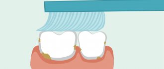
Each dental unit has its own functions, depending on its structure and location on the jaw. Thus, incisors are designed for biting off pieces of food, canines help to hold hard food and “tear off” stubborn pieces, premolars are needed for primary processing, and dense “plump” molars are designed for thoroughly chewing and grinding food. The order of the teeth in the mouth, accordingly, will be as follows: on each jaw there are four incisors, two canines, four premolars (two on each side) and six molars (three on each side).
In dentistry, tooth numbers are assigned according to their location on the jaw and function. Since the incisors and molars are located symmetrically on the right and left, the count starts from the middle of the row, that is, from the central incisors and further to the right and left. If we divide this row into two halves and start counting on one of them, we get: two incisors (teeth numbers 1 and 2), a canine (3rd number), two premolars (4th and 5th), three molar (6th, 7th and 8th, the last one being the wisdom tooth). These are the names and numbers of teeth that are universally accepted in dentistry.
If you have a problem similar to that described in this article, be sure to contact our specialists. Don't diagnose yourself!
Why you should call us now:
- We will answer all your questions in 3 minutes
- Free consultation
- The average work experience of doctors is 12 years
- Convenient location of clinics
Single contact phone number: +7
Make an appointment
But if you simply say “sixth tooth,” then how can you understand whether it is upper or lower, and on which side of the jaw – left or right – it is located? To eliminate confusion in this matter, it is customary to designate a person’s teeth by numbers, indicating the segment of the jaw on which they are located. The segments are considered as follows: the upper right is the first and is indicated by a ten in front of the number of a particular tooth (for example, the 11th is the central right upper incisor, the 16th is the upper right molar, next immediately after the premolars, etc.), the upper left - twenty. Accordingly, the lower left is designated by thirty, and the lower right by the number 40. That is, the segments of the jaws are numbered clockwise, this makes it much easier to remember the order and, if necessary, count.
Thus, the notorious 48th number indicates the location of the wisdom teeth in the lower right segment of the jaw. And insisting on removing the 48th dental unit, the dentist simply indicates what number the wisdom tooth has in the lower right, and does not inform the patient about the supernumerary teeth that came from nowhere in the mouth.
Reasons for creating the formula
Why is numbering of row elements necessary and how does it work when performing dental work? Each tooth has its own structural features, as well as a range of tasks performed. Some elements are involved in the process of biting off large pieces, others grind and crush foods for further better absorption in the body. Only wisdom teeth, which are inherited from ancient ancestors, do not take part in the process of chewing food. Due to the heat treatment of foods and the consumption of softer foods, the size of the jaw of modern humans has decreased, and wisdom teeth have ceased to perform their functions.
However, doctors strongly advise caring for eights, treating them if necessary, like other elements. Wisdom teeth prevent the development of bite pathologies and can be used for prosthetics if, for example, sevens are missing.
The dental formula must be entered into the patient’s outpatient record in order to minimize the risk of errors during further treatment. In dentistry, it is customary to number the dentition from the central part of the jaws. Concise markings allow clinicians to quickly complete the information in the chart so that it is understandable to another dentist or dentist. Doctors cannot use individual calculation schemes, as this will complicate the work of another specialist and change the information in the patient’s hospital record.
And there is also a special formula with which parents can determine the required number of milk units in the baby’s mouth. It looks like this: N = n - 4, where N is the number of milk elements in a child, and n is the baby’s age in months. The result calculated using the formula does not always coincide with the actual number of elements erupted in the child. Mismatches in the formula are not a reason to panic. Each child’s body is individual and minor deviations from the norm are acceptable. This calculation formula only applies to children under 2 years of age.
Symptoms that require consultation with a doctor include:
- complete absence of teeth in the oral cavity in a child older than 12 months;
- the presence of less than 10 teeth in the mouth of a 3-year-old child;
- non-compliance with the pattern of eruption of milk elements (for example, if the canines or premolars erupt first, and not the incisors);
- defects on the enamel.
What does the diagram of a child’s teeth look like with the numbers of each unit?
The pattern of baby teeth in children differs from the pattern with numbers in adults. Doctors specially designate milk teeth with different numbers so as not to confuse themselves and their patients: after all, a child who was treated for a milk bite in childhood will soon grow up and begin to go to the dentist for treatment of already permanent dental units. And if the same numbering is used in both cases, then there will be incredible confusion in the treatment history - but such a history in the dental record is often extremely important for the further competent and effective elimination of problems of the dental system.
Therefore, the numbering order for baby teeth in children is as follows: the serial number remains the same as in adults (1st, 2nd – incisors, 3rd – canine, etc.), but the segments of the jaws are designated by numbers 50 and 60 for the upper right and left segments and numbers 70 and 80 for the lower left and right, respectively. And when the dentist informs parents that their child has caries on the 82nd tooth, you don’t need to immediately imagine a baby shark with several rows of sharp baby teeth, we are only talking about the second lower right incisor. According to the counting scheme, permanent teeth in children are no different from the scheme of adult molars.
Functional properties of elements
Teeth perform several functions at once. Each element has a distinctive shape according to its purpose. The main role of the units is digestive. The quality of assimilation of useful elements by the body depends on the thoroughness of grinding the products. Only after chewing and moistening with saliva will the food be ready for subsequent processing by the digestive organs.
Chewing one piece lasts on average 10-20 minutes. The force exerted by the teeth on food sometimes reaches 80 kg. Incomplete dentition can lead to disturbances in the digestive system.
Another function of the elements is aesthetic. With malocclusion, a person may develop psychological problems, since the first thing others pay attention to during a conversation is a smile. Teeth also play a role in the formation of syllables. In the absence of one or more elements, the pronunciation of syllables changes: a lisp appears, speech is distorted.
International Viola system: a convenient diagram of the arrangement of a person’s teeth by numbers
The described method is very convenient and most common in dentistry. It has received international recognition and has been generally accepted among dentists since 1971, called the two-digit Viola system. The convenience of such a system lies primarily in the fact that there is no need to create a special map of a person’s teeth, the numbering is easily calculated in the mind and information about the condition of certain dental units of the patient can be easily conveyed in an oral conversation, by telephone or by e-mail.
However, many people, having read this far, may say: but our teeth were counted completely differently, and the map contains completely different designations! That’s right, because in addition to the Viola system, there are several other systems that can be used by dentists.
Age-related changes in dentogingival tissues
Over time, the human dentofacial system undergoes the following changes:
- reduction in the height of the enamel layer due to abrasion of the enamel;
- the formation of microcracks, which provokes gradual staining of teeth yellow and brown;
- active formation of secondary dentin and, accordingly, a decrease in the volume of dental pulp;
- age-related thickening of the walls of blood vessels also causes a narrowing of the pulp cavity;
- an increase in the amount of cement, which is observed both with an increase in chewing load and without it;
- thickening of periodontal bundles in the area of teeth that serve as support for bridges.
Age-related microcracks and darkening of enamel
What other options exist for naming a person’s teeth by numbers?
There are three other systems common among dentists that can be used to designate and record dental units:
- A universal alphanumeric system developed by the American Dental Association.
- Zsigmondy-Palmer system. One of the oldest counting systems (it was created back in 1876), it is often used in their practice by maxillofacial surgeons and orthodontists.
- Haderup system.
All of them are convenient and easy to use in their own way, and in all such systems the arrangement of teeth by numbers in adults and children is indicated differently.
Alphanumeric system
It provides an indication of not only the number of the dental unit, but also its functional purpose. Thus, incisors are designated by the letter I, canines by the letter C, premolars by the letter P and molars by the letter M. As for the numbers, they indicate the serial number of a specific functional unit in the segment (whereas in other systems the functions of the dental unit are not taken into account, only its position is taken into account in a row). For example, if according to the Viola system the wisdom tooth is numbered 8, then according to the alphanumeric system it is also designated as M3, that is, the third molar in the segment. Additionally, digital segment designations are also used, which is why the serial number becomes two-digit. The same 48th tooth mentioned more than once with such a scheme will be designated as M43.
For milk teeth, lowercase (small) Latin letters are used in the recording, and instead of numbers, sometimes letter values from A to K are also indicated, counting dental units clockwise from the upper right incisor.
Zsigmondy-Palmer system
In old, and sometimes in new dental outpatient records, you can see a special plate for recording the condition of dental units, based on the Zsigmondy-Palmer square-numeric system. The order of growth of an adult’s teeth is indicated here by the familiar Arabic numerals from 1 to 8; for children, they use Roman numerals from I to V. The numbers of the jaw segments are not indicated here, the data is simply entered into the corresponding parts of the table diagram. The system is quite convenient and visual, but in an oral conversation it can cause difficulties when indicating a specific dental unit.
Regeneration of dental tissues
Restoration of enamel, dentin, pulp and cement is possible, but not in all clinical cases.
Regeneration of the anterior and chewing teeth, as a rule, occurs due to the following processes:
- replenishment of enamel with minerals, which is carried out both from saliva and through the blood vessels of the pulp;
- in the pulp chamber, cellular elements form a special connective tissue capsule around the inflammatory focus, thereby preventing further spread of infection;
Fangs
Upper canine
The upper canine, one per quadrant, erupts at approximately 11-12 years of age and completes its formation after 3-4 years.
This is the longest tooth in the arch (about 27 mm or more).
The crown has a diamond shape with a peculiar sharp cusp, which on the vestibular side divides the mesial and distal sides of the crown. The crown is 10 mm high (length) and has a maximum mesial-distal diameter of 7.5 mm at the point of proximal contact and decreases to 5.5 mm cervically. The buccal-palatal diameter is wider (8 mm), and it remains up to 7 mm at the cervical level due to the presence of a prominent marginal ridge (Fig. 1.16).
Rice. 1.16. Upper canine: palatal view
The long root, like the crown, is narrow in the mesial-distal direction and more pronounced in the buccal-palatal direction. There is almost always only one root canal with lateral canals at different levels in 24% of cases. The apical third is straight in 40% of cases, and is often curved distally (32%) and/or vestibularly (13%) (Fig. 1.17).
Rice. 1.17 Upper canine: graphical representation of the access cavity
The pulp space at the level of the cervical third and the root middle third has an oval shape in the buccal-palatal direction; often two canals are present with a tendency to merge into one common round canal in the apical third (Fig. 1.18a, b).
Rice. 1.18 (a, b) Upper canine: buccal and proximal view
The endodontic access cavity is oval-shaped, extending from the cusp to the marginal ridge of the coronal cervical third to gain access to the radicular space without obstruction that could cause instruments to create steps or transposition of the apical foramen (see Fig. 1.19).
Rice. 1.19. Anatomical pictures from Hess and Keller: the endodontic space of the upper canine at the level of the cervical third of the canal is represented by one oval canal, and in the middle third it is divided into two different canals and often merges into one round canal in the apical third
Lower canine
The lower canine, one per quadrant, erupts approximately 8-10 months before the upper one and completes its formation after 3-4 years.
The crown has a length of 11 mm, and its mesiodistal diameter is narrower than the upper one (7 mm), and decreases to 5.5 mm at the cervical level. The maximum buccolingual diameter is 7.5 mm wide and decreases to a minimum at the cervical level. The cervical lingual marginal ridge is less pronounced than that of the upper canine (Fig. 1.20).
Rice. 1.20 Lower canine: lingual view of the crown
The lower canine is about 27 mm long, and has two separate roots in 6% of cases, or two canals in one root, connected by isthmuses, with a common or two separate apexes (Fig. 1.21). In 20% of cases, the apical third has a distal slope (Fig. 1.22).
Rice. 1.21 Lower canine: graphical representation of the access cavity
Rice. 1.22. Anatomical pictures from Hess and Keller: the endodontic space of the lower canine has two separate canals connected by an isthmus in the coronal third; the canals merge into one opening in the apical third
The endodontic space is oval in shape in the bucco-lingual direction for two-thirds of the root, and then gradually becomes rounded.
The endodontic access cavity should be oval in shape in a buccolingual direction, from the cusp to the marginal ridge (Fig. 1.23a, b).
Rice. 1.23 (a, b) Lower canine: buccal and proximal view
Upper incisors
Central upper incisor
The central upper incisor erupts (one per quadrant) between the ages of six and seven years, and the complete formation of the apical third occurs after 2 or 3 years. The average tooth length is 22-23 mm.
The crown has a triangular shape, about 10.5 mm long, the base extends in the mesial-distal direction, corresponding to the anterior edge of the tooth, up to 9 mm in size, and its buccal-palatal size is 7 mm (Fig. 1.9).
Rice. 1.9 Upper central incisors: palatal view of the crown
The root is usually straight (75%), but according to Ingle, a slight curvature may be present in a small percentage of cases. Lateral canals may be present in more than 20% of cases, and an apical delta is also common (35%).
The coronal pulp space also has a triangular shape, especially in the region of the cervical radicular third, with the base facing the vestibular wall and the apex located palatally, after which it gradually becomes a round canal until the apex.
The access cavity is triangular and follows the shape of the pulp space (Fig. 1.10).
Rice. 1.10 Upper central incisor: graphical representation of the access cavity
Lateral upper incisor
The eruption of the upper lateral incisor (one in the quadrant) occurs 1 year after the central one, and its complete formation takes approximately 3 years.
It is approximately 1 or 2 mm shorter than the central incisor, its mesial-distal size is smaller - about 7 mm, and the vestibular-palatal size is only 5.5-6 mm (Fig. 1.11).
Rice. 1.11 Upper lateral incisor: palatal view of the crown
Normally there is only one root canal, straight in 30% of cases, and often it is curved distally (53%), in a small percentage of cases there is curvature in other directions. The shape of the canal is ovoid in the cervical region, with a tendency to become more rounded in the apical region; the access cavity has a similar structure (Fig. 1.12).
Rice. 1.12 Upper lateral incisor: graphical representation of the access cavity