In what cases is removal of the upper tooth inevitable?
Medical indications for extraction of the upper tooth can be divided into planned and emergency. The planned ones include:
- periodontal inflammation or the formation of a cyst at the roots of a tooth with impenetrable roots;
- tumors of the upper jaw;
- the presence of unerupted, out of place or extra teeth that cause discomfort (damaging soft tissues, causing speech defects, preventing normal chewing of food, increasing the risk of developing inflammatory processes);
- destruction of the crown that cannot be restored;
- periodontal inflammation, one of the signs of which is pathological tooth mobility;
- fractures of tooth roots;
- the presence of teeth that interfere with orthopedic or orthodontic treatment.
Emergency indications are:
- purulent inflammation of the bone tissue of the upper jaw, occurring in an acute form;
- destruction of the crown of a tooth that has no functional value.
In addition, severe toothaches that cannot be eliminated by conservative methods (for example, in case of complex crown fractures) can be the basis for unscheduled extraction.
Atraumatic removal of third molars
Damage to the inferior alveolar and lingual nerves are fairly common complications during extraction of mandibular third molars. However, improved diagnostic approaches and surgical treatments are helping to significantly reduce the incidence of such adverse outcomes. Below is a description of diagnostic and therapeutic techniques aimed at preventing surgical complications during extraction of lower third molars.
Tools and Methodology
When removing an impacted wisdom tooth in the lower jaw, it is extremely important to prevent damage to surrounding anatomical structures such as the inferior alveolar and lingual nerves, as well as periodontal tissues of the second molar. Therefore, the surgical instruments used are of utmost importance. In the presented case, an innovative mechanical periotome Luxator LX (Directa Dental) was used, which directly carried out the extraction procedure. The instrument allows you to cut the Sharpey fibers passing between the root cementum and the alveolar bone, and thereby free the tooth from the periodontal ligament.
Clinical case
A 22-year-old somatically healthy patient sought dental care for pain in the area of the 17th tooth, which spread to the entire lower jaw. The orthopantomogram visualized compression of the mandibular nerve, which was in contact with the roots of the 17th tooth (photo 1).
Photo 1. Orthopantomogram before treatment.
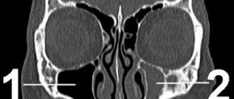
An intraoral examination revealed swelling and redness of the mucous membrane distal to the 18th tooth. There were no sensory disturbances in the lower jaw. The repeat image revealed that the position of the nerve was too close to the distal-lingual root of the tooth, which was also confirmed on CT sections (photos 2 - 3).
Photo 2. CT slices helped evaluate the course of the inferior alveolar nerve.
Photo 3. Localization of the nerve relative to the root based on CT data.
After performing anesthesia with a 2% vasoconstrictor, a single flap was formed, along which an intrasulcular incision was made around the 18th tooth. The releasing distal incision was made at an angle of 45º to the second molar, and at first this incision had a full tissue depth, and at the end it only partially penetrated the tissue. This incision avoids the risk of cutting the lingual nerve and serves as a connector for the flap. After exposing the jaw bone and installing a tongue guard, the Sharpey fibers were cut using a Luxator LX mechanical periotome around the entire circumference of the tooth, freeing it from the periodontal ligament (photos 4 - 5).
Photo 4. Extraction procedure using Luxator LX 360°.
Photo 5. Extraction procedure using Luxator LX 360°.
The tooth was extracted using an elevator without luxation of surrounding tissues or damage to the mandibular nerve. Through a controlled reciprocating motion, Luxator LX penetrates the periodontal ligament space (0.15 mm to 0.38 mm) and separates the fiber bundles from the tooth surface in the least traumatic way. This approach also minimizes the risk of damage to the nerve structures of the jaw. Photo 6 shows an x-ray that demonstrates how the working part of the instrument penetrates into the space of the tooth socket. Photo 7 shows the absence of root remains. The extraction procedure was carried out without segmentation of parts of the molar, so the latter was extracted in its entirety (photo 8).
Figure 6: Visualization of the Luxator LX inside the socket on a radiograph.
Photo 7. X-ray control: absence of root fragments in the hole.
Photo 8. Extracted tooth.
The alveolus was cleaned, irrigated with cold saline, and sutured with 4/0 silk thread. The sutures were removed 7 days after the iatrogenic intervention. The patient reported that the wound was healing without significant pain symptoms, which could be relieved with simple painkillers. The patient was advised to use an innovative gel toothpaste with a mixture of humectant and antibacterial agents (eg, cetylpyridinium chloride, triclosan and essential oils), which helps reduce the risk of plaque formation and reduce signs of bleeding. The level of abrasiveness of this paste was also slightly lower than that of conventional paste. The dental hygienist will further assist the patient in maintaining an optimal level of hygiene, as this is essential to ensure adequate healing. The procedure for professional oral hygiene was recommended to be carried out using glycine powder, which does not irritate the gums (Air Flow, EMS). Since the rough surface of teeth is more susceptible to the formation of bacterial plaque, polishing is an essential step in hygienic cleaning of the oral cavity. Silica-based polishing pastes with a low RDA value, such as ProphyPaste CCS Yellow RDA 40 (Directa Dental), can be used for this purpose.
conclusions
Extraction of the third molars of the mandible is directly associated with the risk of damage to the nervous structures of a temporary and permanent nature. Such complications can even provoke legal problems, so before starting treatment, the doctor must conduct a comprehensive diagnosis of the clinical situation, taking into account all aggravating factors (including the degree of wisdom tooth retention). The surgical approach used allows us to minimize tissue trauma and reduce the risk of damage to nearby nerves. Luxator LX is an effective tool that facilitates efficient molar extraction, significantly reducing the invasiveness of the surgical procedure and subsequent postoperative discomfort.
Authors: Loris Prosper Nicolas Zunica
Operation procedure
The standard (simple) scheme for removing the upper tooth includes the following measures:
- a medical history examination, during which the surgeon makes sure that the patient has no contraindications to the operation;
- referring the patient for an X-ray examination and subsequent study of the resulting images;
- injection of local anesthetics;
- detachment of the gums adjacent to the enamel using a smoothing iron or a rasp;
- grasping the diseased tooth with the cheeks of dental forceps and dislocating it by rocking or rotating around an axis;
- extraction of teeth and movable bone fragments from the alveolus;
- sanitation of the alveolar socket (tissue treatment with antiseptic and anti-inflammatory agents, removal of cystic neoplasms, granulomas, etc.);
- suturing (if necessary).
Simple extraction is carried out in cases where the dental crown is well visualized, and the surgeon has the ability to securely grasp it with the cheeks of dental forceps. In case of incomplete eruption of the tooth, its increased fragility, the absence of its crown part, as well as significant curvature of the tooth roots, surgical (complex) removal is performed. In this case, the doctor can cut the gums, drill out bone tissue, divide the crown into lobes and take other measures designed to facilitate the procedure of removing the upper incisor, canine, molar or premolar from the alveolus.
Contraindications to the procedure
You should temporarily avoid deleting:
- with a decrease in blood clotting, which was caused by taking medications or the onset of menstruation;
- after suffering a hypertensive crisis, heart attack, infectious diseases;
- with severe heart rhythm disturbances;
- during gestation (if there are serious indications, surgical intervention is allowed in the second trimester).
If a patient has hemophilia or mental disorders, the operation is performed only in an inpatient setting.
Postoperative care
After the operation is completed, the surgeon books the patient for a preventive examination and dressing, prescribes anti-inflammatory, antihistamine, antibacterial and painkillers. In addition, the doctor explains to the patient how to properly care for the postoperative wound. In particular, the surgeon may recommend:
- refuse food for 3 hours after surgery;
- ensure that the wound surface is not injured when chewing food, rinsing the mouth, brushing the teeth with a brush, floss or toothpick, etc.;
- refrain from hot drinks, alcohol, cigarettes, excessively salty, sweet, hard or spicy foods for 3 days after the procedure;
- Avoid physical activity, visiting a bathhouse or solarium for a week.
The appearance of heavy bleeding or purulent discharge from the alveolar socket, the development of inflammatory processes and severe pain in the postoperative wound area are signs of complications of the procedure. If they are identified, it is necessary to contact the surgeon who performed the operation and undergo comprehensive treatment according to the scheme drawn up by him.
When should a wisdom tooth in the lower jaw not be removed?
There are no absolute contraindications to removal, but there are relative ones:
- The figure eight does not need to be removed if it is correctly positioned, functions and does not injure surrounding organs;
- The operation cannot be performed without preliminary preparation if the tooth is located in the area of the tumor or puts pressure on a large vessel or nerve;
- For some diseases, it is necessary to prepare the body for surgery (rheumatism, diabetes, blood diseases, etc.)
You can carry out high-quality removal of a wisdom tooth of any complexity at the Berezka dental clinic. Specialists perform various surgical procedures effectively, safely and painlessly. All manipulations are carried out professionally and taking into account the individual characteristics of each patient.
Features of the procedure
Inspection alone is not enough to remove a molar. So that the doctor can see the location of the roots, it is necessary to take an x-ray.
The tactics of the operation are selected depending on the data received:
- if the picture is favorable, the molar is removed with special forceps, which are selected according to the anatomical shape and location of the crown (for the upper or lower jaw, right and left);
- in difficult situations (atypical location, the presence of mucous and bone tissues hiding the crown, the soft structure of the molar, the location of the root deep in the socket), a surgical method is used that involves dissecting the tissues and bones of the jaw.
If removal cannot be carried out only with forceps, then an elevator is used - a tool that reduces the trauma to mucous tissues when extracting single roots.
General anesthesia or local anesthesia?
In most cases, molar extraction can be performed under local anesthesia.
An operation under general anesthesia gives the doctor more time and freedom to remove several roots at once and carry out other surgical and orthodontic measures.
Not every clinic offers this type of pain relief; in addition, the method is not recommended for people with allergic and cardiovascular diseases.
Local anesthesia before molar extraction
Contraindications for removal
If surgery to remove a molar needs to be performed urgently, then it is not possible to take into account contraindications.
There are a number of restrictions for carrying out a planned operation:
- diseases of the heart, blood, nervous system;
- infections of the mouth and gums;
- the first and last months of pregnancy;
- period of menstruation.
Consequences and complications
Complications may arise during the operation:
- when a crown or root splits, there is a danger of soft tissue injury, and unremoved fragments provoke inflammation;
- there is a risk of damage to the jaw nerves and bones.
Such “disasters” do not happen to doctors who have experience and qualifications. You need to remember this and not trust such a complex operation to a random specialist.
Swelling of tissues and dry socket (when a blood clot at the site of an extracted tooth accidentally breaks off or does not form at all) most often occurs due to the fault of the patient who does not perform postoperative hygiene correctly, or due to the individual characteristics of the body (lowered immunity, poor blood clotting).
It is advisable to immediately insert an artificial prosthesis or implant in place of the lost crown, since a “gap” in the dentition entails serious consequences:
- deformation and change in inclination of adjacent molars;
- increased wearability of crowns;
- deterioration of the gastrointestinal tract;
- the occurrence of a cosmetic defect (rapid aging of the skin, sagging cheeks).