Facial nerve, anatomy
The facial nerve, nervus facialis , the 7th pair of cranial (cranial) nerves is a motor nerve.
The facial nerve innervates the facial muscles of the face (except for the musculus levator palpebre superioris), as well as the muscles of the auricle and skull, the posterior belly of the digastric muscle (musculus digastricus), the stylohyoid muscle (musculus stylohyoideus), the stapedius muscle (musculus stapedius), the subcutaneous muscle of the neck (musculus platysma).
The nucleus of the facial nerve is located on the border of the lower part of the pons with the medulla oblongata, outward and anterior to the nucleus of the abducens nerve. The axons of the cells of this nucleus rise in the dorsomedial direction to the bottom of the rhomboid fossa, then loop around the nucleus of the abducens nerve located here, forming the internal knee of the facial nerve in the area of the facial tubercle . The root of the facial nerve exits the base of the brain in the cerebellopontine angle between the pons and the medulla oblongata, lateral to the olive, then, together with its companions and the cochlear part of the 8th pair of cranial nerves, enters the internal auditory foramen (miatus acusticus internus), then the nerve and its companions enter facial canal (canalis facialis) of the pyramid of the temporal bone (fallopian canal). The canal initially has a horizontal direction (parallel to the apex of the temporal bone pyramid), then bends vertically. It then opens at the base of the skull with a stylomastoid opening (foramen stylomastoideum). The bend of the facial nerve in its canal is called the “external genu of the facial nerve.” In this place there is the knee ganglion (ganglium geniculi), where the cells of the first taste sensitivity neuron for the intermediate nerve are located. After emerging from the stylomastoid foramen, the facial nerve penetrates the parotid gland and divides into many terminal branches to innervate the corresponding muscles, forming the pes anserinus. The companions of the facial nerve in the canal are the parasympathetic lacrimal fibers - the greater petrosal nerve (nervus petrosus major) and the intermediate nerve (nervus intermedius), the 13th pair of cranial nerves, the Wriesberg nerve.
Parasympathetic lacrimal fibers originate from the secretory nucleus, which is located near the nucleus of the facial nerve. Together with the facial nerve, they enter the facial canal, then are the first to leave it as part of the nervus retrosus major, which innervates the lacrimal gland. Loss of function of the greater petrosal nerve is accompanied by the following symptoms - dry eye, eye irritation, lacrimation.
The intermediate nerve is mixed, consists of efferent parasympathetic salivary fibers for the sublingual and submandibular salivary glands, afferent taste fibers for the anterior 2/3 of the tongue. Salivatory fibers originate from the superior salivatory nucleus (nucleus salivatorius superior), leave the brain in the cerebellopontine angle and enter the facial canal next to the facial nerve, then leave the canal in its descending part as part of the chorda tympani (chorda tympani). Sensitive taste fibers begin from the cells of the knee ganglion, the dendrites of which go in the facial canal along with the facial nerve, then depart from it in the descending part of the canal, participate together with salivary fibers in the formation of the tympanic chord, then enter the system of the 3rd branches of the trigeminal nerve (nervus lingualis ) and reach the taste buds of the anterior 2/3 of the tongue. The axons of the cells of the knee bridle leave the pyramid of the temporal bone through the internal auditory foramen, pass through the cerebellopontine angle and end in the taste nucleus of the solitary tract (nucleus tractussoliterii), common with the glossopharyngeal nerve, located in the medulla oblongata. Damage to the intermediate nerve and the chorda tympani, which is its continuation, leads to hypofunction of the sublingual and submandibular salivary glands and impaired taste in the anterior 2/3 of the tongue. Between the two companions in the facial canal, motor fibers are separated from the trunk of the facial nerve, which form the stapedius nerve (nervus stapedius). The stapedius nerve penetrates the tympanic cavity and innervates the stapedius muscle, which provides a certain degree of fixation of the stapes and tension of the tympanic membrane, which creates conditions for the best audibility. Damage to the stapedius nerve is accompanied by a symptom such as hyperacusis. Hyperacusis is an increased unpleasant perception of sounds, especially low tones.
The first to leave the facial canal is the facial nerve's companion, the greater petrosal nerve. The second branch of the facial nerve to leave the facial canal is the stapedius nerve. The third to leave the facial canal is the companion of the facial nerve - the intermediate nerve in the form of the chorda tympani.
There are features of connections between the facial nerve and the cerebral cortex - the lower third of the precentral gyrus. The upper half of the nucleus, which provides innervation to the facial muscles of the upper part of the face, has a bilateral cortical representation due to the partial supranuclear decussation of the corticonuclear fibers. Fibers to the lower half of the nucleus, from which the facial muscles below the palpebral fissure are innervated, originate from the opposite hemisphere. This point is an important criterion in making a topical diagnosis.
Facial anatomy
The face, like many other tissues, undergoes sagging processes under the influence of gravity. This process is called gravitational ptosis. I propose to consider the anatomy of the face layer by layer. It is a layer-by-layer description of facial anatomy from the perspective of the concept of Australian surgeon Brian Mendelson that will help to understand the mechanisms of aging.
Essentially, the face consists of 5 layers: skin, fatty tissue, muscles, ligaments, periosteum and facial skeleton.
- The skin is the outermost layer. Traditionally, facial aging is associated with skin changes, so most treatments begin with skin care. Many people use creams, masks, etc. It really has a positive effect. However, up to a certain age. According to various authors, facial aging begins at the molecular level on average at the age of 30. This process occurs deep in the structures of the skin, so all rejuvenation procedures are essentially care and do not have a long-lasting effect.
- Subcutaneous tissue. Lies under the skin and consists of adipose tissue. The thickness of the fat layer varies in different parts of the face. With age, fat sinks and accumulates in certain places, forming bags under the eyes, jowls, etc.
- Muscles . There are 21 facial muscles on the face. With age, muscles weaken and sag, like a hammock in which fat accumulates. In this regard, working exclusively with the skin will not help achieve aesthetic rejuvenation. After all, even after performing a skin tightening, an array of muscles and adipose tissue will stretch the face and the result of skin procedures will disappear in no more than 1 year.
- Ligaments . With the help of ligaments, the soft tissues of the face are attached to the facial skeleton and are located in certain places. Time and gravity have virtually no effect on the ligaments, so weakened muscles and hanging fat are thrown over the ligaments, forming pronounced age-related contours of the face.
- Periosteum and facial skeleton. The bony skeleton determines the shape of the face and the very uniqueness and individuality of our faces. However, over time, the bone tissue slowly collapses, which creates the preconditions for the heavy mass of the face to slide down.
Rice. 1. Layers of the face according to Mendelssohn
Rice. 2 Mendelssohn's ligaments
Rice. 3 Young and old face
Facial nerve: symptoms, lesion syndromes
When the trunk of the facial nerve, the root of the facial nerve or the nucleus of the facial nerve is damaged, paresis of the facial muscles (muscle paresis) of the same half of the face develops - prosoplegia. Facial asymmetry occurs, which is pronounced even at rest. The entire affected side is motionless and mask-like. The forehead folds and nasolabial folds are smoothed. The palpebral fissure is widened. The corner of the mouth is downturned. Due to loss of function of the circular muscle of the eye (musculus orbicularis oculi), the eye does not close. This is lagophthalmos , or hare's eye. When you try to close your eyes, the eyeball on the affected side turns upward, the iris goes under the upper eyelid - Bell's symptom. With mild paresis of the orbicularis oculi muscle, the palpebral fissure closes, but less tightly than on the healthy side, the eyelashes remain visible (eyelash symptom). When the normal function of the lacrimal gland is preserved, lagophthalmos is usually accompanied by lacrimation, which is caused by difficulty in moving tears to the lacrimal canal due to insufficient adherence of the lower eyelid to the eyeball and impaired absorption due to displacement of the opening of the canal. Also, lacrimation is facilitated by the strengthening of the tear reflex due to the constantly open eye. Inflammatory phenomena, conjunctivitis, and keratitis often develop due to irritation of the eye membranes by air flow and dust.
The asymmetry of the face increases sharply when teeth are shown. The corner of the mouth is pulled back and it is skewed towards the healthy side - this is the phenomenon of the exclamation point, it is associated with paralysis of the musculus risorius. Due to the weakness of the orbicularis oris muscle, whistling and stretching of the lips into a tube are impossible. The patient often has difficulty speaking and eating. Liquid food on the affected side spills out of the mouth, thick food gets stuck between the cheek and teeth. When frowning and wrinkling the forehead, folds do not form on the side of paralysis, it is impossible to puff out the cheeks, and there is no tension on the musculus platysma of the neck. The brow reflex, nasopalpebral reflex, corneal reflex, and conjunctival reflex are lost or weakened. If damage to the facial nerve occurs in a child of the first year of life, then the sucking reflex, proboscis reflex, and search reflex are reduced. As with any peripheral paralysis, atrophy of the facial muscles is possible. When studying electrical excitability and myography, a degeneration reaction is noted.
Peripheral paralysis of the facial muscles is sometimes accompanied by pain in the face, ear, and mastoid process. The pain is associated with the phenomenon of repercussion. Repercussion is the irradiation of excitation from the motor branches of the facial nerve to the sensory branches of the trigeminal nerve.
Prolonged peripheral paralysis can lead to the development of contracture of the affected muscles, which is manifested by a narrowing of the palpebral fissure on the affected side and a pulling of the mouth when baring teeth towards the affected affected side. Symptoms of incipient contracture are often pathological synkenesis of the facial muscles - ocular synkenesis. They are characterized by the following symptoms. The closing of the eyes is accompanied by movement of the corner of the mouth or baring of the teeth. Baring your teeth causes the eye on the affected side to close.
In pathological processes that cause irritation of the nucleus or fibers of the facial nerve, facial hemispasm is observed - this is squinting the eye and pulling the mouth and tip of the nose to the affected side with simultaneous contraction of the chin muscles and tension of the subcutaneous muscle of the neck. Signs of irritation of the facial nerve are also tics of facial muscles and Chvostek's sign.
Surgical treatment of trigeminal neuralgia
Patient Sh., 63 years old, was admitted to the 1st neurological department on June 7, 2015.
Upon admission, complaints of paroxysmal, burning, high-intensity pain in the area of the right cheek, chin, ear on the right, in the area of the upper and lower teeth, pain in the temporomandibular joint on the right, with aggravation during talking, eating (chewing), washing, with periodic increases in pain in the form of “lumbago”, hypersalivation (more at night), worsening night sleep due to pain.
History: In the spring of 2011, neuropathic pain first appeared in the zones of innervation of the II and III branches of the right trigeminal nerve. Treatment was carried out, including a course of Finlepsin and Lyrica, and IRT procedures and magnetic therapy were also started, which were canceled after MRI revealed a falx meningioma in the frontal region. The pain syndrome regressed within 3 months.
In the spring of 2014, the pain syndrome relapsed. The examination revealed an increase in anti-VZV IgG (421.2 mU/ml, when the norm is less than 150). Anti-VZV IgM (negative). There were no skin rashes and he denied any history of H.zoster. She underwent 2 courses of treatment with hydrocortisone in tablet form (in April and July 2014), a course of Valtrex, Famvir, and Cycloferon. Dynamics after therapy: increase in anti-VZV IgG titer (496.4 mU/ml), anti-VZV IgM (negative). The pain syndrome regressed after half a year.
For a long time in 2014, she was engaged in dental prosthetics.
The real deterioration of the condition began in mid-May 2015, when facial pain resumed, increasing in intensity, “shooting”, limiting household activity. During the study, the titer of anti-VZV IgG continues to increase - 571.7 mU/ml), anti-HSV (types 1 and 2) IgG is detected - 23.5 (positive), anti-HSV (types 1 and 2) IgM - negative, anti-EBV IgG-EBNA (white caps.) – 385.0 U/ml (positive), anti-EBV IgM-VCA (white caps.) <10 (negative), anti-EBV IgG-VCA (white caps.) (positive ). I started taking Valtrex, Cycloferon, Famvir, Lyrica. I couldn’t sleep at night because of the pain. Hospitalized on an emergency basis.
History: surgical interventions: NAM for endometriosis, takes hormonal therapy (in gel form); appendectomy. In 2009 - a circular facelift.
As a child, she suffered from chickenpox, mumps, and rubella; did not suffer from frequent ARVI; in adulthood - double lacunar tonsillitis.
Photos provided upon admission:
CT scan of the TMJ with functional tests and multiplanar reconstructions dated November 26, 2014: Condition after secondary partial edentia, after prosthetics. CT picture of degenerative changes in the right TMJ with signs of subluxation during a functional test.
MRI of the TMJ dated 12/15/14: MR signs of TMJ asymmetry. Picture of partial reducible dislocation of the right TMJ. Severe degenerative changes in the right articular disc. MRI signs of osteoarthritis and synovitis of the right TMJ. MRI signs of functional overload of the superior head of the right lateral pterygoid muscle.
MRI of cerebral vessels dated 06/04/14: no pathological changes in the main arteries and veins of the brain were detected.
MRI of the brain dated 06/03/14: a picture of a space-occupying falx formation in the frontal region (12*10 mm), most likely meningiomas. Fine-focal changes in the brain substance of a vascular nature.
CT scan of the skull bones dated November 7, 2014: no bone-destructive or space-occupying formations of the upper and lower jaw were detected. Condition after secondary partial adentia, after prosthetics. CT picture of degenerative changes in the right TMJ. Signs of a space-occupying formation in the area of the falx of the frontal region of the brain, most likely meningioma. Local thickening of the left frontal bone (exostosis? Compact osteoma?)
Objectively: The condition is satisfactory. The skin is of normal color. There are no rashes in the area where the pain is localized and in the area of the ear canal on the right. There is no swelling. In the lungs there is vesicular breathing, no wheezing. NPV up to 14 per minute. Heart sounds are muffled, rhythmic, heart murmurs cannot be heard, heart rate is 62 per minute. Blood pressure 120/80 mm Hg. Art. The abdomen is soft, painless on palpation, there are no symptoms of peritoneal irritation, peristalsis can be heard. The liver does not protrude from under the edge of the costal arch. Controls pelvic functions.
Neurological status: Conscious, contactable, oriented. There is no decrease in background mood. There are no meningeal signs. Palpebral fissures D=S. Pupils D=S. Photoreactions are live. Movement of the eyeballs is not limited. There is no nystagmus. There are no violations of superficial sensitivity on the face. Pain is provoked by palpation of the exit point of the 2nd branch of the trigeminal nerve on the right. The face is symmetrical. Swallowing and phonation are not impaired. Tongue in the midline. There are no paresis. Tendon reflexes are of medium vivacity, D=S. No sensory disturbances were detected. Romberg's posture is unsteady. Coordinator tests are performed satisfactorily on both sides.
In the tests - moderate leukocytosis: 11.38 10e9 /l (N: 4-9), without a shift in the formula; ESR acceleration: 20 mm/hour
ECG: Sinus rhythm with heart rate 76/min. Horizontal position of the EOS.
X-RAY examination of the chest organs : No recent focal and infiltrative changes were detected.
Consulted with an infectious disease specialist: there is no evidence of an acute infectious process or signs of a persistent viral infection. The condition can be regarded as postherpetic trigeminal neuralgia. The existing increase in the titer of antibodies to VZV and EBV is of an anamnestic nature (previous infection). Active antiviral therapy is not indicated. Complex therapy is necessary to correct neuropathic pain syndrome.
The dentist suggested possible compression of the terminal branches of the second branch of the trigeminal nerve by a foreign body (filling material) at the apex of the 1.4 tooth, which was confirmed by CT scan of the upper jaw.
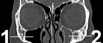
When performing a high-field MRI of the brain, a formation measuring up to 13 mm (meningioma) is determined in the projection of the anterior parts of the falx. During a targeted examination of the trunk, a vein with a blood flow diameter of up to 2 mm is adjacent to the lower parts of the right trigeminal nerve, slightly deforming them in the area of the grubber notch. Picture of meningioma of the anterior parts of the falx. MRI signs of neurovascular conflict of the right trigeminal nerve (see Fig. 1)
Thus, the polyetiological genesis of the pain syndrome was revealed:
- Postherpetic neuropathy
- formation of trigeminal pain syndrome due to irritation of the second branch of the trigeminal nerve from the periphery (as a result of contact with filling material),
- trigeminal neuralgia due to compression of the nerve root by the vein in the area of the cerebellopontine angle,
- The influence of falx meningioma could not be excluded.
With EEG: Epileptiform activity and signs of interhemispheric asymmetry at the time of the study were not recorded either in the background recording or during afferent tests.
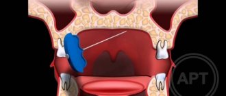
Considering the possibility of the formation of trigeminal pain syndrome both due to irritation of the second branch of the trigeminal nerve from the periphery (as a result of contact with filling material), and trigeminal neuralgia due to compression of the nerve root by the vein in the area of the cerebellopontine angle, surgical treatment for both nosologies is indicated. A decision was made on a staged intervention: first of all, cystectomy with resection of the apex of the 14th tooth, then microvascular decompression of the trigeminal nerve root on the right.
The patient received treatment: no-spa + analgin + novocaine + relanium; Perfalgan IV, Tramal, Milgamma IM, Lyrica, Valtrex, Amitriptyline, Augmentin, Glycine, PC-Merz, Hydrocortisone, Cycloferon, Femivir, Finlepsin in the table. During the treatment, moderate positive dynamics were noted in the form of a decrease in the intensity of the frequency of painful paroxysms: the patient could talk and eat.
A surgical intervention was performed - cystectomy with resection of the apex of the 1.4 tooth. The course of the postoperative period was smooth, fluctuations in the pain syndrome were noted, and after a week there was a persistent increase. Finlepsin was added to therapy at a dose of 300 mg/day, which resulted in a good analgesic effect.
The patient had doubts about the need for neurosurgical intervention. In order to resolve the issue of the need to decompress the trigeminal nerve root, Finlepsin was discontinued, after which the pain syndrome resumed. There was no doubt that the patient was indicated for surgical intervention - microvascular decompression of the trigeminal nerve root on the right in order to resolve the vasoneural conflict and achieve stable remission of trigeminal neuralgia.
Neurosurgical intervention was performed (see Fig. 2-3), as a result of which the pain syndrome was completely relieved.
Diagnosis of the level of damage to the facial nerve
Isolated lesions of the nucleus of the facial nerve are observed quite rarely. It is manifested by total paresis of the facial muscles and occurs in the pontine form of polio.
More often, pathological foci localized in the pons are more common and lead to the involvement of the nucleus of the facial nerve, radicular fibers, and pyramidal tract in the process, which is manifested by alternating Millard-Hübler syndrome. Simultaneous damage to the nucleus of the abducens nerve is manifested by alternating Foville syndrome.
When the pathological process is localized in the cerebellopontine angle, the symptoms of damage to the facial nerve are combined with damage to its companions (the intermediate nerve and the greater petrosal nerve) and the vestibulocochlear nerve. Paralysis of facial muscles in these cases is accompanied by dry eyes - xerophthalmia, impaired taste in the anterior 2/3 of the tongue on the affected side. Xerostomia may be felt - dry mouth, but more often it does not occur due to the functioning of other salivary glands (parotid, sublingual, submandibular on the healthy side). Hyperacusis does not occur due to concomitant damage to the cochlear nerve. Hearing loss or deafness is more common. There may be signs of dysfunction of the trigeminal nerve and abducens nerve located in the immediate vicinity, as well as cerebellar disorders.
When the facial nerve is damaged in the facial canal above the origin of the greater petrosal nerve, dry eyes, taste disorders and hyperacusis develop simultaneously with paralysis of the facial muscles.
The lesion after the departure of the greater petrosal nerve is accompanied by increased lacrimation, taste disturbance, and hyperacusis.
When the facial nerve is damaged below the origin of the stapedius nerve, but above the origin of the chorda tympani, paralysis, lacrimation, and taste disturbance are observed.
Damage to the nerve in the bony canal below the origin of the chorda tympani or after exiting the stylomastoid foramen causes only paralysis with lacrimation.
When the process is localized in the area of the outer knee of the facial nerve with involvement of the knee node, Hunt's syndrome can be detected - this is paresis of the facial muscles (facial muscles), severe pain and herpetic rashes in the area of the auricle (ear).
Sometimes there are cases of bilateral damage to the facial nerve. Bilateral damage to the facial nerves is called diplegia facialis. The patient's face is mask-like, his eyes are half-open, it is impossible to form his lips into a tube or close his mouth.
Facial nerve: treatment in children
Damage to the facial nerve in children is more common than damage to other cranial nerves, which is due to its anatomical features. The facial nerve is supplied with blood from the external carotid artery system, therefore, when the head is hypothermic, spasm of the external carotid artery leads to nerve ischemia, swelling and compression of the facial nerve. Compression of the facial nerve develops especially easily when the process is localized in the narrow canal of the temporal bone pyramid. The facial nerve canal is connected to the tympanic cavity and the pneumatic cells of the mastoid process. The outflow of lymph from the trunk of the facial nerve occurs in the cervical lymph nodes. In childhood, damage to the cervical lymph nodes is often observed.
Causes of inflammation of the trigeminal nerve
There are several reasons leading to inflammation of the nerve ducts:
- Poor blood supply associated with physical compression of the nerve. First of all, this is swelling caused by diseases of the ENT organs. The resulting tumor can also pinch the nerves.
- Inflammation associated with dentistry. This includes gingivitis, periodontitis, caries, pulpitis, and eruption of wisdom teeth. Each of these diseases can lead to suppuration, abscesses, swelling and bacterial infections of tissues.
- Medical error by an anesthesiologist - if the injection was given unsuccessfully and the needle got into a nerve, pain cannot be avoided.
- Hypothermia causes muscles to lose their elasticity, which leads to pinching of the nerves passing between the fibers.
- Bacterial infections, in particular tetanus and polio.
- The cause of inflammation of the trigeminal nerve, which is difficult to diagnose, is the psychological state of a person - frequent experiences, stress, and nervous disorders.
To determine the cause of inflammation, you need to consult a specialist.
Facial nerve: causes of damage, causes of neuritis, neuralgia, neuropathy, paresis, paralysis
The main causes of damage to the facial nerve a are inflammatory diseases (diseases) leading to primary damage to the facial nerve and its involvement in the process secondary to pathological changes in adjacent formations. Meningitis, arachnoiditis of the cerebellopontine angle, inflammatory processes in the area of the eustachian tube (eustacheitis) and mastoid process (mastoiditis), inflammation of the ear (otitis), jaw arthritis, lymphadenitis, mumps can cause the development of neuritis, paresis, paralysis, inflammation of the facial nerve. Also the cause of damage to the facial nerve are primary and secondary polyradiculoneuritis. Traumatic damage to the facial nerve occurs during traumatic brain injury with a fracture of the base of the skull in the area of the temporal bone, during surgical operations on the ear. The cause of paralysis and paresis of the facial nerve in children can be birth trauma, the application of obstetric forceps, or facial presentation. The facial nerve suffers from various tumors of the cerebellopontine ganglion region. Neuroma of the facial nerve, Recklinghausen's neurofibromatosis, tumor of the parotid salivary gland, infiltration due to leukemia are also causes of damage to the facial nerve.
In rare cases, congenital aplasia of the facial nerve nucleus and congenital narrowness of the facial nerve canal occur.
Central palsy - facial nerve
Central paralysis of the facial muscles (facial muscles) is observed as a result of damage to the corticonuclear fibers going to the nucleus of the facial nerve. Central paralysis is characterized by dysfunction of the muscles of the lower half of the face, which have unilateral cortical innervation. The main symptom of central paralysis is the smoothness of the nasolabial fold on the side opposite to the lesion. In some patients with central paralysis of the facial muscles, mild deficiency of the orbicularis oculi muscle can be detected. Central paresis of the facial muscles is usually observed in combination with central hemiparesis, or hemiplegia. In contrast to peripheral paralysis, with central paralysis of the facial muscles, the conjunctival reflex, brow reflex, and corneal reflex are preserved, and there is no degeneration reaction.
How to slow down facial aging?
Facial aging is a multifactorial process manifested in bone resorption, changes and sagging of the soft tissues of the face. Procedures for external influence on facial skin are essentially care products that do not bring the proper rejuvenating effect.
- Superficial treatments: creams, ointments, gels and other procedures moisturize and maintain skin condition.
- Injection techniques: fillers and botulinum toxin. Fillers based on hyaluronic acid help fill the volume of tissue that has undergone atrophy, and botulinum toxin relaxes the muscles that form wrinkles. The absolute advantage of these techniques is their reversibility of action. Typically, fillers and botulinum toxin preparations dissolve within 6 months.
- Surgical facial rejuvenation: the most effective method. There are different types of face lifting. It is necessary to approach the choice of face lifting method from the standpoint of existing facial changes, their severity, the patient’s wishes and the surgeon’s capabilities.
Fundamentally, the following types are distinguished: skin lifting; SMAS lifting; MACS-lifting; spacelifting.
- Skin lifting
In the mid-20th century, it was believed that it was skin changes that led to facial aging. However, the era of skin tightening showed its ineffectiveness due to the short-term results - the result lasted no more than 1 year. Modern cosmetology is again resorting to skin tightening using threads. Thread lifting gives short-term results and makes sense for initial facial changes. Pronounced age-related changes are not properly corrected with the help of threads, so their use must be strictly dosed.
- SMAS lifting
SMAS is a complex acronym that stands for the entire muscular aponeurotic complex of the face. After separating the skin layer from the muscle layer, an incision is made and the facial muscles are tightened and fixed in a new position. At the same time, the neck is lifted and jowls are eliminated. After the muscles are moved to the new position, excess skin is created, which is removed and the skin is sutured along the incision around the auricle. Therefore, this type of lifting is suitable for pronounced changes in the middle zone of the face, the presence of jowls and a flabby neck.
- MACS-lifting
It is considered a low-traumatic technique. Through a small incision near the auricle, the skin is peeled off and the muscles are tightened using purse-string sutures. The difference from SMAS is that the muscles do not move and are fixed, but are stretched like a drum, which will soon lead to drooping of the face. However, the commercial attractiveness of this method is very high. It is worth remembering that this method can only be good for initial changes - just like thread lifting.
- Spacelifting
The essence of the operation developed by Dr. Mendelson is to eliminate the facial spaces that stretch under the influence of gravity. Since these spaces exist only in certain areas of the face, spacelifting does not provide a comprehensive lift and does not allow for significant rejuvenation of the neck area. Therefore, spacelifting gives a good rejuvenating effect in the middle zone of the face with unexpressed changes in the neck.
Primary neuritis of the facial nerve
Primary facial paralysis is caused by herpes viruses, mumps, enteroviruses, and adenoviruses. Bell's palsy can develop with general or local hypothermia of the face. Bell's palsy is characterized by acute development within 3 to 24 hours. The leading role in the development of Bell's palsy belongs to ischemia, which develops due to vasospasm, or vasodilation with the development of swelling of the facial nerve and compression of the nerve.
Often, neuritis of the facial nerve occurs for no apparent reason against the background of absolute complete health. Sarklinik observed many cases where the first symptoms of facial neuritis appeared after a person slept, at the moment of awakening. This may be due to the activation of chronic infection with weakened immunity, the development of various allergic reactions.
Treatment methods
Depending on what caused the inflammation, a course of treatment is prescribed. For bacterial lesions, the emphasis is on antibacterial therapy through systemic administration of drugs.
However, regardless of the reasons, the doctor prescribes painkillers to relieve pain and reduce inflammation. It could be:
- ibuprofen;
- paracetamol;
- analgin;
- ketorol;
- diclofenac.
All of the listed drugs can be prescribed either in the form of tablets for oral administration, or prescribed in the form of solutions for intramuscular administration.
When conservative methods are not possible, the help of a surgeon may be needed. This primarily concerns abscesses due to the eruption of wisdom teeth, pulpitis or other dental diseases. In this case, the abscess will be opened, pus will be removed, the wound will be treated with antiseptic, and the tooth will be removed, if necessary. If a pinched nerve occurs as a result of pathologies in the structure of the skull, the surgeon will perform an operation to correct the situation and free the nerve bundles.
As a complex therapy, massage, heating or exposure to a magnetic field and electric current can be prescribed. You cannot massage or warm the inflamed area yourself, because this can lead to complications associated with rupture of the purulent capsule, blood poisoning and paralysis of the facial nerve.
Separately, you may need to consult a neurologist who will determine the cause of the inflammation if other specialists have not found obvious foci of infection and abscesses.
Traditional methods of treatment are permissible only as an addition to the main therapy. For example, rinsing with chamomile decoction will relieve inflammation and reduce swelling. But you can resort to such procedures only with the permission of the attending physician.
Neuritis of the facial nerve secondary
Secondary neuritis of the facial nerve is most often of otogenic origin, observed after or during otitis, mastoiditis, eustacheitis. In these cases, reactive inflammation of the nerve trunk develops. Also, the inflammatory process can penetrate directly into the facial nerve itself due to the penetration of infectious agents into it. This is often observed with purulent epidemic partitis, lymphadenitis of the stylomastoid area. Secondary neuritis of the facial nerve can occur with infectious mononucleosis, toxoplasmosis, typhus, tuberculous meningitis, acute leukemia, cranial polyneuritis, polyradiculoneuritis.
Neuritis of the facial nerve occurs with fractures and cracks of the base of the skull passing through the pyramid of the temporal bone (post-traumatic neuritis of the facial nerve). Hereditary factors and congenital developmental anomalies play a major role in the occurrence of paresis of the facial muscles. There are aplasia of the facial nerve trunk and aplasia of the nucleus of the facial nerve. In this case, there may be unilateral and bilateral violations.
The occurrence of neuritis of the facial nerve is facilitated by a narrowing of the fallopian canal and an increase in the size of the styloid process.
Damage to the facial nerve can be one of the signs of such syndromes as Melkersson-Rosenthal syndrome, Mobius syndrome.
Neuritis of the facial nerve diagnosis
Diagnosis of facial nerve neuritis is based on an analysis of the symptoms preceding and accompanying diseases of the nerve damage. Neurologists, neuropathologists, and reflexologists distinguish such forms of damage as neuritis of the facial nerve and neuropathy of the facial nerve. According to pathogenesis, neuritis is divided into primary and secondary.
Neuritis of the facial nerve must be differentiated from isolated damage to the motor nucleus of the nerve, a variety of common pathological processes in the area of the cerebellopontine angle. Damage to the nucleus of the facial nerve is accompanied by isolated paresis of the facial muscles of the same half of the face without autonomic and sensory disorders, which occurs mainly in the pontine form of poliomyelitis or poliomyelitis-like diseases, tick-borne encephalitis.
When the lower parts of the bridge are damaged, which occurs with tumors, encephalitis, vascular diseases, along with the nucleus of the facial nerve, the pyramidal tract is involved in the pathological process. In such cases, peripheral paresis of the facial muscles is combined with central hemiparesis of the opposite side (Millard-Hübler syndrome). If the nucleus of the abducens nerve is affected, then paresis of the external rectus muscle of the eye (Fauville syndrome) is added to the above symptoms.
Isolated neuritis of the facial nerve is differentiated from processes in the area of the cerebellopontine angle, which develop, for example, with arachnoiditis, tumors of the vestibulocochlear nerve, which is manifested by decreased hearing, deafness, paresis of the facial muscles, sometimes dysfunction of the trigeminal nerve, contralateral spastic hemiparesis.
Polyneuritis and polyradiculoneuritis often involve lesions of the facial nerve. Usually the lesion in these cases is bilateral, often asymmetrical, accompanied by limited or diffuse damage to other parts of the human peripheral nervous system.
Prevention of inflammation
To prevent the risk of developing inflammation of the trigeminal nerve, it is recommended to follow a number of measures:
- monitor oral hygiene and consult a dentist in a timely manner;
- do not stay in the cold for a long time or protect your face from freezing with a scarf;
- do not self-medicate otitis media.
At the first manifestations of pain on the face, you should immediately consult a doctor. This will stop the development of inflammation. In addition, early diagnosis allows for conservative treatment methods.