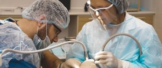
When is suturing necessary in dentistry?
Surgical intervention is performed if:
- cystectomy - removal of a medium to large cyst that extends over several dental units. Resection of the apex of the previously sealed roots occurs;
- cystomy - partial removal of the cyst in order to reduce the pressure inside the formation and its gradual flattening;
- eliminating certain inflammatory processes - opening abscesses, treating osteomyelitis, periodontitis;
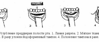
- gingivectomy - excision of overgrown gum tissue;
- gingivoplasty - tissue transplants to improve the aesthetics of the gums;
- removal of dental units - resection of a wisdom tooth is especially often accompanied by suturing;
- preparation for prosthetics - alveoloplasty, sinus lift, bone block transplantation.
A deep incision in the soft tissues of the oral cavity always requires sutures.
Types of suture material
Material for suturing is divided into two large groups - absorbable and non-absorbable.
Absorbable suture materials include:
- catgut is a thread obtained from purified connective tissue; when left in the oral cavity for a long time, the possibility of an inflammatory process arises due to rejection of the material;
- vicryl - synthetic threads containing polyglycomic acid, polyclatin, do not provoke the development of inflammatory processes, which makes it possible to use them where there is a high risk of postoperative complications.
Non-absorbable suture material:
- monofilament - a synthetic thread with a rigid structure that can injure the mucous membrane;
- silk is an elastic, durable material, but requires timely removal of sutures (after 10 days it causes inflammation of the soft tissues);
- polyster is a durable woven thread that requires special care when fixing; in order to make it more smooth, it is often coated with silicone.
Before surgery
- Prepare an ice pack at home.
- On the day of surgery, eat a light meal 2-3 hours before your scheduled time.
- The day before surgery, the consumption of alcoholic beverages is strictly prohibited.
- Be sure to inform your dentist about all the individual characteristics of your body and any allergic reactions, in order to accurately select an anesthetic that is safe for you.
- Visit the toilet before surgery.
- It is better to come to the operation in loose clothes without a collar.
Main types of sutures in dentistry
The most commonly used seams are:
- single - used for minor incisions in the soft tissues of the patient’s oral cavity;
- nodal - a reliable way to fix the edges of the wound;
- supporting - used when flaps (buccal, lingual) are located at different levels;
- continuous - its advantage is good adaptation of the wound edges and is highly aesthetic.
Instruments used for suturing in dentistry:
- needle holder - there are many models of the instrument, the choice depends on the preferences of the surgeon;
- two-pronged hook, eye surgical tweezers - necessary for maintaining the edges of the wound;
- eye scissors - needed for cutting suture material;
- suture needles - made of medical stainless steel, having different sizes, shapes, diameters.
Prevention of complications after suturing
It is important to complete the medication prescribed by your dentist.
The following will help speed up and relieve pain from wound healing:
- applying cold on the first day for 10-15 minutes helps relieve swelling and reduces the risk of bleeding;
- baths with antiseptics - rinses, herbal decoctions with antimicrobial, anti-inflammatory properties;
- Three days after surgery, you can clean the suture material with a soft toothbrush.
After surgery it is prohibited:
- take aspirin and other anticoagulants - they thin the blood and can cause bleeding;
- eating hard, spicy foods can damage soft tissues and cause irritation;
- carry out warming procedures - heat compresses, visiting saunas, baths leads to inflammatory processes;
- rinse your mouth - active rinsing of the mouth is prohibited until the stitches are removed;
- active physical activity.
A visit to a dental surgeon is a prerequisite for preventing and timely eliminating the causes that contribute to the occurrence of postoperative complications.
Before surgery:
Prepare several days off after the date of the planned operation.
Do not smoke or reduce the number of cigarettes you smoke.
If you are sick on the eve of the operation, please notify the implantologist.
Ask your implantologist about the medications you will need immediately after surgery.
Patients suffering from compensated diabetes mellitus must follow a strict diet 2 weeks before surgery and 2 weeks after it.
Get a good night's sleep the night before surgery.
Make sure you are accompanied if you plan to undergo anesthesia, sedation or a complex operation, do not plan to be behind the wheel.
If you have herpetic rashes on the mucous membrane, the operation should be rescheduled.
How does the procedure work?
The suture should be applied from the side of the moving edge of the wound towards the fixed one. The free edges of the tissue flap must be carefully held with special anatomical tweezers. In the process of suturing the skin and mucous membranes, the needle grasps the flap from the edge of the wound by about 3 mm, and then the wound is tightened along the edges with the ends of the suture material, tightening the loops.
It is very important that this procedure is done professionally; mistakes and carelessness are unacceptable. For example, you should not tighten the knot too much, as this disrupts the blood supply at the edges of the wound, as a result of which the tissue quickly turns pale at the suture site. However, it is also unacceptable for the seam to be too loose.
Damage to the soft tissues of the maxillofacial area occurs in 70% of all maxillofacial trauma. Defects and deformations of the face cause not only anatomical and functional disorders, but also cause severe psychological distress for patients. The degree of these disorders and the nature of reconstructive operations depend on the size and location of the defect, as well as on the combination of damage to individual organs and tissues of the face. Depending on the severity of the damage, destruction of the skeleton of the maxillofacial area, soft tissues, muscles, nerves, as well as organs of the oral cavity, victims need various methods of reconstructive interventions: from small local plastic surgeries to long-term and multi-stage plastic surgery with Filatov stems, bone grafts, adipose tissue and allo- and xenoimplants. Surgical treatment of wounds in the maxillofacial area must be carried out in the early stages. This allows you to reduce the risk of developing wound infection and achieve primary wound healing. Depending on the time factor, primary surgical treatment of wounds is divided into early (in the first 24 hours), delayed (after 24-48 hours) and late (after 48 hours).
According to the general principles of suturing wounds in the maxillofacial area, surgical interventions include: careful treatment of the edges of the wound being sutured; precision - exact comparison and adaptation of the same layers of the wound being stitched; slight elevation of the wound edges to prevent scar retraction during contraction; providing prolonged dermal support to prevent scar expansion in the postoperative period; exclusion of strangulation marks from ligature pressure sores on the skin surface.
There is a primary suture, applied immediately after surgery or injury, and a secondary suture, applied to a granulating wound. A delayed primary suture is applied 2-4 days after the initial surgical treatment of the wound. Removable sutures are placed on the skin, which are removed after the wound has healed. Surgical sutures made of non-absorbable material placed in deep tissue are usually left in the tissue permanently.
Types of seams
Interrupted seams.
The technique for performing them requires passing the needle in two stages. Stitching both edges of the wound in one movement is only possible if small superficial wounds are closed. It is necessary to bring the edges of the wound together atraumatically, using your fingers. If the surgeon uses ophthalmic surgical tweezers for this purpose, they should not press on the edges of the wound, but can only lift the edges from the inside, or support the skin from the outside opposite the needle insertion.
Requirements for tying a knot: 1) each surgeon must know the basic methods of tying knots; the ends of the ligatures in the hands of the surgeon must be constantly and evenly stretched. If the pulling force on one end predominates, a slip knot will result that may come undone; 2) the knot should be tightened until the thread stops sliding, but not too much, since the thread may break or ischemia of the tissue being stitched will occur, which will lead to excessive scarring and a decrease in the aesthetic effect; 3) when using the fingerprint method of tying a knot, it is necessary to help its movement with the index finger; 4) the node should not be left on the line of compared tissues, as it can provoke additional ischemia; 5) the ends of the ligatures on the skin should be no more than 0.5-0.8 cm. If their ends are shorter, the knot may come undone; if they are longer, the surrounding tissue may be injured; 6) the number of nodes is determined by the manipulative properties of the suture material. As a rule, suture material manufacturers indicate the optimal number of knots.
Surgical knot
is a combination of two horizontal crossings of threads and one vertical crossing. The application of this knot is necessary when there is some tension in the tissues, since the first cross prevents the knot from weakening before the second cross.
A simple (female) knot is a combination of two threads crossing vertically.
Knot
- quite reliable, however, in the case of increasing swelling of the tissue, it is delayed, which leads to severe ischemia of the connected edges of the wound.
The application of interrupted sutures to the oral mucosa has some features. Thus, if a surgeon connects mucoperiosteal flaps, he is faced with the problem of tissue tension, even when mobilizing the periosteum. In this case, it is optimal to apply U-shaped sutures, and in areas without tension - interrupted ones.
When suturing areas such as the tongue, palate, buccal area along the line of closure of the teeth, leaving the knot and the ends of the ligatures directed into the oral cavity can lead to trauma to the suture line (biting with teeth) and the contacting surfaces (tongue-palate). Therefore, in such cases, it is necessary to give preference to the use of absorbable suture material and a screw-in suture.
When applying a conventional interrupted suture to a deep wound, a residual cavity may be left. Wound discharge can accumulate in this cavity and lead to suppuration of the wound. If it is difficult to compare the edges of the skin wound, a horizontal mattress U-shaped suture can be used. This can also be avoided by suturing the wound in several layers. Stage-by-stage suturing of the wound is possible with both interrupted and continuous sutures.
Continuous seam
. A continuous suture is applied with one thread. First, a simple knotted suture is placed on one edge of the wound. The knot is tied. Then the entire wound is sutured, making sure that the edges of the wound fit carefully and tightening the thread after each stitch. Having reached the end of the wound, tie the end with a loop formed from incomplete tightening of the last stitch. Sutures are removed on the 7-8th day, and on the face - after 4-6 days.
In addition to floor-by-floor suturing of the wound, a vertical mattress suture is used (according to Donatti). In this case, the first injection is made at a distance of 2 cm or more from the edge of the wound, the needle is inserted as deep as possible to capture the bottom of the wound. A puncture on the opposite side of the wound is made at the same distance. When passing the needle in the opposite direction, the injection and puncture are made at a distance of 0.5 cm from the edges of the wound so that the thread passes through the layer of skin itself. When suturing a deep wound, the threads should be tied after all the sutures have been applied - this facilitates manipulation in the depths of the wound. The use of the Donatti suture allows the edges of the wound to be compared even with their large diastasis.
Surgical sutures applied to the wound, but not tightened, are called provisional. They are tied on the 3-4th day after application in the absence of an inflammatory process in the wound.
An extradermal continuous suture is not used to bring the edges of the wound closer together, but only to accurately align them without tension on the suture line. When applying such a suture, thin suture material and optical magnification are used.
Double row continuous seam
. Deep wounds can be closed with double-row continuous sutures. The first row runs in the subcutaneous fatty tissue, approximately in the middle of the plane of the cut of the adipose tissue, the second row in the skin itself (dermis). The ends of the threads of each row of sutures are brought to the surface of the skin and tied to each other.
When applying an intradermal suture, the needle is injected into the area of the middle of the dermis. In the future, to obtain an optimal postoperative scar, the stitch radius is maintained to 2 mm. You should always prick the needle opposite the point where it was inserted so that when the thread is tightened, these two points coincide. Having completed the seam, the two ends of the thread are grabbed with a tool and pulled until the edges of the wound are completely brought together. A necessary condition when applying such a seam is to avoid tension on the edges. Intradermal suture is not recommended for wounds less than 2 cm in length.
Stitching wounds with staples
. The brackets consist of metal plates several millimeters wide and 1 cm or slightly longer in length. The ends of the staples are bent in the form of rings and equipped with a tip on the inside, which penetrates the tissue when applied and prevents the staples from slipping.
Bracketing technique. To apply staples, grab the edges of the wound with special tweezers, bring them together, fitting them well together, then hold the edges of the wound with one tweezers, and with the other, in the right hand, grab the staple, place it on the suture line and forcefully compress the ends of the tweezers, resulting in the staple bends and grasps the edges of the wound. The staples are placed at a distance of 0.5-1 cm or more from each other.
Removing brackets. The staples are removed, just like the sutures, after 6-8 days using special tweezers or hooks. If special tweezers are not available, the staples can be removed using surgical tweezers by grasping the rings of the staples.
The most important role in the wound healing process is the absence of significant tension on the suture line. Neglect of this principle leads to impaired blood circulation in the edges and walls of the wound, causing their necrosis, which is a prerequisite for wound suppuration. Rough and traumatic surgical technique, extensive detachment of the wound edges to reduce the tension of the suture line, also causes the formation of marginal necrosis. All this largely depends on the training of the surgeon and on the availability of the necessary equipment. Maintaining sufficient blood circulation in the tissues forming the walls of the wound ensures primary healing of the wound with the formation of a thin, delicate scar.
Maintaining all layers of the wound in a position of close contact during the formation of a durable scar largely depends on the correct choice of suture material. An optimal scar is achieved by using special types of sutures, applied with suture material, the biodegradation of which occurs at a later stage.
Seam removal and post-processing
If the wound healing process proceeds normally, then already on the seventh day it is possible to perform an operation to remove the sutures. This is a simple procedure; special surgical threads are simply removed using surgical tweezers, sharp scissors, or a special suture cutter.
You need to grab the knot with tweezers, and then simply cut one side of the seam. Carefully remove the suture material from the tissue. In modern dentistry, materials are often used that dissolve themselves and do not need to be removed at all. Some remnants of materials from the wound can also be removed later using a chlorhexidine solution (if necessary).