Home / Articles / Impacted fang
It has long been known that teeth can fall out too early and humanity is trying to combat this problem in every possible way. Recently, another problem of a slightly different nature has appeared - teeth began to erupt ahead of time, and this leads to another problem - an impacted tooth. Most often, this happens in childhood, while adults are faced with impacted teeth at the time of the eruption of the so-called “wisdom teeth” or eights. What is an impacted molar or canine tooth, why does it need close monitoring, and should it be removed?
Causes of impacted teeth
- Due to injury or damage to the jaw;
- If the child’s primary fangs were pulled out prematurely;
- Incorrect location of the tooth germ;
- Dental crowding, which means teeth are too close together;
- Inflammatory diseases of baby teeth and gums;
- The presence of “extra” teeth (supercomplete);
- Poor nutrition;
- Rickets;
- Exhaustion or weakness of the body.
The main prerequisite for the appearance of retention is the peculiarities of the child’s development in the embryonic period (lack of minerals in the body, thickening of the mucous membranes, low fetal growth rate).
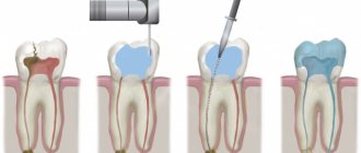
How is canine implantation performed?
- Diagnostic stage – the condition of the bone tissue, the general condition of the body is assessed, and the absence of contraindications is checked.
- Implant installation is performed under local anesthesia. The doctor opens the area of mucous membrane above the fang, removes the bone flap, forms a bed and inserts a titanium implant into it.
- The wound is sutured.
- After a week, the stitches are removed.
- Within 3-5 months, the implant takes root without load (a temporary prosthesis is not installed).
- After making sure that the implant has fused well with the bone, the doctor opens the mucosa above the top of the implant (local anesthesia is used).
- An individual abutment is made or a universal adapter is used.
- A gum former is installed, since the canine is located in a visually open area of the row.
- Impressions are taken from the abutment, a crown is made, which will exactly repeat the canine in shape and match the color of the neighboring teeth.
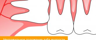
Types of location of an impacted tooth
| Fine | The tooth has a vertical position and grows upward very slowly. Baby teeth and canines can grow properly. “Wisdom teeth” are rarely positioned normally. |
| Horizontally | The impacted tooth forms a right angle to the healthy one, located horizontally. This situation is detrimental to a healthy “neighbor”. In such a situation, it is necessary to remove the impacted tooth, which is performed only surgically. |
| Obliquely | An impacted tooth grows at an angle toward the cheek or tongue. If such an anomaly does not cause inconvenience to a person, then removal is not necessary. |
Contraindications to the procedure
The list of contraindications for removing a fang on the upper jaw includes:
- Cardiovascular pathologies – myocardial infarction (suffered earlier six months ago), bacterial endocarditis, hypertension.
- Diseases of the urinary system - pyelonephritis in the acute stage, glomerulonephritis, renal failure.
- Diseases of an infectious and viral nature - hepatitis, acute respiratory infections, influenza and other pathologies transmitted by airborne droplets.
- Exacerbation of psychological problems - manic psychosis, epilepsy.
- Pregnancy at 1, 2 and 9 months.
- Poor clotting .
- Cancer – acute leukemia.
- Malignant tumors located in the maxillotemporal zone.
Symptoms of tooth impaction
In most cases, an impacted tooth is detected on an x-ray at the dentist or at an appointment with an ENT doctor. Retention in a child can be determined if one of the elements of the jaw row is absent for a long time or if a baby tooth is not replaced by a permanent “brother” for too long. The eruption of an impacted tooth is usually accompanied by inflammatory processes. The child may be bothered by swelling of the gum tissue and jaw, pain and aches, numbness and tingling in the retention zone.
On palpation of the gums, thickening and hardening are noted, reminiscent in shape and outline of an unerupted tooth.
What to choose?
If in the case of other teeth in the dentition, you can always discuss prosthetics or implantation, then when it comes to restoring the troika, the answer is almost clear - only implantation! Why is that? Due to the shape, size and functionality of the canine, it undergoes the greatest stress during the process of biting and chewing food. Therefore, it is important to ensure reliable and sustainable results. In this case, bridge prosthetics is not suitable due to the fact that the canine is located at the intersection of two zones - the frontal and chewing. Here the jaw arch bends, which means that even the strongest bridge will fail very quickly. In addition, the use of a bridge in this area of the jaw poses serious risks to the supporting teeth.
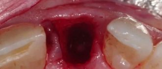
Removal of impacted teeth
If there are no complaints at all about the impacted tooth, and it does not cause any inconvenience (cysts do not form near it, the mucous membranes of the mouth are not injured), then removal may not be necessary. But if the impacted tooth is not removed, then it is necessary to undergo an X-ray examination of the mouth 2 times a year.
However, there are a number of cases when it is necessary to remove an impacted tooth:
- Retention causes constant inflammatory processes in the oral cavity;
- The occurrence of various paradental or follicular carpal formations, abscess, osteomyelitis in the gum tissue;
- Injuries and scratches of the oral mucosa that are caused by retention;
- An impacted molar or canine is also dystopic (improperly positioned in the dental arch) towards the cheeks or tongue.
The main factors on which the decision is made to remove impacted teeth are:
- The likelihood of injury during surgery,
- The location of the canine or molar and ease of access to them,
- Possibility of complications.
In most cases, the operation is painless and eliminates the possibility of pain even after the anesthesia wears off.
The removal of an impacted tooth itself takes place in several stages:
- pain relief by local anesthesia,
— an incision in the gum above the impacted canine and removal of a flap of tissue of the mucous membrane and periosteum,
- sawing out the bone to the dental crown using a drill,
- removal of a fang or molar from the bone using forceps (special biomaterials are left in its place).
Removal of premolars located on the upper jaw, canines and molars of the lower jaw has its own characteristics:
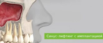
- Due to the fact that the roots of the teeth of the upper jaw are sometimes located close to the sinus, their removal must be carried out with the utmost care.
- In the case where the maxillary sinus has nevertheless been opened, medical treatment of the hole is not carried out, and the place where the bone has been broken is immediately sutured.
- Then a biomaterial is placed inside, which will promote rapid and painless overgrowth of bone tissue.
- The previously removed flap of mucous tissue and periosteum is returned to its place and sutures are applied.
- When removing impacted teeth in the lower jaw, the dentist must determine their location as accurately as possible, calculating their proximity to the mandibular canal. The safest way to access such teeth is through the vestibule of the mouth.
Features of the procedure
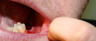
The canines on the upper jaw have one root located laterally. In cross section, part of the element resembles the outline of a triangle. In 30% of cases, the apex of the canine root has a curved structure. The outer part of the root is thicker than the inner one, but both sides of the alveoli are thicker in width than the incisors. Due to the listed features of the structure of the fangs, some difficulties arise when removing them from the socket.
During the operation, the doctor should place the fingers of the left hand in the same way as when removing incisors on the upper jaw. When removing an element from the right side, the patient's head should be slightly turned to the left and vice versa. This position of the patient will be more comfortable at the time of surgery.
The canines on the upper jaw are removed from the socket using wide forceps. During the operation, the element swings alternately towards the lips and palate with rotation around the longitudinal plane of the fang. The doctor makes the first movement of the instrument towards the outer wall of the alveoli, since it has a thinner structure.
Preparation and stages of tooth extraction
The next dislocation is performed towards the palatine alveolus, and then the specialist performs rotations. The procedure consists of successive rocking and rotation of the element until the nerve fibers and tissues surrounding it are completely ruptured.
Due to the anatomical features of the canines on the upper jaw, when extracting them, a specialist has to make a lot of effort.
Consequences of deletion
Eye teeth probably began to be called because the branches of the facial nerve are located next to them. Their irritation leads to long-term painful sensations that can radiate to the eyes, ears, and spread throughout the face. For this reason, tooth extraction requires severe local anesthesia or general anesthesia. The prejudice that you can lose vision after the procedure is unfounded. However, doctors rarely resort to it because of the danger of possible complications:
- long healing period, bleeding of the hole due to the deep root,
- violation of diction,
- inflammation of the maxillary sinuses (the roots of the eye teeth reach them),
- soft tissue injury,
- alveolitis,
- jaw dislocation,
- swelling in the eye area,
- temporary visual impairment,
- increased load on other teeth,
- damage to the optic nerve
- chaotic displacement of teeth (in the absence of rapid prosthetics),
- sagging cheeks,
- for the worse, a change in the shape of the face.
Molar removal
The lower molars have branched roots. One of the roots has a long, wide shape with a curved line. The second one is thin and has a deviation towards the back. There is a possibility that the roots will move closer to each other or move in different directions. Removing such teeth can be a difficult procedure. The surgeon uses forceps equipped with wide cheeks. To ensure a secure grip, the tongs have protrusions or spikes. Cases of tooth crushing and parts being extracted separately cannot be ruled out. It is not advisable to perform rotational movements during this procedure.
From medical practice, it is worth noting that wisdom teeth can sometimes have more than three roots. The roots may diverge to the sides and be twisted. In most cases, wisdom teeth are abnormal, which makes them difficult to remove.
Why is it dangerous to pull out fangs?
Removing the upper teeth, which interfere and cause aesthetic problems, seems to be a way out only at first glance. No specialist will remove it without good reason.
INTERESTING: what is atypical tooth extraction? If you can get rid of eights (wisdom teeth) without serious consequences, then with eye teeth the situation is unpredictable. They play an important role in chewing and the proper functioning and development of the jaw.
Removing these units is painful and dangerous for the following reasons:
- They play a significant role in chewing food, so their absence will complicate the process. Additionally, when removing the upper teeth, problems with diction are possible.
- If some of the eye teeth are missing, other units begin to perform their task, experiencing an unnatural load. This leads to rapid wear, weakening, disruption of the symmetry of the face, bite, and serious health problems.
- The deep position and unique shape virtually eliminate their susceptibility to caries. This means that the infection will not spread to neighboring teeth.
- These are the most stable teeth. They do not allow the incisors and molars to grind down when the jaws are compressed.
Before removing the upper tooth, the surgeon thinks through the tactics of manipulation, based on an X-ray of the patient’s jaw (we recommend reading: how is the nerve of the 6th tooth in the upper jaw removed?). He looks at how deep the root is planted and whether there is any curvature (even the slightest curvature makes the procedure even more difficult). The patient is positioned in a chair with the back slightly reclined, the doctor's position is in front and to the right of him. Extirpation is carried out in the following sequence:
- separation of the gums from the alveolar margin,
- separation of the neck of the tooth and the circular ligament,
- advancement of the forceps, their fixation,
- gentle rocking of the tooth, during which the periodontal tissue connecting its root with the walls of the socket is broken,
- tooth extraction,
- inspection of the hole, removal of fragments,
- wound treatment.
The success of removal largely depends on the experience and qualifications of the surgeon. When the specialist is convinced that the bleeding from the hole has stopped, he will give instructions on how to care for the operation site and schedule a routine examination. Despite proper care of the wound after canine removal, complications are possible. If pain intensifies, bleeding from the socket resumes, or general malaise occurs, there is no need to wait for a scheduled visit to the doctor. You need to see a dentist urgently.