- What are root canals?
- What are roots, how many roots do teeth have?
- How many canals in a tooth
- Indications for dental canal treatment
- Canal diseases
- Pus in the dental canal
- Endodontics
- Stages of root canal treatment
- Canal filling methods
- Canal treatment under a microscope
- Possible complications after endodontic treatment
- Prevention of dental canal diseases
- Choosing a paste for prevention
- Tooth canal treatment: price
What are roots, how many roots do teeth have?
The roots of the teeth are located in the jaw under the gum, bone formation, 70% of the entire tooth, thanks to them the teeth are firmly held in the jaw and cope with chewing everyday loads.
The number of roots does not equal the number of tooth crowns. The total number of roots is different for each person, it is individual. It differs depending on the location in the oral cavity, race, age, and load taken.
Number of roots in different teeth:
- 1 root has incisors and fangs,
- 1-2 roots have premolars,
- 3-4 roots at molars,
- Wisdom teeth can have up to 5 roots.
Roots are also present in baby teeth; they are the attachment of the teeth to the jaw; one tooth can have three roots. During the change of teeth, they dissolve, giving way to growing permanent ones, so the fallen baby teeth do not have “roots”.
Lateral upper incisor
Average age of teething: 8-9 years
Average age of root formation: 11 years
Average length: 22.0 mm
The crown of the upper lateral incisor, approximating an oval shape, is almost ideal for endodontic access, as is the case with the central incisor. Fiber optic illumination is also helpful when accessing this tooth.
The initial opening using a fissure bur is made immediately above the enamel tubercle in the equatorial third on the palatal surface of the tooth. The access cavity is oval in shape. When performing the initial opening, the fissure bur often occupies the entire narrow cavity of the coronal pulp. After removing the roof of the pulp chamber, a ball-shaped bur No. 4 or 6 is used to clean it from carious dentin, pigmented areas and calcifications.
A fissure bur may again be required to finalize the oval shape of the access cavity.
Adequate expansion is then created using spherical burs. Care must be taken to ensure that probes, endodontic cutting instruments and condensation instruments do not come into contact with the walls of the access cavity.
To ensure the cleanliness of the canal walls and their hermetically sealed filling, all carious tissues and leaky fillings must be removed and replaced with temporary filling materials.
The cross-section of the canal varies from oval in the cervical part to round in the apical foramen. The root is slightly cone-shaped and can bend at the apical part, usually in a distal direction. The apical foramen is often located closer to the anatomical apex than the central incisor, but can be located laterally within 1-2 mm of it.
In rare cases, access is complicated by the presence of a “tooth-in-tooth” developmental anomaly, invagination of part of the lingual surface of the tooth into the crown. This creates a space in the tooth that is surrounded by enamel and communicates with the oral cavity. Tooth-in-tooth is most common in the upper lateral incisors, but can occur in other teeth. Due to anatomical developmental defects, these teeth are prone to caries and the pulp may die before the apex is fully formed. This formation (“tooth within a tooth”) is localized in the crown; it must be processed mechanically and removed or bypassed.
Indications for dental canal treatment
Root canal treatment
- a necessary procedure in case of inflammation, helps to save the tooth. Since tooth canals are treated endodontically, a number of indications and contraindications must be taken into account. You should immediately contact a dentist for treatment when the tooth hurts when pressing on it, if there is pain in the cheek, swelling of the gums, and the tooth reacts to cold and hot.
You should consult a doctor if you have symptoms:
- Inflammation, there is a risk of pulp damage
- Deep caries, there is a risk of developing pulpitis, periodontitis, and abscess.
- Pulp chamber injury
- Toothache and swelling of the gums.
- After dental treatment, after a few days, painful sensations arise and do not go away.
Contraindications:
inflammation of the tooth root and maxillary sinus, root fracture, narrowing of the canals, destruction of the alveolar process.
If it is impossible to pass through the canal, it is removed or treated with mummifying paste.
Second upper premolar
Average age of teething: 10-12 years
Average age of root formation: 12-14 years
Average length: 21.5 mm
Similar to the first premolar in crown shape, the second premolar differs mainly in its root shape. Its crown is narrower in the bucco-palatal direction and somewhat wider in the mesial-distal direction. The mouth of the canal is located in the center, but it is more slit-like than oval. In the presence of a slit-like orifice, the physician should assume the presence of two canals until proven otherwise.
The external shape of the tooth is slightly oval, but wider in the mesial-distal direction than that of the first premolar.
All infected dentin and leaking fillings should be removed and replaced with temporary fillings.
The root may have two separate channels connecting into one, or two channels interconnecting in the form of a “web”. Accessory or lateral canals are possible but are less common than in incisors. Vertucci et al found that 75% of upper second premolars had one foramen at the apex, 24% had two foramina, and 1% had three foramina. Of all the teeth studied, 59.5% had additional canals. These investigators reported that when two canals are joined into one, the palatine canal is often directed toward the apex in a straight line. They further stated that “if, on a direct periapical photograph, the root canal sharply narrows or even disappears, this means that at this point it is divided into two canals, which either remain separate (type V) or, before reaching the apex, merge again (type II)".
The length of the root of the second upper molar is comparable to that of the first premolar. Apical bending is common, especially with a large volume of the maxillary sinus.
To prevent vertical coronal or crown-root fractures after endodontic treatment, complete closure of the occlusal access cavity is necessary.
Canal diseases
Canal diseases make themselves felt by inflammation and pain, so you need to see a dentist. If there is internal inflammation, but there is a possibility of soft tissue necrosis.
- Pulpitis
- inflammation of the dental pulp. With pulpitis, blood vessels collapse and die.
- Periodontitis
- inflammation of connective tissues resulting from complications of pulpitis.
- Abscess
- the presence of pus in the gums, which arose as a complication of pulpitis
- Advanced caries
leads to inflammation of the tooth canals.
Pus in the dental canal
Pus can accumulate in the dental canal in case of improper therapeutic treatment, or as a complication of this treatment, tooth trauma. Pus in the dental canal
appears as a consequence of deep caries, when the carious process has reached the canal and pathogenic microorganisms began to develop, causing pain.
Pus accumulates due to infection and the development of bacteria in the dental canal, and has two exits: through the carious cavity of the tooth or through the gum. It is very important to seek help in time, since the presence of pus can lead to the following complications: spread to the tissues of the oral cavity, spread to nearby organs. The presence of pus over time can lead to a fracture of the jaw and dissolution of bone tissue.
Treatment boils down to removing the pus and rinsing the canal cavity through the tooth tissue or gum. The canal is washed daily for several days and done in the dentist's office. The patient is taking antibiotics to prevent the infection from spreading. It is important to see a doctor in a timely manner because of the danger of an acute condition becoming chronic.
Second upper molar
Average age of teething: 11-13 years
Average age of root formation: 14-16 years
Average length: 20.0 mm
The shape of the crown of the second upper molar is very similar to the first upper molar, although it is not as rectangular and massive. Adequate access on both teeth can usually be achieved without disturbing the transverse enamel ridge. The second molar is often easy to prepare due to the straightforward approach to the orifices.
A distinctive feature of the morphology of the upper second molar is the closely spaced and sometimes fused three roots. The shadows of parallel root canals often overlap on the radiograph. Its roots are usually shorter than those of the first molar and not as curved.
The three orifices may form a blunt triangle, and sometimes they are located almost in a straight line. The floor of the pulp chamber is noticeably convex, creating a slightly funnel-shaped shape of the canal orifices. Sometimes the canals extend from the bottom of the pulp chamber at an acute angle, resulting in the need to remove the edge of the dentin in order to create a straight line of access.
Complications during access occur if the molar is tilted distally. Initial exposure is performed with a fissure bur with a cutting tip, and then a short ball-shaped bur is used, which is best suited for opening the pulp chamber and forming an access cavity. Then, small hand instruments are used to establish the patency of the canal and its working length. After this, the bulk of cleaning and shaping can be done by machining the files in the endodontic handpiece.
To improve radiographic visibility, especially when layering the shadow of the process of the zygomatic bone, photographs can be taken in perpendicular and distal projections at an angle.
All infected dentin, leaking fillings and denticles must be removed before endodontic treatment begins. To prevent vertical coronal or crown-root fractures, it is necessary to completely close the access cavity. If indicated, immediately after endodontic treatment it is necessary to apply internal reinforcement with intraradicular pins.
Endodontics
Endodontics
is a branch of dentistry that studies the methods and techniques of prevention and treatment in the root canals of human teeth. Here we can draw a non-trivial analogy: a tooth is like a tree - if the roots are deep and powerful, then both the stem and crown will be strong and beautiful. Similarly, the tooth - its aesthetic beauty and health largely depend on the condition of the root canals!
Root canal treatment
necessary for complications of caries disease, namely pulpitis and periodontitis. This is the so-called primary root canal treatment. When re-treating a tooth that has been manipulated - re-canal treatment, or re-treatment.
Upper canine
Average age of teething: 10-12 years
Average age of root formation: 13-15 years
Average length: 26.5 mm
As the longest tooth, the canine has an imposing shape designed to withstand strong occlusal forces. Its long crown with a thick layer of enamel is subject to abrasion by the cutting edge. As she ages, she often has deep cervical erosions.
The access cavity corresponds to the shape of the lingual surface of the crown and is oval. To obtain direct access, the cavity must be expanded incisally, but not so much as to weaken the actively functioning cusp. The initial access is made in the middle part of the crown from the palate. If the pulp cavity is deeper, a No. 4 or 6 ball-shaped extended bur may be required. A sweeping motion with this bur will open up the oval pulp cavity.
As it moves through the cervical portion and down apically, it remains oval. Thorough cleaning of this oval-shaped canal is difficult, so attention must be paid to targeted file processing.
The root canal is quite straight and long. Most canines require instruments 25 mm or longer in length. The last 2-3 mm of the top often bends in some direction.
The morphology of the canines rarely changes radically, and lateral and accessory canals are less common than in the upper incisors.
The vestibular cortical plate over the apex of the tooth root is often destroyed to form a fenestration. The apical foramen is usually located close to the anatomical apex, but can be located laterally, especially if there is an apical bend of the root.
Stages of root canal treatment
- The preparatory stage is when a diagnosis is made, a treatment plan and a method of pain relief are thought out. According to X-ray
- Preparing a carious cavity, it is opened, the dentin is removed, and access to working with the canals is opened.
- If the pulp is inflamed, a devitalizing paste is applied and a temporary filling is placed.
- The tooth cavity is opened and the pulp chamber arch is removed.
- The pulp of the crown is removed.
- Pulp extraction. The endodontist treats the canal up to the apical foramen.
- Treatment of the canal using instruments and medications.
Central upper incisor
Average age of eruption: 7-8 years
Average age of root formation: 10 years
Average length: 22.5 mm
The crown of the central upper incisor, close to rectangular on the vestibular side and wedge-shaped on the proximal side, allows for convenient endodontic access and is ideally positioned for direct examination using a mirror. The tooth is especially suitable for the novice doctor, since in it the canal is directly visible for a third of its length. With fiber optic illumination, the view of the channel can be improved.
Primary opening using a fissure bur is made immediately above the enamel palatal tubercle of the equatorial third of the crown on the lingual surface of the tooth. The instrument is directed along the long axis of the root. Based on the final shape of the access cavity, a triangular hole is made. Trephination of the tooth cavity often occurs during the first implantation. After the feeling of “falling” into the pulp chamber, the fissure bur is replaced with a spherical bur No. 4-6 with an extended shank.
A ball bur is used to widen the hole towards the incisal edge. You need to make sure that the pulp cavity is completely open. A fissure bur may again be required to widen the access cavity and give it its final shape. At this time, all carious dentin, which has significantly changed its color and pulp calcifications, is removed. It is necessary to remove leaking fillings and treat proximal carious cavities with adequate temporary filling.
The root is quite characteristically cone-shaped and sharply tapering towards the apex. The cross-section of the root canal approaches triangular in the cervical part, gradually becoming rounded closer to the apical foramen. Multiple canals in the root are rare, but accessory and lateral canals are common. The apical foramen is rarely located exactly at the apex, but is usually within 2 mm laterally.
Canal filling methods
Canal filling
helps prevent the development of dental canal diseases. Canal treatment is a painstaking process, complicated by the fact that the dental canals themselves are narrow, the shape can be curved, which requires painstaking work to fill the entire canal. Today, dentistry has several methods for filling canals.
1. Heated gutta-percha
Gutta-percha is a hard material that becomes elastic when heated and ideally fills the canal cavity. Several methods are used to treat a tooth canal using gutta-percha:
1) liquid injectable gutta-percha;
2) continuous wave;
3) vertical condensation;
4) syringe administration of gutta-percha.
2. Lateral condensation - cold gutta-percha
A gutta-percha pin is inserted into a canal filled with sealer paste, compacted, and sealed.
3. Thermofil - volumetric filling with hot gutta-percha
A plastic rod is inserted into the canal and the canal cavity is filled with hot gutta-percha, penetrating into all branches, leaving no free space.
4. Depophoresis technology
It is used in cases of difficult access to a curved canal that was previously filled, as well as in cases where the canal contains a part of an instrument broken during treatment.
All methods are painless for the patient. After treatment, pain is possible for two weeks if the root is removed.
Canal treatment under a microscope
In endodontics, the method of treating tooth canals under observation of the process through a microscope is widely used. This method allows you to see much more and provide better treatment. Root canal treatment under a microscope is a convenient method for the patient and the doctor. A magnification of 30-40 times under a microscope makes it possible to see all the branches of the canals, clean the canal perfectly and seal it.
The microscope allows you to see cracks in the canal, find all the branches of the canal, remove foreign objects, precisely treat hard-to-reach areas of inflammation, and remove the nerve. A microscope helps the dentist determine the condition of the filling, avoid damaging healthy tissue, and fill all the voids in the canal, leaving no room for infection to develop.
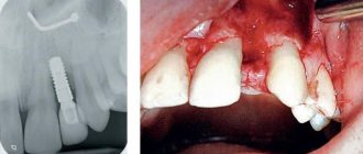
Possible complications after endodontic treatment
- The walls of the tooth cavity and the bottom are perforated, subject to the presence of dentin, if the instrument penetrates too deeply.
- The contents of the root canal are not completely removed in cases of obstruction, lateral branches, denticles or bleeding.
- The lumen is clogged with dentin filings, pulp residues, or an instrument that has broken in the canal.
- If the canal is bent, perforation of the root walls is possible
- The canal was not sealed thoroughly enough.
- The canal lumen was not expanded correctly.
- Filling falling out.
Complications may not appear immediately. After root canal treatment, the patient has slight sensitivity, pain, and discomfort in the area of the treated tooth and gums. If these sensations do not go away after two weeks and the pain intensifies, you should contact your doctor for an examination to determine the cause.
Upper first premolar
Average age of teething: 10-11 years
Average age of root formation: 12-13 years
Average length: 20.6 mm
The first upper premolar is a transitional tooth between the incisor and molar and most often has two roots.
When molars are lost, the main chewing load falls on the premolars. In removable prosthetics, these teeth are used as supporting teeth, which increases the impact of torque on them. Additional torque forces, together with deep carious lesions, can cause severe calcification of the pulp cavity. Early molar loss often causes rotation of the premolars, which can make identification of the pulp chamber difficult.
The mouths of the canals are located below and somewhat to the center of the tops of the mounds. The initial opening is made in the central fissure, giving it an oval shape in the bucco-palatal direction. After identifying the mouth, the doctor must accurately determine the presence of an anastomosis leading to the mouth of another canal. The direction of the roots can be determined using an endodontic probe. Root bifurcation visible on a routine periapical photograph may indicate tooth rotation. With divergent roots, less expansion of the occlusal approach is required, and with parallel roots, on the contrary, it may be necessary to remove the crown tissue towards the tops of the cusps. All infected dentin and leaking fillings should be removed and replaced with suitable temporary fillings.
Options for root anatomy include fused roots with separate canals, interconnecting canals or a “web,” a common apical foramen, and the possible presence of three roots, which is rare but should always be kept in mind. In the latter case, the mouths of the buccal canals will not be clearly visible using a dental mirror. An endodontic probe or a thin file will help determine the structure of the canal. Cams and Skidmore report that maxillary premolars with three roots and three apical foramina are found in 6% of cases. The length of the root is much shorter than that of the canine, and a distal bend is not common. The apical foramen is usually located close to the anatomical apex. The length of the roots when using intact tubercles as reference points is usually the same. The apical part of the roots often tapers sharply, ending in very narrow and curved tips.
Given the possibility of vertical mesial-distal fractures of the crown or root of the first premolar, before endodontic treatment, all fillings should be removed and the crown should be carefully examined under fiber light.
To prevent vertical fractures of the crown or root after endodontic treatment, it is necessary to completely close the occlusal access cavity.
Prevention of dental canal diseases
Prevention of dental canal diseases begins with the prevention of diseases that precede them. It is necessary to take care of the condition of the teeth, preventing the progression of carious processes. Regular dental hygiene: brushing with a toothbrush with the right toothpaste, using dental floss, and rinsing your mouth reduces the number of bacteria and slows down the development of caries and inflammation.
Regular preventive visits to the dentist will allow you to detect the carious process in the initial stage. Do not wait until the tooth makes itself known with painful sensations. Compliance with preventive measures will not only preserve teeth for many years, but also significantly save on treatment and avoid complications caused by inflammatory processes in the dental canals.
Choosing a paste for prevention
Dental health should be taken care of from early childhood. The most effective way is prevention and regular teeth cleaning.
For daily use you need to choose a toothpaste. With a variety of options to choose from, your dentist can recommend the right one for your teeth type. To avoid the addictive effect, it is recommended to periodically change the toothpaste.
Hygienic toothpastes do not have medicinal properties and are intended for everyday use. Daily use of hygienic pastes ensures cleansing of the oral cavity from food debris after eating, removes surface plaque, and has a short-term refreshing effect.
Medicinal pastes should be used as prescribed by a doctor, since they contain high concentrations of medicinal substances, and it is not recommended to use them constantly. The use of medicinal pastes helps fight the development of caries and inflammation in the gums.
Therapeutic and prophylactic are suitable for regular use. They contain active medicinal substances and natural ingredients, but in smaller quantities.
A small amount of toothpaste (about the size of a pea) is enough to effectively brush your teeth and prevent large amounts of toothpaste from ending up in your stomach. It is important to remember that to get results from toothpaste, you must use your toothbrush correctly and change it every three months. The toothbrush is selected depending on the condition of the teeth and oral cavity.
Tooth canal treatment: price
Tooth canal treatment price
differs depending on how many roots are in the canal, whether access to them is difficult or not. The price is influenced by the technique used in the treatment process and the equipment involved (microscope).
Dental canal treatment cost
consists of many components: consultation, examination, opening access to the canal, treatment with medications. The patient needs to expect an amount of 5,000 rubles above. On our portal you can navigate prices, find great deals and trusted clinics near your home.