Palate cancer is a malignant neoplasm that forms from the mucous membrane of the hard and soft palate. This is a fairly rare disease. It is more common between the ages of 40 and 60 years. Men get sick 4 times more often than women.
Diagnosis of cancer of the soft and hard palate at the Yusupov Hospital is carried out using modern research methods. The oncology clinic is equipped with modern equipment from leading American and European manufacturers, which allows you to quickly establish an accurate diagnosis. To treat malignant tumors of the palate, surgical interventions, chemotherapy drugs and innovative methods of radiation therapy are used.
The most malignant tumor of the palate occurs due to metastases of cancer of the nasopharynx and nose. The neoplasm affects the periosteum of the hard palate, lower and upper jaws, muscles and tissue of the oral cavity, and tongue. Atypical cells spread to the submandibular, mental and cervical lymph nodes.
Classification of oncological pathology of the palate
In half of all cases, malignant tumor of the hard palate is squamous cell carcinoma.
Nonsquamous cells, including minor salivary gland cancer, sarcoma, and melanoma, account for the other half of hard palate neoplasms. In 80% of cases, a tumor of the soft palate is squamous cell carcinoma. The remaining 20% are benign neoplasms.
The histological distribution of malignant neoplasms of the hard palate is as follows:
- squamous cell carcinoma - 53%;
- adenoid cystic carcinoma - 15%;
- mucoepidermoid carcinoma - 10%;
- adenocarcinoma - 4%;
- anaplastic carcinoma - 4%;
- other - 14%.
The histological types and frequency of neoplasms of the minor salivary glands of the palate are as follows:
- Benign - 26%
- Malignant - 74% overall Adenoid cystic carcinoma - 30%
- Mucoepidermoid carcinoma - 16%
- Adenocarcinoma - 18%
- Malignant mixed tumor - 8%
- Other - 2%
Causes of palate cancer
The causes of this disease have not been established, but it is known that certain factors may increase the risk of developing malignant tumors of the palate.
Smoking and chewing tobacco
Active smoking, especially unfiltered cigarettes and cigars, at least doubles the risk of developing malignant neoplasia of the oral cavity. Their development is also facilitated by the use of chewing tobacco mixtures - betel nut, gutka, snus, etc.
Alcohol
Strong alcohol is a recognized risk factor for palate cancer. This is mainly associated with the direct damaging effect of ethyl alcohol on the oral mucosa.
Human papillomavirus
HPV infection has been significantly associated with an increased risk of developing oral squamous cell carcinoma in recent studies.
Chronic inflammatory diseases of the oral cavity
Sluggish inflammatory processes, such as chronic gingivitis or periostitis, increase the likelihood of the formation of palate tumors.
Precancerous diseases
In the presence of pathologies of the mucous membrane such as leukoplakia or erythroplakia, the likelihood of developing cancer becomes extremely high - up to 90%.
Reasons and factors
The cause of the disease can be any damage to the oral mucosa.
This may occur if exposed to:
- Nicotine;
- Alcohol;
- Narcotic drugs.
Damage and inflammation of healthy cells due to prolonged contact with carcinogenic substances is a path to the appearance and development of palate cancer.
In addition, factors contributing to the transformation of healthy cells into cancer cells may include:
- Hereditary predisposition;
- Eating excessively hot food;
- Eating sour and spicy foods;
- Lack of vitamin A in the body;
- Mechanical injuries.
Of course, precancerous diseases should not escape attention, primarily leukoplakia and papillomatosis.
Symptoms of the disease
Squamous cell carcinoma appears as ulcerative superficial lesions. However, there are often no signs of palate cancer in the initial stages. The first manifestations appear, as a rule, already in the advanced stages of the disease.
Palate cancer - symptoms and first signs:
- bleeding;
- difficulty swallowing;
- speech disorders;
- bad breath;
- wounds in the mouth that do not heal for a long time;
- loosening of the teeth of the upper jaw;
- pain when swallowing;
- weight loss;
- ear pain;
- swelling of the neck;
- white spots in the mouth that do not disappear for a long time.
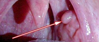
A swelling on the roof of the mouth, bleeding, foul odor, or loose teeth may be symptoms in patients with hard palate cancer. Patients with advanced soft palate cancer may have difficulty swallowing, altered speech, trismus, or neck swelling.
Because the area is easily visualized, tumors are often discovered in the early stages by chance by the patient or physician.
Stages of development of palate cancer
The stages of malignant neoplasms of the palate are determined based on the international TNM system. With its help, the characteristics of the tumor are described according to three main criteria.
Primary tumor stage (T)
- TX—primary tumor not evaluable
- T0 - no evidence of a primary tumor (T - carcinoma in situ.)
- T1—tumor 2 cm or less in greatest dimension
- T2 - tumor larger than 2 cm but not larger than 4 cm in greatest dimension
- T3 - tumor larger than 4 cm in greatest dimension
- T4 - Tumor invades adjacent structures (eg, cortical bone, soft tissue of the neck, maxillary sinuses)
Regional lymph node involvement (N)
- NX—regional lymph nodes not evaluable
- N0 - Absence of regional metastasis in the lymph node
- N1 - Metastasis in one ipsilateral lymph node, 3 cm or less in greatest dimension
- N2 - metastasis in one ipsilateral lymph node, more than 3 cm, but not more than 6 cm in greatest dimension; in several ipsilateral lymph nodes, not more than 6 cm in greatest dimension; or in bilateral or contralateral lymph nodes, not more than 6 cm in greatest dimension
- N3 - Metastasis in a lymph node greater than 6 cm in greatest dimension
Distant metastasis (M)
- MX - The presence of distant metastases cannot be assessed
- M0 - no distant metastases
- M1 - distant metastases
Based on the TNM characteristics, the so-called clinical stage is determined.
| Stage | Characteristics |
| 0 | Tis N0 M0 |
| I | T1 N0 M0 |
| II | T2 N0 M0 |
| III | T3 N0 M0 |
| (T1, T2, T3) N1 M0 | |
| IVA | T4a ( N0 or N1) M0 |
| (T1, T2, T3 or T4a) N2 M0 | |
| IVB | Any T N3 M0 |
| T4b Any N M0 | |
| IVC | Any T Any N M1 |
Tumors of the oral mucosa
SPECIALIZED ONCOLOGY CARE www.OncoClinic.com
Precancerous diseases
The development of cancer of the oral mucosa in 20-50% of cases is preceded by various precancerous diseases . Most often, precancer develops on the mucous membrane of the tongue (50-70%) and cheeks (11-20%). Processes with a high incidence of malignancy (obligate precancer) include Bowen's disease. Facultative precancers are represented by verrucous leukoplakia, papillomatosis, keratoacanthoma, erosive-ulcerative and hyperkeratotic forms of lupus erythematosus and lichen planus, post-radiation stomatitis.
Bowen's disease occurs more often in men aged 40-70 years, mainly in the area of the soft palate, uvula, and tongue. Clinically, it is characterized, as a rule, by the appearance of a single lesion in the form of a limited, slowly enlarging spotty-nodular lesion of a congestive red color measuring more than 1 cm in diameter. Its surface can be eroded and covered with small papillary growths. The diagnosis of the disease must be confirmed morphologically.
Verrucous leukoplakia differs from the flat form of leukoplakia by a greater degree of keratinization. The affected area of the mucous membrane protrudes above the surrounding tissues, its color is grayish-white, and there is a slight thickening upon palpation. Verrucous leukoplakia has plaque and warty clinical varieties. The warty variety has the greatest likelihood of malignancy. With this form, against the background of flat leukoplakia, dense-lumpy warty growths appear, which form mainly on the mucous membrane of the cheek, closer to the corners of the mouth.
Papillomatosis is characterized by the appearance of papillary growths on the mucous membrane. With the increased development of leukokeratosis, the surface of papillomatous growths acquires a whitish color. Inflammation often develops at the site of mucosal lesions. Superficial ulceration of the formation is possible.
Treatment of localized forms of precancerous diseases of the oral mucosa is surgical - excision, cryo- and laser destruction, electrical excision. Treatment of chronic ulcers on the oral mucosa includes the elimination of traumatic factors and conservative therapy. If there is no positive effect, a cytological examination of the prints or an excisional biopsy is performed within 2-3 weeks.
Cancer of the oral mucosa
The group of tumors of the oral mucosa includes malignant neoplasms of the mucous membrane of the tongue (excluding the root of the tongue), floor of the mouth, cheeks, hard and soft palate, alveolar process of the lower and upper jaw.
In the structure of incidence, cancer of the oral mucosa makes up 1.5% of the total number of malignant neoplasms. The highest rates are observed in southern and southeastern Asia, France and some countries in South America. The incidence of malignant neoplasms of the oral cavity in Russia (1997) is 2.7 per 100,000 population. In men, cancer of the oral mucosa develops 4-5 times more often than in women. Most patients are in the 5th-7th decade of life. The main factor contributing to the occurrence of cancer of the oral mucosa is smoking, which increases the risk of developing the disease by 2.5 times. When smoking and drinking strong alcohol are combined, the risk increases 15 times. Eating nasa and chewing betel leaves have a similar carcinogenic effect. Occupational hazards (prolonged contact with soot) and lack of vitamins B and A play a certain role. Other important etiological factors are chronic injuries and inflammatory processes of the oral mucosa.
The tongue (50-60%) and the mucous membrane of the floor of the mouth (20-35%) are most often affected by malignant tumors. Cancer of the buccal mucosa is observed in 8-10% of patients. It is extremely rare that tumors develop on the mucous membrane of the hard palate (1.3%). The vast majority of malignant neoplasms of the oral mucosa are represented by squamous cell carcinoma. A feature of its course is a tendency to lymphogenous metastasis to the superficial and deep lymph nodes of the neck (40-75%).
International histological classification of malignant tumors of the oral cavity and oropharynx
I. Tumors arising from stratified squamous epithelium:
1. Intraepithelial carcinoma (carcinoma in situ). 2.Squamous cell carcinoma. 3. Types of squamous cell carcinoma (verrucous carcinoma, spindle cell carcinoma, lymphoepithelioma). II. Tumors arising from soft tissues:
1. Fibrosarcoma. 2. Liposarcoma. 3. Leiomyosarcoma. 4. Rhabdomyosarcoma. 5.Chondrosarcoma. 6. Malignant hemangioendothelioma (angiosarcoma). 7. Malignant hemangiopericytoma. 8. Malignant lymphangioendothelioma (lymphosarcoma). 9. Malignant schwannoma. III. Tumors arising from the melanogenic system:
1. Malignant melanoma. IV. Tumors of controversial or unknown origin:
1. Malignant granular cell tumor. 2.Alveolar soft tissue sarcoma. 3. Kaposi's sarcoma. International classification according to the TNM system
T - primary tumor: Tx - insufficient data to evaluate the primary tumor, T0 - primary tumor not determined, Tis - preinvasive carcinoma (carcinoma in situ), T1 - tumor up to 2 cm in greatest dimension, T2 - tumor up to 4 cm in greatest dimension , T3 - tumor more than 4 cm in greatest dimension, T4 - tumor spreads to adjacent structures - bone, deep (external) muscles of the tongue, maxillary sinus, skin.
Notes:
1.The deep muscles of the tongue include the hypoglossus, genioglossus, and palatoglossus. 2. Superficial erosion of the osteodental cavity by a primary gingival tumor is not sufficient to designate the process as T4. 3. If there is doubt about the spread of the tumor to the bone, choose a less common category. If scintigraphy identifies a focus of pathological accumulation of radiopharmaceuticals, then the tumor is classified as T4. N/pN - regional lymph nodes: N/pNx - insufficient data to evaluate regional lymph nodes. N/pN0 - there are no signs of metastatic lesions of regional lymph nodes. pN0 - histological examination of material from a selected area of neck tissue includes 6 or more lymph nodes; histological examination of the material obtained using radical or modified radical lymphadenectomy includes 10 or more lymph nodes, N/pN1 - metastases in one lymph node on the affected side, up to 3 cm or less in the greatest dimension, N/pN2 - metastases in one or several lymph nodes on the affected side, up to 6 cm in greatest dimension, or metastases in the lymph nodes of the neck on both sides or the opposite side, up to 6 cm in greatest dimension: N/pN2a - metastases in one lymph node on the affected side, up to 6 cm in greatest dimension, N/pN2b - metastases in several lymph nodes on the affected side, up to 6 cm in greatest dimension, N/pN2c - metastases in lymph nodes on both sides or on the opposite side, up to 6 cm in greatest dimension, N/pN3 - metastasis in a lymph node more than 6 cm in greatest dimension.
Note. Lymph nodes on the affected side also include lymph nodes located in the midline.
M - distant metastases: Mx - the presence of distant metastases cannot be assessed, M0 - no distant metastases, M1 - distant metastases.
The requirements for determining the рТ category correspond to the requirements for determining the T category.
Grouping by stages
Stage 0 TisN0М0 Stage I Т1N0М0 Stage II Т2N0М0 Stage III Т3N0М0 Т1-3N1М0 Stage IVA Т4N0-1М0 Any ТN2М0 Stage IVB Any ТN3М0 Stage IVC Any ТAny NM1
Clinic. The anatomical forms of oral cancer are very diverse and do not have a generally accepted classification. Most often, cancer of the oral mucosa is a dense exophytic, easily bleeding neoplasm or ulcer with an uneven, tuberous bottom covered with a gray coating and raised edges, clearly demarcated from the surrounding tissues (ulcerative form). Sometimes an infiltrate is detected at the base of the ulcer, going deep into the tissue (ulcerative-infiltrative form). Tumors with an endophytic type of growth are more aggressive. The clinical course of oral cancer is divided into three periods of development. The early period is characterized by minor subjective unusual sensations of thickening of the mucous membrane, the appearance of a white spot of papillary neoplasms or a superficial ulcer, etc. As the tumor develops, it disintegrates, infection occurs, local pain of varying intensity occurs, salivation increases, and foul breath appears (advanced period). The period of neglect is characterized by rapid spread of the tumor to surrounding tissues, leading to significant impairment of the function of swallowing and speech.
Diagnostics includes examination and bimanual palpation, laryngoscopy, fiberoscopy of the oropharynx and nasopharynx, radiography of the facial skeleton (if indicated), morphological and cytological verification of the tumor, cytological examination of punctate enlarged lymph nodes. The clinical minimum examination includes chest x-ray and fibrogastroscopy (high probability of developing synchronous and metachronous damage to the mucous membrane of the upper respiratory and digestive tract).
When making a differential diagnosis, it is necessary to keep in mind nonspecific inflammatory processes, actinomycosis, tuberculous and syphilitic lesions of the oral mucosa, and goiter of the root of the tongue.
Prevention of oral cancer consists of timely treatment of precancerous diseases, sanitation of the oral cavity and rational prosthetics, elimination of bad household habits.
Cancer of the mucous membrane of the tongue
The predominant localizations of the tumor are the lateral edges of the tongue (72-84.4%) and the lower surface of the tip of the tongue (4-15%). The frequency of occurrence of the main clinical and anatomical forms of tongue cancer (exophytic, endophytic, mixed) is approximately the same. The vast majority of malignant tumors of the tongue are squamous cell carcinoma. In the anterior 2/3 of the tongue, cancer of the I-II degree of malignancy is more common; in the posterior third, carcinoma of the III degree of malignancy predominates. About 40% of patients at the time of treatment have clinically detectable metastases in regional lymph nodes. Cancer of the tip of the tongue most often metastasizes to the submental lymph nodes, and from there to the submandibular and deep cervical lymph nodes, cancer of the lateral surface of the tongue - to the submandibular and middle deep lymph nodes of the neck. When the tumor is located at the tip of the tongue or along the midline, bilateral metastasis is observed (5-20%). The biological aggressiveness of malignant tumors of the tongue is reflected in high rates of latent metastasis (30-40%).
Treatment . For stage I cancer of the movable part of the tongue, the surgical method is effective. For tongue cancer of stages II-III with an infiltration depth of no more than 4 cm without deep transfer of the tumor to the mucous membrane of the alveolar process of the mandible, the method of choice is combined radiation therapy. During implantation of intrastats, the issue of applying a temporary tracheostomy should be decided (high risk of tongue swelling and asphyxia). In other cases, treatment is combined. Remote gamma therapy is carried out in the preoperative period (SD 40-45 Gy). The irradiation field includes the primary focus and the area of stage I regional lymph nodes (in the absence of metastases) or all lymph nodes of the neck (in the case of clinically detectable metastases). The interval between the end of radiation therapy and surgery should not exceed 2-3 weeks. Hemiglossectomy is performed when the tumor diameter is no more than 4 cm using the electrosurgical method. Locally advanced tumors of the tongue are an indication for combined or extended operations. To perform combined operations, approaches through the mouth are used, with additional dissection of the tissues of the cheek or lower lip along the midline and various types of osteotomy. In case of advanced tongue cancer, the volume of resection is increased due to partial removal of the root and the opposite part of the tongue (subtotal and total glossectomy), all muscle structures of the tumor-affected area, the contents of the submandibular (required) and mental (if indicated) triangles. The spread of the tumor to the lower jaw is an indication for its segmental resection. In this case, it is necessary to apply a temporary tracheostomy and provide enteral nutrition to the patient. Total glossectomy or intersection of both hypoglossal nerves leads to the need for long-term tube feeding of the patient or the application of a gastrostomy. To improve functional and cosmetic results in penetrating resections of the mandible, vascularized complex bone-soft tissue free flaps, titanium implants, pedunculated musculocutaneous regional flaps, and free bone grafts are used.
Postoperative irradiation is carried out in the presence of a residual tumor (R1-2) up to a total focal dose of 70 Gy (taking into account previously performed radiation therapy). The volume of irradiated tissue includes the bed of the removed tumor and areas of its local spread.
Since the frequency of latent regional lymphogenous metastasis of tongue cancer is high, prophylactic bilateral lymphadenectomy is performed 2-3 weeks after surgery on the primary lesion or after completion of a course of radiation therapy. In the presence of clinically detectable metastases, cervical lymph node dissection on the affected side is performed simultaneously with removal of the primary lesion. After 3-4 weeks, prophylactic lymphadenectomy is performed on the opposite side.
The main types of surgical interventions on the cervical lymphatic system are the Krile operation and fascial-sheath excision of the tissue of the neck. The Crail operation is performed in the presence of multiple or single non-displaceable metastases in regional lymph nodes. With this type of intervention, the tissue and lymph nodes of the mental, submandibular and lateral triangles of the neck are removed. The boundaries of excision are the midline of the neck, the clavicle, the anterior edge of the trapezius muscle, and the lower edge of the mandible. The fiber is removed along with the sternocleidomastoid muscle, internal jugular vein, accessory nerve, submandibular and lower pole of the parotid salivary gland. Fascial-sheath removal of neck tissue is performed for enlarged lymph nodes that are suspicious for metastatic lesions, as well as for single metastases that are not fused with adjacent anatomical formations. The principal feature of fascial-case lymphadenectomy is the preservation of the internal jugular vein, sternocleidomastoid muscle and accessory nerve.
The prognosis depends on the degree of infiltration and the presence of regional metastases. The five-year survival rate for stage I-II cancer is 50-70% and stage III-IV cancer is 15-30%. Local-regional relapses in tongue cancer lead to death in 60-70% of cases. The mortality rate from distant metastases is 15%, from primary multiple tumors - 20-40%.
Cancer of the mucous membrane of the floor of the mouth
Clinic . The clinical course of tumors of the floor of the mouth is characterized by early spread of the process to surrounding tissues (gums, periosteum of the lower jaw). In turn, the tissues of the floor of the oral cavity are infiltrated secondarily by malignant tumors of the tongue, gums, lower jaw, and submandibular salivary glands, which must be taken into account in the differential diagnosis. Tumor involvement in the lower jaw can be determined by palpation - fixation of the tumor to the lower jaw is one of the signs of involvement of the periosteum or bone growth in the process. The immobility of the tongue indicates deep invasion of the tumor.
The first lymphatic barrier to cancer of the mucous membrane of the floor of the mouth is the submandibular and jugular-digastric lymph nodes (level I and II). The submental lymph nodes are rarely affected. Clinically recognizable metastases in regional lymph nodes occur in 30-40% of patients, in 20% of cases hidden metastases are observed. Distant metastases occur in 10-15% of patients.
Treatment of small tumors (T1) can generally be carried out by transoral resection with a margin of 2 cm from the macroscopically defined tumor edges, cryodestruction or radiation therapy. A number of foreign clinics are currently studying the possibilities of electrochemotherapeutic and electrochemical treatment. For exophytic tumors of the mucous membrane of the floor of the mouth with an infiltration depth of no more than 3-4 mm, the method of choice is application radiation therapy. Interstitial radiation therapy as an independent method can be used in the treatment of clearly demarcated tumors (T1-2) with an infiltration depth of 2.5-3 cm (SD 60-70 Gy). In other cases, preference is given to combined radiation therapy with external gamma therapy at the first stage at a dose of 30-40 Gy (T1-2) or 40-50 Gy (T3). If tumor regression is less than 50%, treatment is completed with surgery. Treatment of radiosensitive tumors is completed with interstitial radiation therapy at a total focal dose of 30-50 Gy. For medium-sized tumors (T2) of the anterior part of the floor of the mouth and deeply invasive neoplasms, bilateral irradiation of the first lymphatic barrier is mandatory. Contraindications to combined radiation therapy are the involvement of bone, large vessels, trismus in the tumor process, the presence of regional metastases, and the inaccessibility of the tumor for the introduction of sources.
Treatment of tumors corresponding to T3-4 local spread is combined: resection in combination with pre- or postoperative irradiation (40-50 Gy). For locally advanced tumors of the mucous membrane of the floor of the mouth, combined and extended operations are performed. In this case, the external carotid artery is first ligated and a temporary tracheostomy is applied for 1-2 weeks. The volume of excised tissue is determined by the extent of the tumor process. The most typical operations are:
1.resection of the floor of the mouth with simultaneous resection of a third or half of the tongue in a single block; 2.resection of the floor of the mouth with simultaneous marginal resection of the alveolar edge of the lower jaw or resection of the inner part of the lower jaw, to which the tumor adjoins; 3.resection of the floor of the mouth with simultaneous through resection of the lower jaw; 4. similar operations with simultaneous excision of the cervical tissue in a single block. Large postoperative soft tissue defects are eliminated with skin autografts, local flaps, and regional musculocutaneous flaps on a pedicle. In some cases, transplantation of complex tissue complexes (musculocutaneous, musculoskeletal, etc.) using microsurgical techniques is required. For through resections of the lower jaw, primary or delayed bone grafting or endoprosthesis replacement with titanium structures is indicated. If the continuity of the lower jaw is not restored simultaneously with the removal of the tumor, it is necessary to fix the bone fragments with special splints.
For stage I-II cancer, if there are no clinically detectable metastases and it is possible to regularly examine the patient, you can refrain from prophylactic regional lymphadenectomy. In common forms of the disease (stages III-IV), preventive irradiation of regional lymph nodes is indicated, which is carried out simultaneously with irradiation of the primary lesion, or bilateral cervical lymph node dissection. Fascial-sheath lymphadenectomy is performed 3-4 weeks after completion of treatment of the primary lesion. The treatment tactics for established regional metastases are similar to those for tongue cancer.
Drug treatment for cancer of the oral mucosa is used as neoadjuvant chemotherapy or for palliative purposes as part of chemoradiotherapy. The following combinations of drugs can be used:
1.cisplatin - 100 mg/m2 intravenously on day 1, fluorouracil - 1000 mg/m2 intravenously on days 1-4 with cycles every 3-4 weeks; 2. methotrexate - 40 mg/m2 intravenously on days 1 and 15, bleomycin - 10 mg/m2 intravenously on days 1, 8 and 15, cisplatin - 50 mg/m2 intravenously on day 4, with cycles every 3-4 weeks. If chemotherapy is insufficiently effective, monotherapy with taxanes or polychemotherapy using taxanes can be performed (taking into account cross-resistance to previously used drugs).
Treatment results depend on the size of the primary tumor, the presence of regional metastases, the degree of damage to the lower jaw and the radicality of the operation. According to summary literature data, the five-year survival rate for localized stage I-II tumors of the floor of the mouth is 60-80%. If the tumor crosses the midline of the fundus or spreads to the tongue and lower jaw, the five-year survival rate is within 50-60%, for tumors of stages III-IV - less than 50%. The presence of regional metastases reduces its rates to 25%.
Cancer of the mucous membrane of the gums and cheeks
Gum tumors are most often (80%) localized in the lower gum area behind the small molars. Exophytic neoplasms are more often papillary or verrucous and in the initial period of development may be similar to benign hyperkeratosis. Regional metastasis is observed in 15-30% of cases with cancer of the gum mucosa, and is less common with tumors of the cheeks. Latent metastases are detected in 10-20% of patients.
Treatment. Small superficial gum tumors can be treated with surgery or radiation therapy. When bone is involved in the process and large tumors (T3-4), combined treatment is performed (external gamma therapy + surgery). The volume of irradiated tissue includes the primary lesion and cervical lymph nodes on the affected side. Preventive irradiation of the lymph nodes of the neck is limited to the level of the first regional barrier. 2-3 weeks after completion of radiation therapy, segmental resection of the upper or lower jaw is performed. If there are metastases in the lymph nodes, cervical lymph node dissection on the affected side is performed simultaneously with excision of the primary tumor.
The spread of the tumor to the mucous membrane of the cheek and floor of the mouth is an indication for combined operations. A prerequisite for extended operations when the tumor is localized on the alveolar process of the lower jaw is the removal of the contents of the submandibular triangle on one side in a single block with the primary lesion, in case of damage to the anterior parts of the alveolar part of the lower jaw - the mental triangle, in case of tumor spread to the anterior parts of the floor of the oral cavity - submandibular triangles on both sides.
Malignant neoplasms of the cheek of stages I-II can be cured by surgery or combined radiation therapy. At the first stage, external beam radiation therapy is performed to reduce the biological activity of the tumor and reduce its volume; at the second stage, contact or interstitial radiation therapy is performed. For superficial tumors, contact radiation therapy can be used as an independent treatment method. In other cases, treatment is combined. Surgery is performed 2-3 weeks after completion of preoperative radiation therapy. Surgical access is carried out by crossing the lower lip along the midline with the continuation of the incision into the submandibular region. The block of tissue removed includes the tumor with surrounding tissues, a fragment of the lower jaw and masticatory muscles (if they are involved in the tumor process), and the contents of the submandibular triangle. Tumor growth into the mandibular canal is an indication for removal of half of the lower jaw. The resulting defect in the mucous membrane, skin and bone is eliminated by one of the types of plastic surgery. Cervical lymph node dissection is performed in the presence of clinically detectable regional metastases. Preventive irradiation of the regional lymphatic system is not carried out.
Forecast. Survival rates for tumors of the gums and cheeks depend on the size of the tumor, bone damage, and the presence of regional metastases. The average five-year survival rate of patients with gum tumors ranges from 78% (for stage I) to 15% (for stage IV). The results of combined treatment when bone is involved are significantly better than with radiation therapy.
According to summary data, the five-year survival rate for stage I-II cheek cancer is 65-74%, stage III-IV varies from 20 to 30%.
Cancer of the mucous membrane of the retromolar triangle
Tumors arising in the retromolar triangle are rarely limited to the gingival mucosa; the process may involve the nearby mucous membrane of the cheek, the anterior palatine arches, the floor of the mouth, and the back of the gums. A tumor of the retromolar triangle that extends to the anterior palatal arches may appear more similar in appearance to nasopharyngeal cancer.
The risk of clinically positive and latent metastases in the lymph nodes with these tumors is significantly higher than with gum tumors of other locations.
Treatment of superficial tumors, including those spreading to the soft palate and buccal mucosa, is radiation. If the periosteum is damaged, a partial (marginal) resection of the lower jaw is performed, even with small tumors. Moderately advanced and infiltrative tumors are treated in combination. At the first stage, surgical intervention is performed (resection of the lower jaw and cervical lymph node dissection), then external beam radiation therapy is performed. For locally advanced processes, treatment begins with preoperative radiation therapy, which in some cases allows the tumor to be transferred to a resectable state.
Cancer of the mucous membrane of the hard palate
On the hard palate, malignant tumors from the minor salivary glands (adenocystic carcinoma, adenocarcinoma) often develop. Adenocarcinomas tend to grow encapsulated for a long time. Rarely developing, squamous cell carcinoma tends to ulcerate quickly.
Treatment. The method of choice for treating squamous cell carcinoma without damage to the periosteum is intracavitary contact therapy (if it is technically possible to bring the stomastat to the lesion). In other cases, treatment is combined. At the first stage, preoperative radiation therapy is carried out, electrosurgical removal of the tumor is performed, retreating from the visible lesion of the mucous membrane by 3-4 cm. Postoperative defects are covered with obturating prostheses or eliminated using plastic surgery.
SIGN UP FOR A CONSULTATION by phone +7 (812) 951 - 7 - 951
Share link:
Diagnosis of palate oncology
In most cases, detection of oncological pathologies of the palate occurs during a routine examination by a dentist. The diagnosis is then confirmed by biopsy findings.
However, to assess the extent of the tumor in the structures of the upper jaw, pharynx, etc. Additional diagnostic tools are required:
- MRI (magnetic resonance imaging) - to detect tumor growth in the sinuses of the upper jaw or the nasal cavity.
- PET (positron emission tomography) - to detect metastasis in regional lymph nodes and separated metastasis.
Probability of disease recurrence
After intensive therapy, the patient is discharged home. He must be observed by an oncologist on an outpatient basis for a year. It is recommended to visit a doctor for a preventive examination every three months. This will allow you to recognize the symptoms of relapse in time and carry out treatment again.
In case of relapse, a cancerous tumor arises either in the same place or in other organs. Distant metastases are difficult to diagnose if the cancer cells are dormant in the organ they entered through the blood or lymphatic system. The period of remission in this case can be long.
Treatment of the disease
Radiation therapy
Radiation therapy to treat palate cancer can be used in several ways.
- As the primary treatment for small, early-stage tumors.
- As an adjunct to surgery or ongoing chemotherapy. In this case, it is possible to use adjuvant radiation therapy as after surgery to destroy tumor remnants. So is the use of radiation before surgery to shrink the tumor and make it easier to remove—neoadjuvant radiation therapy.
- Radiation therapy can also be used to relieve symptoms of advanced, inoperable cancer, such as pain, bleeding, problems swallowing, and problems caused by bone metastases.
Modern radiotherapy for palate cancer in Belgium
Radiation in cancer centers in Belgium is carried out using techniques that help doctors focus the radiation more precisely.
There are two main such methods.
- Three-dimensional conformal radiation therapy (3D-CRT);
- Intensity modulated radiation therapy (IMRT).
These methods rely on the results of imaging tests, such as MRI, and special computer programs to pinpoint the location of the tumor. The beams are then shaped and directed at the tumor from multiple directions, reducing damage to healthy nearby tissue.
Another modern way of delivering radiation is to place radioactive materials directly into the tumor. This is called brachytherapy.
To do this, a small grain of radioactive material is placed into the tumor. In this case, the radiation spreads only over a very short distance and is limited to the tumor.
Surgery
The type of operation is selected depending on the location and extent of the tumor.
For small to medium-sized tumors up to T3, an oral approach is often used without trauma to the mandible. For most tumors that do not have regional spread, surgery to remove the hard or soft palate is used - palatectomy.
For tumors that have grown into the paranasal sinuses or nasal cavity, a unilateral or bilateral maxillectomy (removal of the upper jaw) may be required.
Chemotherapy
Treatment of palate cancer with chemotherapy is considered as an additional method in combination with radiation therapy and surgery.
The chemotherapy drugs most often used for cancer of the oral cavity and oropharynx are:
- Cisplatin;
- Carboplatin;
- 5-fluorouracil (5-FU);
- Paclitaxel (Taxol);
- Docetaxel (Taxotere).
Targeted and immunotherapy
As a rule, targeted and immunological drugs for this disease are used for metastatic inoperable forms, or for relapses.
Cetuximab (Erbitux®) is a monoclonal antibody that targets the epidermal growth factor receptor (EGFR), which helps cells grow and divide. Palate cancer cells often have higher than normal amounts of EGFR. By blocking EGFR, cetuximab can help slow or stop cell growth.
Pembrolizumab (Keytruda®) and nivolumab (Opdivo®) are drugs that target the PD-1 checkpoint. It is a protein on the membranes of T cells that normally helps keep these cells from attacking other cells in the body.
By blocking PD-1, these drugs boost the immune response against cancer cells, which can shrink some tumors or slow their growth.
Diagnosis of cancer of the soft and hard palate
The resulting cancerous tumor of the palate is difficult to determine independently in the early stages. If the pathological process has affected large areas of the soft or hard palate, a preliminary diagnosis can be made after a visual examination of the oral cavity.
To confirm the diagnosis, oncologists at the Yusupov Hospital perform the following diagnostic procedures:
- X-ray – finds pathological changes in bone tissues adjacent to the oral cavity;
- Biopsy – taking a piece of tissue for histological examination (analysis is necessary to identify altered tumor cells and its type);
- Blood tests - signs of anemia;
- Radioisotope examination - allows you to examine the structure of the neoplasm.
Ultrasound examination is carried out to detect cancer metastases in distant organs. Patients of the Yusupov Hospital can undergo complex diagnostic procedures in partner clinics and receive advice from leading dental oncologists in Moscow.
Rehabilitation after treatment
Treatment of malignant tumors of the palate is always very traumatic. The consequence of the operation is, at a minimum, a violation of the process of swallowing and speech. In multidisciplinary clinics in Belgium, rehabilitation doctors and speech therapists begin working with the patient immediately after the operation - this greatly shortens the rehabilitation period and increases its effectiveness
Reconstructive surgery - palate surgery in Belgium
Palatectomy and maxillectomy operations require subsequent prosthetics.
Small defects of the hard palate can be covered with local flaps from the rest of the hard palate or from the buccal mucosa. Larger defects are adequately and effectively treated with obturators. Because the hard palate is not dynamic, obturators are very effective and well tolerated.
The soft palate is a dynamic structure and its optimal functioning requires muscular action to lift and contract the palate during swallowing and relax it during nasal breathing. Therefore, any reconstruction of the soft palate using flaps and prosthetics does not reproduce its functionality - it will be limited even with the most successful outcome.
How is inflammation of the palate treated?
How to treat inflammation on the roof of the mouth, a specialist will tell you after determining the diagnosis. The following therapy methods can be used:
- If the cause of inflammation is minor damage to the mucous membrane, rinsing with decoctions of medicinal herbs is usually sufficient. Decoctions and infusions based on calendula, sage and chamomile have an antiseptic and healing effect. The herbal decoction for rinsing should be used warm.
- When the palate is affected by a fungus, patients are prescribed topical agents. The most effective are the solution and gel “Chlorhexidine”, “Rotokan” or “Stomatofit”.
- In case of severe pain symptoms, regardless of the cause of inflammation, drugs of combined action are prescribed. “Cholisal-gel” and “Kalgel” have proven themselves well.
- If a purulent inflammatory process is detected in the oral cavity, the patient is prescribed antibiotics. The minimum course of treatment is usually 7 days, but there are also stronger drugs that only need to be taken for 3 days. Along with local agents, antibacterial drugs for internal use - Sumamed, Flemoxin Solutab - can be prescribed.
- The inflammatory process of the palate is easily treatable. The earlier therapy is carried out, the less likely it is to develop complications.
Complications
Complications of surgical resection of the soft palate may include:
- velopharyngeal insufficiency (most often);
- hypernasal speech;
- dysphagia (impaired swallowing);
- effusion in the middle ear due to scarring at the opening of the eustachian tube.
The extent and likelihood of these complications depend on the extent of the resection, the size of the defect, and the method of reconstruction. The larger the resection and defect, the greater the likelihood of these complications occurring.
With soft tissue resection preserving only the bony palate and soft palate, the resulting defect heals with granulation and epithelialization, and no complications are expected. If patients have previously had radiation therapy in this area, healing may be delayed.
Palate cancer and its treatment can sometimes cause problems such as loss or changes in taste, dry mouth, or even tooth loss. This can make it difficult to eat, leading to weight loss and weakness due to poor nutrition.
Radiation therapy may also cause long-term or permanent side effects.
- Damage to the salivary glands. It may cause dry mouth. This leads to problems with eating and swallowing. Lack of saliva can also lead to tooth decay (cavities).
- Damage to the jaw bone. This problem, known as osteoradionecrosis of the jaw, can be a serious side effect of radiation therapy. This condition is difficult to treat. Osteoradionecrosis is manifested by pain in the jaw, and sometimes by fractures, which require surgical treatment.
Newer radiation treatments such as IMRT may help reduce this side effect. The drug amifostine (Ethyol®) may also help reduce this side effect by limiting radiation damage to normal tissue. It is injected into a vein over 15 minutes before each radiation treatment.
Advantages of treatment in Israel
- The opportunity to be treated by world-class specialists at reasonable prices.
- Availability of modern diagnostic and treatment equipment in the clinic.
- Using the latest and most effective treatment protocols.
- Caring care during the stay for treatment and the services of a case manager, simplifying the patient’s stay in a foreign country.
- The ability to pay for services in any convenient form upon receipt of them.
If signs of the disease are detected in you or your loved ones, and you are interested in treatment in Israel, fill out the application form on our website or request a call back. Our consultant will contact you shortly and help organize your trip. There's no reason to put off applying, do it today!
- 5
- 4
- 3
- 2
- 1
(1 vote, average: 5 out of 5)
Prevention
Limit smoking and alcohol
Tobacco and alcohol are the most important risk factors for these cancers. Not starting to smoke is the best way to limit your risk of cancer. Quitting tobacco also significantly reduces the risk of developing these cancers, even after years of use. The same applies to alcohol - limiting its amount helps reduce the risk of developing palate cancer.
Preventing HPV infection
Vaccination against HPV reduces the risk of mouth and throat cancer. But vaccines are only effective if they are given before someone gets HPV, given at a young age, before they become sexually active.
Correctly selected dentures and proper oral hygiene
Avoiding sources of oral irritation (such as dentures that don't fit properly) can help reduce your risk of developing oral cancer.
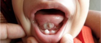
Prerequisites for the occurrence of a lump
Modern medicine has not created a complete list of reasons why bumps may appear. Doctors' reasoning is based on hypothetical cause-and-effect relationships. The appearance of a lump on the palate is caused by:
- Bad habits (smoking, alcohol, oral drugs);
- The presence of micro and macro injuries to the oral cavity (surgeries, scratches of the upper parts of the oral cavity);
- Availability of dentures;
- Viral infections;
- Intrauterine disorders (hemangioma is a congenital disease and acquired from the mother);
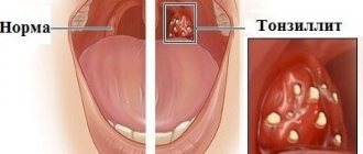
- Violation of the activity and integrity of the mucous membrane (typical of angina);
- Congenital and acquired dysfunction of the glands (the main cause of cysts).
There are two more hypothetical reasons for the appearance of malignant tumors:
- Eating too hot and spicy foods, constantly disrupting the structure of the cells of the oral cavity;
- The presence of papillomatosis or leukoplakia - diseases that are precancerous lumps that can develop into an oncological disease.