Oral tuberculosis is an infectious disease that, like other types of tuberculosis, is caused by mycobacteria. This pathology is classified as chronic. As a rule, the inflammation characteristic of tuberculosis, affecting the lining of the mouth and the red border around the lips, is secondary: that is, it occurs as a consequence or the most characteristic manifestation of other forms of tuberculosis. For example, these include tuberculous lesions of the lymph nodes and bones.
The mucous membrane of the oral cavity is far from the most favorable environment for the development and reproduction of mycobacteria that cause tuberculosis. As a rule, when entering this environment, mycobacteria die. However, in the presence of favorable circumstances (microtraumas, mechanical damage in the oral cavity, opening the gates for infection), bacteria penetrate inside and provoke the formation of a tuberculosis ulcer.
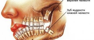
Oral tuberculosis is a relatively rare form of tuberculosis, usually diagnosed only in children. This is due to the characteristics of teething in children.
In an infant, such a disease can be extremely severe and includes the generalization of tuberculosis infection. Secondary tuberculosis infection begins to develop as tuberculous lupus or miliary ulcerative tuberculosis.
Tuberculosis is transmitted by airborne droplets. The incubation period lasts from eight to thirty days, and after this the formation of an uneven, blurred ulcer begins, which is severely painful. After a few days it begins to gradually increase. At the same time, the adjacent lymph nodes swell. There are no other signs of inflammation yet
Classification of types of oral tuberculosis
Oral tuberculosis is a disease that appears and develops mainly in patients with reduced immunity. The disease is caused by Koch's bacillus. The source of inflammation located in the patients' oral cavity is secondary in most cases. It develops as a result of the spread of infection from the main focus, and spreads through the circulatory or lymphatic systems. In the case when the patient is affected by the pulmonary form of tuberculosis, the infection spreads to the oral cavity due to the penetration of mycobacteria contained in the sputum.
There are four forms of oral tuberculosis. Among them:
- Primary tuberculosis of the oral cavity. Transmitted by the respiratory or fecal-oral route, this disease almost never occurs in adults. Most patients with this diagnosis are infants.
- Tuberculous lupus. It is the lupus form of oral tuberculosis that is most often encountered in dental practice. Erosive elements are localized on the mucous membrane of the gums. If treatment is not started on time, these tumors can develop into a malignant form.
- Miliary-ulcerative form. This form usually develops in patients who have already been weakened by tuberculosis. Along with their cough, they produce sputum, which is a source of bacteria that cause oral tuberculosis. The lesions are on the palate and tongue, in rare cases - in the area of marginal gums or on the side of the cheek.
- Scrofuloderma. This form of the disease is diagnosed in most cases in children. The clinical signs of scrofuloderma are somewhat different from those of other forms of oral tuberculosis. In the first stages - the appearance of not a tubercle, but a rather large nodule. After its softening and necrosis occurs, the stage of formation of fistula tracts begins. The healing process of the surface of the resulting ulcers takes a long time, and after it characteristic fringed scars form.
Symptoms and first signs of tuberculosis in children
First of all, tuberculosis in children is manifested by such clinical signs as:
- weakness;
- stopping weight gain;
- irritability;
- fatigue during school hours;
- absent-mindedness;
- lagging behind peers in studies;
- body temperature – up to 37.5°C;
- enlarged lymph nodes;
- sweating;
- chills.Source: N.M. Koretskaya Tuberculosis in children and adolescents in modern conditions // Siberian Medical Review, 2010
Forms of tuberculosis and early signs
| Form | Symptoms |
| Tuberculosis of the bronchial glands | Cough, elevated body temperature for a long time, lethargy, fatigue, pallor, thinness. |
| Pulmonary form | Cough, shortness of breath, weakness after sleep, apathy, decreased performance, absent-mindedness, unhealthy blush, thinness, pale skin, sweating, chills, dry (and then “wet”) cough for more than 3 weeks, sputum with blood. |
| Lymph node involvement | An increase in the size of the lymph nodes, their softening, suppuration (the contents leak out), the formation of fistulas in places where pus comes out, scrofuloderma (skin lesions in the form of tumors with subsequent release of the contents). |
| Involvement of bones and joints in the process | Slow development, pain when moving, changes in gait, lameness. |
| Damage to the meninges (tuberculous meningitis) | Anxiety, lethargy, loss of appetite, bad mood, headaches, fever, vomiting, convulsions. |
| With damage to the gastrointestinal tract | Constipation or diarrhea, increased body temperature, blood in the stool, pain. Source: A.V. Mordyk, E.A. Tsygankova, L.V. Puzyreva, A.A. Turitsa Tuberculosis in children of the Russian Federation at the present stage // Pediatric pharmacology, 2014, v. 11, no. 3, pp. 27-30 |
Clinical manifestations by age of children
| Age | Symptoms |
| Infancy (up to one year) | Low mobility, apathy, weakness, attacks of suffocation or coughing, retraction of part of the chest, weight loss (including muscle mass), loss of appetite, cessation of crying, insomnia. Early detection and diagnosis are extremely important, because at this age this pathology is most dangerous. |
| 5-8 years | Decreased activity, weakness, lack of sleep and appetite, loss of body weight, cough, condensation of the chest. |
| 8-15 years | Rapid appearance of pain in the lungs against a background of apathy and weakness, active urge to cough, shortness of breath even at rest, thinning or discoloration of the skin, the appearance of cracks, wounds, hemoptysis, changes in lymph nodes in size and structure, intoxication of the lungs (at the last stage) . |
Manifestations of the chronic form:
- sleep disorders;
- liver enlargement;
- lagging behind peers in physiological development;
- dry and pale skin;
- mild euphoria.
Diagnostics
Primary tuberculosis of the oral cavity is diagnosed during an external examination. Thus, it is characterized by the presence of lacerations and ulcers in the corners of the mouth with yellow, gray and bluish layers. Areas with ulcers tend to gradually enlarge.
At the same time, the lymph nodes begin to enlarge.
Diagnosis is carried out on the basis of the collected anamnesis, as well as using histological and bacterioscopic studies. During examination, dense painful lumps or ulcers are found on the mucous membrane, depending on the severity of the condition.
There is minor inflammation. The process of formation of adhesions begins between the lymph nodes and the surrounding surface.
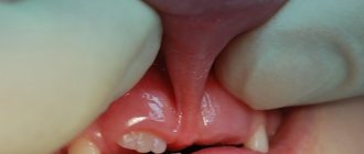
Perforation of the hard palate in tuberculosis
Tuberculosis is a chronic granulomatous disease caused by Mycobacterium tuberculosis. WHO reports more than 8 million new cases each year and approximately 3 million deaths from the disease worldwide. So recently, antibiotic resistance of bacteria has become a huge problem. Oral tuberculosis is quite rare and accounts for less than 1% of all cases. With the increasing incidence of the disease, rare and unusual forms are increasingly observed, which often become malignant. In this clinical case, we describe a rare case of perforation of the hard palate during the development of tuberculosis.
Description of a clinical case
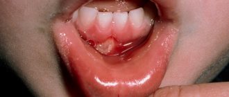
A 7-year-old boy sought help because he had problems swallowing solid food, low-grade fever, and weight loss for the last six months. His mother was on anti-tuberculosis treatment due to pulmonary tuberculosis. There was no history of cough, abdominal pain, vomiting, diarrhea, or urinary problems. On examination, the pulse was 104 beats/min, the respiratory rate was 28/min. and blood pressure 98/60 mm Hg. The child's weight is 13 kg and his height is 104 cm (both figures are below the norm for this age and gender). There was slight pallor of the skin. Examination of the oral cavity revealed a perforation of the hard palate measuring 3x3 cm with irregular, torn edges and necrotic contents (Photo 1). Bilateral lymphadenopathy of the cervical lymph nodes was noted. Auscultation of the lungs revealed crepitus on the right side above. The remaining organ systems are without pathology. The Mantoux test is positive (28 mm). Laboratory examination: hemoglobin 8.8 g/dl, total leukocyte count 9000/cumm (neutrophils 40%, lymphocytes 60%), ESR 80 mm/h. Liver and kidney function is not impaired. Serological test for HIV is negative. Urine and blood cultures revealed no growth of microorganisms. A CT scan of the oral cavity revealed erosion and a defect in the posterior part of the hard palate on the right (Figure 2). X-ray examination of the chest revealed a homogeneous opacity in the upper right zone. Lymph node puncture showed granular caseous necrosis and granuloma composed of epithelioid cells and histiocytes (Figure 3). The child's parents did not consent to a biopsy of the palate lesion. However, taking into account the clinical manifestations, as well as the presence of pulmonary tuberculosis and caseous necrosis in the lymph nodes, it is highly likely that perforation of the palate also occurred due to tuberculosis. The patient was prescribed antituberculosis therapy (isoniazid, rifampicin, pyrazinamide and ethambutol). The boy was registered with maxillofacial surgeons for subsequent elimination of the perforation of the palate.
Photo 1: Examination of the oral cavity, revealing a perforation of the hard palate measuring 3x3 cm with uneven, torn edges and necrotic contents
Figure 2: Axial CT scan with 3D reconstruction showing erosion and defect in the right posterior hard palate
Figure 3: Biopsy of cervical lymph nodes showing granular caseous necrosis and garnulloma consisting of epithelial cells and histiocytes.
Discussion
Tuberculosis in the oral cavity can be either primary or, more often, secondary to pulmonary tuberculosis. With secondary damage to the oral cavity, bacteria reach the mucosa by hematogenous or lymphogenous route. In primary oral tuberculosis, mycobacterium penetrates the mucosa directly due to disruption or loss of the natural barrier due to injury, inflammation, leukoplakia, tooth extraction or poor hygiene. Other predisposing factors include odontogenic cysts, periapical granulomatosis, gingival abscesses, periodontitis and jaw fractures. Abbot was able to detect mycobacteria in oral lavage from 44.9% of patients with active pulmonary disease, confirming the importance of intact mucosa for oral resistance to tuberculosis infection. The incidence of tuberculosis in the oral cavity varies from 0.8% to 3.5%. Oral involvement is rare, even in populations with a high incidence of tuberculosis. Factors responsible for oral resistance include the protective properties of saliva, the presence of saprophytes, muscle resistance to microbial invasion, and the thickness of the protective epithelial cover. The most common location of tuberculous lesions in the oral cavity is the tongue. Other localizations may include the soft and hard palate, lips, cheeks, tonsils, gums, floor of the mouth, uvula and alveolar mucosa. Tuberculosis in the oral cavity usually appears as single, painless ulcers with hardened, irregular edges and necrotic contents. The lesions may also appear as nodules, grooves, plaque, vesicles, tubercles, or granulomas. Tuberculosis of the palate can be expressed in granulomatosis, ulceration or perforation, and damage to the hard palate occurs somewhat more often than the soft palate. According to Baruah, palate involvement occurs in patients with a reactive immune response and hypersensitivity to acidic bacteria that cause tissue destruction.
In addition to tuberculosis, cases of perforation of the hard palate can be associated with various infections (syphilis, leprosy, leishmaniasis or fungal infection), granulomatosis, sarcodia, neoplasms (glandular and epithelial tissue), drug use (cocaine) and median granuloma. In this case, the differential diagnosis was based on clinical manifestations, the presence of pulmonary and lymphatic lesions by mycobacteria, and improvement in condition with anti-tuberculosis therapy. Treatment of palatal tuberculosis should be carried out on the basis of instructions for the treatment of any form of extrapulmonary tuberculosis.
Conclusion
Tuberculosis of the palate is a relatively rare phenomenon, but should be included in the differential diagnosis in the presence of perforation of the hard palate. A thorough examination of the patient is also necessary to determine the primary source of mycobacterial infection.
Authors: Syed Ahmed Zaki, Swapnil Bhongade, Shailesh S. Vartak Department of Pediatrics, Lokmanya Tilak Municipal General Hospital and Medical College, Sion, Mumbai, India
Treatment of oral tuberculosis
Treatment of this form, like all others, is carried out in specialized institutions - tuberculosis dispensaries. First of all, the underlying disease is treated. To prevent the bacterial infection from spreading further, antiseptic baths are prescribed.
After the acute condition has been relieved, it is necessary to take care of timely and high-quality sanitation of the oral cavity. It is important to promptly consult a specialist at the first symptoms: advanced oral tuberculosis leads to serious consequences for the patient’s health.
Causes
Koch's bacillus has a shell that is resistant to acidic environments. It is able to survive freezing, drying, exposure to alkalis, and much more. etc. Characterized by the ability to form so-called L-forms with increased adaptability. The most pathogenic microbacteria for humans are:
- Mycobacterium tuberculosis humans;
- Mycobacterium bovis.
Microbacteria enter the body through contact, air, mixed and other routes. This is how the first inflammatory focus is formed. A child can also be infected during the mother's pregnancy - through the placenta or during childbirth, if it swallows amniotic fluid. Source: https://www.ncbi.nlm.nih.gov/pubmed/24548085 Marais BJ Tuberculosis in children J Paediatr Child Health . 2014 Oct;50(10):759-67. doi: 10.1111/jpc.12503. Epub 2014 Feb 19
Children at risk include:
- not vaccinated with BCG;
- long-term treatment with antibiotics, hormonal or cytostatic agents;
- having HIV status;
- living in unfavorable conditions (social and/or sanitary);
- having diabetes mellitus;
- with weakened immunity;
- under 2 years of age;
- and etc.
Types and forms of pathology
According to the period of occurrence in medicine, stages are distinguished:
- infiltration;
- decay;
- contamination;
- resorption;
- seals;
- the appearance of scars;
- calcification.
| Form | Peculiarities |
| Chronic and early | It occurs against a background of body temperature up to 40℃, cough, pain in the lungs, uneven breathing, wheezing, decreased appetite, and loss of strength. |
| Respiratory damage | Includes pathologies:
|
| Other localizations | There may be lesions:
|
Treatment
Chemotherapy, antibiotics
Bactericidal and bacteriostatic drugs are used to achieve complete recovery. In this case, the correct combination of drugs is important, taking into account the resistance of some bacteria to this type of therapy. First, treatment is aimed at suppressing the growth of bacteria and eliminating their resistance to medications. Residual infection in the cells is then eliminated. Duration of therapy is 6-12 months.
Surgery
Pulmonary resection is practiced as a radical technique. The operation is performed for bronchial stenosis, fibrous-cavernous lesions, pleural empyema, abscesses in the lungs, tuberculomas prone to progression, etc. In addition, decortication is used, that is, removal of fibrous layers, and cavernotomy - cleansing of the opened cavity.
DOTS
A treatment system consisting of several levels: bacterioscopic examination, chemotherapy, anti-tuberculosis treatment.
Sources:
- N.M. Koretskaya. Tuberculosis in children and adolescents in modern conditions // Siberian Medical Review, 2010.
- A.V. Mordyk, E.A. Tsygankova, L.V. Puzyreva, A.A. Turitsa. Tuberculosis in children of the Russian Federation at the present stage // Pediatric pharmacology, 2014, v. 11, no. 3, pp. 27-30.
- https://www.ncbi.nlm.nih.gov/pubmed/24548085 Marais BJ. Tuberculosis in children // J Paediatr Child Health. 2014 Oct;50(10):759-67. doi: 10.1111/jpc.12503. Epub 2014 Feb 19.
- V.N. Krivohizh. Modern methods of early detection of tuberculosis among children and adolescents // Health is the basis of human potential: problems and solutions, 2013, pp. 570-585.
The information in this article is provided for reference purposes and does not replace advice from a qualified professional. Don't self-medicate! At the first signs of illness, you should consult a doctor.
Prices
| Name of service (price list incomplete) | Price |
| Appointment (examination, consultation) with a pulmonologist, primary, therapeutic and diagnostic, outpatient | 1750 rub. |
| Consultation with a candidate of medical sciences | 2500 rub. |
| Professor consultation | 4500 rub. |
| Consultation (interpretation) with analyzes from third parties | 2250 rub. |
| Prescription of treatment regimen (for up to 1 month) | 1800 rub. |
| Study of external respiratory function (RPF) with drug tests | 1800 rub. |
| X-ray of the chest organs (survey) | 1900 rub. |
| X-ray of the chest organs in 2 projections | 2900 rub. |