Home > Laser surgery / Removal of tumors / Removal of hemangiomas > Treatment of hemangiomas of the tongue
Tongue hemangioma develops in people of different ages. The appearance and symptoms of such a tumor depend on the type of tumor.
Hemangioma of the tongue is a benign formation that grows very slowly. One thing about hemangioma is that it can grow very quickly after a period of slow development. The tongue disease affects young children, adults and elderly patients. The method of treating the disease is removal.
Consultation on the day of the procedure is free
Symptoms and causes of formation
Hemangioma of the tongue can be:
- cavernous;
- capillary.
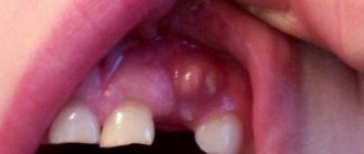
A simple capillary hemangioma grows in breadth without affecting the tissue. It consists of capillary tissue and is often affected during conversation or eating. Externally, the tumor looks like a spot or bruise. The formation must be removed - damage to the hemangioma can lead to infection.
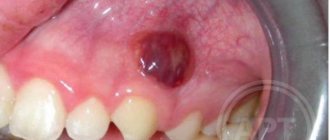
Cavernous hemangioma of the tongue penetrates deep into the tissues; it protrudes noticeably above the surface of the tongue. This formation consists of many vessels and causes serious discomfort. The tongue may lose its mobility and increase in size due to the tumor.
The causes of hemangiomas have not been fully established. In children they occur mainly due to the growth of vascular tissue.
Causes and mechanisms
The development of lumps on the roof of the mouth always indicates pathology. However, they can have different origins. Among the diseases that have this manifestation may be:
- Benign tumors (myxoma, angioma, fibroma, papilloma, osteoma, cyst).
- Viral diseases (herpes).
- Exostoses (bone protrusions).
- Lichen planus.
- Syphilis (chancre or gumma).
- Leukoplakia of the oral cavity.
- Oncological processes (cancer).
Thus, tubercles on the mucous membrane are most often of inflammatory or proliferative origin. Various factors contribute to the appearance of such changes:
- Mechanical trauma (with dentures, after operations).
- Burns (hot food, chemicals).
- Bad habits (smoking, alcohol, drugs).
- Congenital anomalies, etc.
Most often we are talking about local disorders in the oral cavity, but disorders of a systemic nature cannot be ruled out, against the background of which growths form on the palate. And each case requires an individual approach to diagnosis.
Pathological formations on the oral mucosa can arise due to various conditions that require timely detection.
Danger of hemangiomas
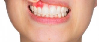
If a hemangioma appears on the tongue, you should urgently consult a doctor. A benign tumor can be dangerous. Capillary and cavernous types of hemangiomas are damaged during the absorption of food. With cavernous hemangiomas, bleeding is possible. This threatens the penetration of infections.
A simple capillary hemangioma may not cause discomfort. Some patients don't even feel it. But this does not mean that the disease can be ignored. The unexpected can happen at any moment. There are a lot of bacteria on the mucous membrane of the tongue, and it is only a matter of time before infection penetrates into the damaged tumor. Do not delay contacting a specialist.
At the clinic, a patient with a hemangioma will be prescribed treatment. The tumor must be removed. When removing, several methods are used:
- laser;
- surgical method;
- cryotherapy;
- removal by radio wave equipment.
Laser is the most popular method for removing hemangiomas. However, this type of treatment is not always possible. In some cases, doctors are able to solve the problem with a simpler surgical method.
Symptoms
You can find out about the reasons for what is happening based on an analysis of clinical manifestations. Any diagnostic program begins with a medical examination, regardless of the nature of the pathology. First, a survey and examination are carried out, followed by physical methods (for example, palpation), and the information obtained, if necessary, is supplemented with laboratory and instrumental studies.
Benign tumors
Growths on the palate are often benign. The most common tumors of the oral cavity are hemangiomas. They can be capillary, cavernous and mixed. The tubercle on the mucous membrane has a soft consistency and a red-bluish color, and collapses when pressed. Very often, hemangiomas are injured, which leads to bleeding.
Fibromas consisting of connective tissue often form on the palate. Their shape is round or oval, the consistency is densely elastic. The tumor is not fused with the surrounding tissues and is surrounded by a capsule, often growing on a thin stalk. The color of fibroma does not differ from the normal mucous membrane. Often, a fibroma can transform into a myxoma, a mucous soft tumor of a whitish color.
Cost of hemangiomas removal
| Laser tumor removal | Prices, rub. |
| Laser removal of papillomas, single warts - Cat. I. difficulties | 1200 |
| Laser removal of papillomas, multiple warts - Cat. I. difficulties | 350 |
| Laser removal of moles, papillomas, warts - Cat. II. difficulties | 700 |
| Laser removal of moles, papillomas, warts - Cat. III. difficulties | 1500 |
| Laser removal of moles, papillomas, warts - IV category. difficulties | 3000 |
| Laser removal of moles, papillomas, warts - Cat. V. difficulties | 4500 |
| Laser removal of moles, papillomas, warts - Cat. VI. difficulties | 6100 |
| CO2 Laser callus removal (per unit) | 6100 |
| Removal of atheroma, lipoma, fibroma, xanthelasma with laser - Cat. I. difficulties | 6 900 |
| Removal of atheroma, basal cell carcinoma, lipoma, fibroma, xanthelasma with laser - Category II. difficulties | 9 400 |
| Removal of atheroma, basal cell carcinoma, lipoma, fibroma, xanthelasma with laser - Cat. III. difficulties | 16 900 |
| Removal of atheroma, basal cell carcinoma, lipoma, fibroma, xanthelasma with laser - IV category. difficulties | 22 400 |
| Removal of atheroma, basal cell carcinoma, lipoma, fibroma, xanthelasma with laser - Cat. V. difficulties | 33 400 |
Sign up for laser removal of tongue hemangioma
- Full name
- Telephone
Angiomas in children
Angioma in children is usually diagnosed at birth or during the first weeks of life. Girls get sick twice as often as boys. Angioma in a child goes through several stages. In the first few months, the formation actively grows, the size of the spot increases. At the next stage, growth stops. By the age of 9, in 90% of cases, self-healing occurs - spontaneous involution of the pathology. The stain disappears, leaving no traces in its place.
Unfortunately, in some cases, benign formation is accompanied by the following complications:
- ulceration and bleeding of the skin as a result of trauma, for example, due to rubbing with a diaper when the formation is located on the leg or buttock;
- formation of scars at the site of healing ulcers after ulceration of the skin at the site of formation;
- dysfunction of internal organs due to the germination of the formation or compression of them by its boundaries;
- When the formation is located on the forehead, ophthalmological diseases develop.
Important: Very often, angioma in a child is located on the face, neck, or chest. This leads to a pronounced cosmetic defect.
It is recommended to observe uncomplicated disease in children. To do this, during the appointment, photographs of the formation are taken using a dermatoscope. At subsequent appointments, images can be compared to analyze the behavior of the formation.
Removal methods
When treating a tumor, the patient is prescribed removal. The method of removal depends on the complexity of the disease. If there is no risk of damaging surrounding tissues, then a surgical method is used. This treatment is used if the tumor has not penetrated inside. In this case, the surgeon will be able to remove all the affected cells.
Surgical treatment is not suitable if the tumor has penetrated deep into the tissue. There is a risk of damaging the tongue and not removing damaged cells completely. In difficult cases, the radio wave method is prescribed. It is used to treat cavernous hemangiomas. Affected cells are removed at ultra-high temperatures.
Treatment with ultra-low temperatures - cryotherapy - is also possible. During treatment, applications are applied to the tongue, which freeze the affected cells. The tumor tissues die and separate.
Angiomas: causes of appearance
Until now, scientists cannot name the exact reasons for the appearance of vascular formations. Doctors agree that vascular pathology begins during embryonic development. Infectious diseases suffered by the mother can affect this. Toxicosis, anemia of a woman during pregnancy, and serious hormonal fluctuations also contribute to the appearance of abnormalities. Acquired angiomas occur due to sunburn, liver disease, skin injuries, and increased fragility of blood vessels.
Laser treatment
The easiest way to remove a hemangioma is with a laser. This treatment method has several advantages:
- no risk of infection;
- This is a non-contact method of therapy;
- tumor cells are completely removed;
- the procedure is painless;
- It is possible to cure even cavernous tumors;
- the wound heals quickly.
Laser treatment does not take much time. The procedure uses a laser with a short beam length. Removal does not cause serious discomfort to the patient. The laser is absolutely safe, the procedure does not lead to side effects. After treatment of hemangioma of the tongue, no bleeding is observed.
Complete laser removal leads to rapid restoration of healthy tissue. The tongue heals and the patient returns to normal life. A special laser is used in clinics where they offer the procedure for removing hemangioma in this way.
Removal of tumors at Lazmed Clinic
Types of angiomas
According to the etiology and pathogenesis, formations caused by the dilation of small vessels are divided into hemangiomas, formed from a cluster of capillaries, and lymphangiomas, consisting of small lymphatic vessels. Formations of the lymphatic system are less common. Their peculiarity is that the skin with this pathology does not change color. Flesh-colored nodules simply appear on the skin. Angiomas on the body are another matter. This disease always causes red spots to appear. And according to the type of structure, such formations come in several types.
Simple angiomas
Vascular formations of this type are, as a rule, congenital pathologies. They can be smooth or protruding above the surface of the skin. The color of the new growths can be scarlet, burgundy, sometimes with a bluish tint. Vascular angioma grows on any part of the body, but mainly on the upper part of the body. The formation of dilated capillaries reaches a size of 10 cm in diameter. The anomaly is not dangerous, but its presence on the skin can lead to emotional imbalance. After all, the stain greatly spoils the appearance and becomes the reason for the close attention of others and their ridicule. Capillary hemangioma can especially ruin life when it is located on the face, neck, or arms.
Cavernous angiomas
A benign formation of this type looks like a pulsating purple spot. The structure of the pathology is characteristic. Blood enters the cavernous chambers through narrow arteries and is drained from the formation through wide venous channels. The spot is soft on palpation, and after squeezing it quickly restores its shape and appearance.
Branched angioma
The pathology often affects the limbs, less often appears on the scalp. The structure of the formation looks like intertwined dilated vessels. The skin over such a pathology is often affected by simple angioma. This is why capillary hemangioma must be deeply investigated. After all, a branched angioma may be hidden under it.
Intraosseous angiomas
These formations develop on the bones and are detected when they spread to nearby tissues. Bleeding becomes a symptom of the pathology. Accurate diagnosis is possible after obtaining radiographic images. The photo clearly shows the boundaries of the pathology.
Of all the listed types of angiomas, it is simple capillary formations that affect the skin.
Our specialists
- Kiani Ali
Candidate of Medical Sciences, laser medicine specialist, dermatocosmetologist.
Sign up
- Stepanova Inna Igorevna
Candidate of Medical Sciences, maxillofacial surgeon, specialist in laser medicine.
Sign up
- Fedotova Marina Andreevna
Surgeon, dermatocosmetologist, laser medicine specialist
Sign up
- Popovkin Pavel Sergeevich
Surgeon, oncologist, laser medicine specialist.
Sign up
Angiomas: treatment
Angioma in adults and children is easily diagnosed. The doctor performs a dermatoscopy to examine the formation. A digital dermatoscope repeatedly magnifies the skin, allowing you to determine the depth, nature, and boundaries of the pathology. In some cases, angiography, blood flow analysis, and histology may be prescribed.
Important: Many patients are interested in why angiomas are removed if the formation is benign and non-aggressive. The main reasons have already been mentioned above. A large angioma on the face disfigures the appearance and there is a risk of complications. In this case, it is enough to observe congenital small spots, since there is a high probability of their disappearance without a trace.
When an angioma on the body or face is perceived as a serious aesthetic defect, if the risk of complications is high, in case of bleeding the formation must be removed. Today, several methods of treating vascular formations are practiced: radio wave therapy, cold treatment (cryotherapy), electrocoagulation, surgery and laser treatment. Each method has its own advantages. But there are also disadvantages. For example, removing part of the skin with a scalpel leaves scars and does not allow you to work with pathology accurately. Cold treatment can negatively affect adjacent tissues. And electrocoagulation is not suitable for neoplasms of significant depth and large area.
The most effective treatment for pathology of any complexity is laser removal of angioma. The laser beam removes abnormally dilated capillaries without affecting adjacent tissue. The doctor controls the intensity and depth of the impact, so he works with high precision. It is important that the treatment is bloodless, painless, and safe. The beam also cauterizes blood vessels, preventing infection. There are no scars left on the skin.
Important: Many people try to get rid of senile moles and red spots on their legs, arms, and face with dubious means at home. Aggressive agents and various ointments, including those containing hormones, are used. Doctors strongly do not recommend doing this, since you most likely will not be able to get rid of the pathology. But serious harm to health can be caused. Moreover, it is not difficult to treat skin lesions professionally today.
In each case, the dermatologist practices an individual approach to the patient. This allows you to get optimal results.
Important: If the formation does not bother you and is located in an inconspicuous place, you can replace treatment with a wait-and-see approach. Because there is a high chance that it will disappear.
Prevention of angioma
It is impossible to prevent the occurrence of congenital vascular pathologies, so effective prevention does not exist. There are several ways to reduce the risk of abnormalities such as red spots. Women are advised to carefully plan their pregnancy, and while carrying a child, follow all the recommendations of the obstetrician-gynecologist.
As for senile vascular anomalies, their occurrence can be prevented by avoiding prolonged exposure to the sun and cold, and by building a healthy diet rich in minerals and vitamins. It is also recommended that older people lead a healthy lifestyle and do not ignore physical activity agreed upon with their family doctor.