Ball in mouth - what is it?
When the inflammation begins to gradually progress, a blister appears on the gum. The main reason for the formation of a lump is poor oral hygiene and prevention. But the ball can also form under the influence of other factors: inflammation occurring in the roots of the teeth, gums, mucous membranes, and periosteum.
The problem is that the disease can be diagnosed in the later stages, with the development of a purulent process. This happens because the patient does not visit the dentist. For patients who experience fear while in a chair, the doctor can give sedative anesthesia, in which the receptors are turned off and the patient falls into a shallow sleep.
Attention! It is better to diagnose the disease early in order to prevent more dangerous consequences, including blood infection and inflammation of the entire jaw.
Home therapy
Treatment can be started at home in the case of a single manifestation, when the tendency for the lump to grow in size does not persist. Here are a few recipes that can help you cope with this disease on your own without seeking medical help:
- If the manifestation is completely fresh, then you should try using raw chicken egg white several times a day.
- You should take a small piece of cotton wool and soak it in castor oil, then apply it to the bump in the cheek area.
- The procedure should be repeated twice a day.
- It is necessary to cut a clove of garlic into slices, and then lubricate the growths with them three times.
- You need to prepare a tincture, for which you will need the peel along with walnut leaves. You should pour some alcohol into the dry ingredient and let it sit for two weeks, and then lubricate the formation with it once or twice daily.
Why does a lump appear on the gum?
The accumulation of conditionally pathogenic and pathogenic microflora due to improper oral care leads to inflammation of the mucous membrane. Other reasons why a ball appears on the gum include:
- chemical, thermal or mechanical trauma to the periodontium;
- carious formations and their complications: periodontitis, pulpitis;
- inflammation of the wisdom tooth;
- eruption of molars;
- jawbone overgrowth;
- weak immune system;
- infectious pathologies of the oral cavity: herpes, candidiasis, stomatitis.
A lump in the mouth also occurs under other conditions: epulis, exostosis, fibroma, focal fibromatosis, malignancy, cyst and flux. The main thing is to promptly establish the causes of the pathology and begin treatment.
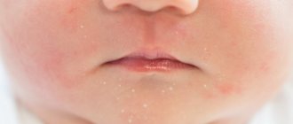
What can cause lumps on a child's cheeks?
Any redness or hardness on the baby’s cheeks causes a lot of trouble for parents. The reasons for the appearance of cones may be different, and, accordingly, the methods of getting rid of them also vary:
- Most often, children receive facial injuries as a result of falling or while participating in active games.
- With mild frostbite while walking, lumps may appear on the skin, which can be eliminated by rubbing in a rich cream.
- Candidiasis, cysts and biting injuries can also appear in a child, in some cases even in a very small one.
Without a medical examination, nothing should be done. The local pediatrician will be able to determine which specialist to show the baby to.
Only a doctor can make a diagnosis and give prescriptions for a speedy recovery.
If the ball in your mouth does not cause discomfort
The color of the bubble will help the doctor make a diagnosis. A white lump on the gum indicates the presence of exostosis or purulent exudate. A red or bloody ball indicates the development of inflammation. If the growth matches the shade of the tissue, it is the initial stage of epulis, flux, or a malignant tumor. When the tumor does not hurt, this indicates the presence of one of the pathologies:
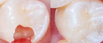
A fistula is a white ball on soft tissues that appears under or on a tooth. There is a hole on the surface for the release of pus. If, when pressed, suppuration flows out of the bladder, the patient does not feel pain. If the hole is closed with pus or bloody clots, the patient will experience discomfort with any impact.
A fistula is often formed due to advanced periodontitis, accompanied by periodontal hyperplasia. Overgrown tissue is good soil for the proliferation of microorganisms. In this case, the patient urgently needs treatment for periodontitis.
In the absence of therapy, the fistula enters a chronic stage, which can only be eliminated through surgical treatment. The progression of the disease must not be allowed, otherwise you may lose healthy molars.
Hematoma is a round lump on the inner surface of the cheek. Sometimes it occurs in the form of a dark bluish swelling on top of the gums. Blood accumulates in or around the root of the molar. The mucous membrane grows, the patient experiences discomfort and cannot completely close the jaw. The main reasons: consequences of filling or tooth extraction, gum damage, poor blood clotting.
Hematomas are generally not dangerous. Processes take place in the body through which soft tissues are cleared of bloody clots. After some time, the bubble disappears, but if the seal remains, you need to visit a dental clinic.
Exostosis is a hard blister that is an abnormality in which the bones protrude and protrude from the jaw. Gradually, the lump increases in volume, which causes discomfort and pain. Exostosis can be provoked by various reasons:
- jaw damage;
- heredity;
- congenital disorders;
- tissue diseases after molar removal.
An examination by a dentist or an x-ray will help detect the disease. The formation will need to be removed if the development of a malignant tumor is suspected.
Epulis is a pedunculated bubble in the form of a mushroom-shaped growth on the periodontium. The tumor may be the same color as the gums or red. The reasons causing the development of pathology include:
- improper filling of the molar or too large a filling;
- dental plaque, stone;
- jaw damage;
- malocclusion;
- hormonal imbalance;
- poor prosthetic material or incorrect prosthetics.
The symptoms of epulis resemble gingivitis; for diagnosis, the dentist prescribes radiography and histology to the patient. With their help, the degree of destruction of bone tissue at the site of the epulis lesion is determined. Mostly, pathology occurs in children during the growth of primary molars and in women.
Papilloma or fibroma is a bubble on the gum, sometimes a benign formation that does not pose a threat to the health or life of the patient. They are formed in people of different genders and ages. Predisposing factors to the appearance of a lump can be: damage to the mucosa, stress, systemic pathologies, heredity.
Papilloma is an enlargement of the papillary layer of skin. The bubble grows gradually, but with reduced immunity, systemic pathology, or stress, growth accelerates, but without turning into a malignant tumor. A papilloma neoplasm looks like a smooth, soft lump on the mucous membrane of a white or pink shade on a thin stalk.
Papilloma often does not create any discomfort. But after some time it may increase in size. You should consult a dentist and get tested.
Causes of cheek cancer
Cancer of the buccal mucosa develops under the influence of the following provoking factors:
- Use of tobacco in any form (cigarettes, cigars, pipes, chewing tobacco);
- Alcohol abuse (the risk of developing cancer increases when the use of alcohol and tobacco is combined);
- Infection with carcinogenic forms of human papillomavirus.
One risk factor is exposure to sunlight. Both family history and genetic predisposition, as well as exposure to mutagenic environmental factors, play a role in the development of cheek cancer. The formation of a malignant tumor occurs in several stages. The most important is the disruption in the functioning of oncogenes and genes that inhibit tumor growth. The development of malignant neoplasms of the cheek is associated with inactivation of the p16 gene, mutations in the p53 gene, and the introduction of the human papillomavirus.
If the formation on the gum causes discomfort
If a bubble near a molar causes pain, this indicates an infectious inflammatory process. Discomfort is also typical with gum injuries and hematomas. Painful formations can be caused by a cyst, cancer, fibroma, periostitis, periodontitis.
Gingivitis - the disease affects only one gum, and the periodontium near the molar remains uninjured. The main symptoms of the pathology are swelling, bleeding, and peeling of the epithelium. Gingivitis often occurs against the background of halitosis. Sometimes pathology develops as a result of endocrine and metabolic disorders.
When treating, it is necessary to eliminate the cause of gingivitis. Professional oral hygiene, diagnosis and treatment of metabolic disorders are performed. To eliminate inflammation and prevent further spread of bacteria, antibiotic therapy is administered. For pain and severe discomfort, analgesics are prescribed.
Periodontitis - purulent bumps on the gums appear as a result of degeneration of the alveolar process, through which the tooth root is held in the alveolus. Tissues can be destroyed due to poor oral hygiene and internal pathologies.
In the initial stages, periodontitis is treated by teeth cleaning at the dental clinic and further proper care. An advanced form of the disease, in which the teeth become loose, pus accumulates, requires surgical intervention - restoration of the alveolar processes or removal of molars.
Periostitis or flux is a dense formation near a problematic molar with carious lesions. Patients complain of pain radiating to the temple, chest, neck, ear. The condition gradually worsens and the temperature can rise to 38 degrees. In the oral cavity, lesions due to periostitis are hyperemic. A purulent fistula forms. When the discharge is removed, the discomfort decreases.
Causes of periostitis: trauma, periodontitis, osteomyelitis, weak immune system, infectious pathologies occurring in the body and vitamin deficiency.
The mechanism of development of cheek cancer
Malignant tumors of the cheek occur against the background of precancerous changes in the epithelial and subepithelial layers (leukoplakia or erythroplakia). The risk of leukoplakia degenerating into invasive cheek cancer is about 4-6%, with erythroplakia it reaches 30%. Subsequently, dysplasia transforms into “cancer in situ”, which penetrates into surrounding tissues and metastasizes to local and regional lymph nodes.
In some patients, even those cells that do not initially raise suspicion of dysplasia upon initial microscopy may gradually become malignant. As the tumor process progresses, distant metastases occur in the bones, lungs, and liver. Buccal cancer can grow through the skin.
Treatment of neoplasms in the oral cavity
A lump under the jaw or on the gum can be detected upon examination. For an accurate diagnosis, a biopsy or x-ray is prescribed. Treatment is selected taking into account the cause of the formation of a bubble in the oral cavity. It will be necessary to stop the further spread of infection and eliminate discomfort.
The doctor will determine how to remove the lump after a complete diagnosis, but the main measures include:
- fistula - pus is removed by rinsing with a disinfectant solution;
- epulis - surgical intervention (removal using diathermocoagulation, cryodestruction or a scalpel);
- periodontitis - removing fillings, cleaning canals, removing pus, rinsing with herbal decoctions and soda solution;
- periostitis - placement of special drugs under a temporary filling (if there is no result, the tooth will have to be removed);
- gingivitis - cleaning of periodontal tubules, antiseptic and antibacterial therapy, removal of lumps.
When a formation appears on the gum, you can rinse your mouth with vodka, diluted alcohol or juice from fresh Kalanchoe leaves.
Timely diagnosis allows you to prevent further spread of infection and save teeth.
Only an experienced dentist will help you get rid of a lump in your mouth, minimizing the risk of developing serious complications. At the first signs of pathology, you should immediately consult a doctor. The doctor's consultation
Doctors' advice and recovery period
According to the advice of experts, after excision of the affected area (in the event that the internal bump on the cheek was removed surgically), the patient should be very careful and take his condition seriously. For example, you should completely review your diet, eliminating roughage from it.
At first, if a bandage has been applied to the patient, you must be extremely careful not to touch the seam; it is important to allow it to tighten efficiently and painlessly. In addition, in case of excision of areas on the cheeks, the risk of scar formation remains.
We must not forget that the operation itself is additional stress for the body; after it, it is very important to strengthen all your strength from the inside. To do this, you will need to eat right, in addition, good sleep is important along with giving up bad habits, and if possible, you need to avoid any worries and stress. You should not self-medicate; seeking medical help will help you quickly cope with the disease.
Bisha's lumps: before and after surgery
The procedure for eliminating fatty lumps of Bisha helps to achieve a pronounced effect, radically changing your appearance. If previously the girl was dissatisfied with the oval of her face and was worried about chubby cheeks and undefined cheeks, then after the operation you can relax, since Bish’s lumps do not form again.
The effect after the procedure is very noticeable, it consists in improving the proportions of the face, reducing the volume of the cheeks, middle and lower thirds of the face, the appearance of more pronounced cheekbones, reducing the depth of the nasolabial folds and visual rejuvenation of the entire face. It is worth noting that the result will not appear immediately, since in the first days there will be slight swelling. The preliminary result will become noticeable after 7-10 days, when all sutures are removed (if non-absorbable ones were used). The final effect will be achieved 5-6 months after surgery.
Where to have surgery for Bisha's lumps (before and after photos)
According to statistics, every third woman wants to remove Bisha's lumps (before and after photos are presented on the Internet and also in a special section on the website). Bichectomy is not the simplest surgical procedure. Its poor quality can lead to various complications. In order not to make a mistake in choosing a clinic, it is recommended to consider the following parameters:
- rating;
- pricing policy;
- quality of equipment and materials used;
- qualifications and experience of specialists;
- guarantees;
- patient reviews.
The selected clinic must be fully equipped with modern equipment and high-quality materials. The safety of the procedure directly depends on these factors. It is also important to choose a medical center where doctors with extensive experience work. This criterion is the main guarantee of a positive result.
Prices for the service may vary in different private clinics in Moscow. The final cost includes the doctor’s work, materials used and diagnostic measures. As a rule, this price is specified in the concluded contract. Conscientious medical centers provide guarantees for the services provided. After surgery, most patients leave reviews on the official website of the clinic. If everything suits you, the feedback is positive. When a patient is dissatisfied with the quality of treatment, he leaves a negative review, warning other potential clients. Based on the number of positive and negative reviews, conclusions can be drawn about the quality of the services provided.
Inflammation of the salivary gland
The appearance of a ball or cyst on the cheek inside the oral cavity can occur as a result of infections or injuries to the salivary gland. The Stenon duct (located near the human ears) is blocked by a plug consisting of organic particles - bacteria, blood, pus.
A person experiences a noticeable deterioration in health, one of the manifestations is the growth of a lump. When touched, her body has a soft structure; manipulation is not accompanied by pain. Such a pathological process cannot be left without medical attention.
After confirming the diagnosis, the patient is prescribed treatment - antiseptic treatment of the internal surfaces of the mouth, antibiotic injections (intramuscularly or into the salivary gland itself) or surgery to restore the salivary duct.
Exercises for jowls
If you are not lazy and devote only 15-20 minutes a day to yourself, then gymnastics will return a blooming, fit look to young people. People with pronounced age-related changes, unfortunately, will not be able to remove jowls with exercises, but the complex proposed below will help slow down further sagging of the skin:
- - puff out your cheeks strongly for 30 seconds every day and roll the air in all directions,
- - pursing your lips like a tube, “blow out” an imaginary candle 50–55 times daily,
- - press your index fingers firmly to your cheeks and try to smile, overcoming resistance. This is a great exercise to strengthen your facial muscles!