Every person has several nevi on the surface of the skin. A mole on or near the lip is a fairly rare occurrence. Some consider pigment formations to be a highlight of appearance, while others consider them to be an undesirable defect.
Nevi occur due to the accumulation of melanocytes - cells responsible for the production of melanin. The bulk of moles are formed during global changes in the body. This includes puberty, pregnancy, and menopause. The process of secretion of hormones by the pituitary gland is accompanied by excessive production of pigment. Many doctors believe that the algorithm for the development of nevi is embedded in the genetic code. Members of the same family often have similar numbers and locations of pigment formations.
A mole on the lip can appear throughout life. This is due to exposure to ultraviolet radiation. Visiting a solarium and prolonged exposure to the sun lead to intense melanin production. The reasons for the formation of nevi also include:
- radiation and x-rays,
- pathologies of internal organs,
- taking hormonal drugs,
- inflammatory skin diseases,
- allergic reactions.
Causes of red moles
The exact reason why red moles appear on the body is unknown; it may be a predisposition due to a genetic factor. But there are other reasons:
- pregnancy;
- exposure to chemicals;
- some diseases, including infectious ones;
- unsuitable climate (too hot or cold)
- mechanical damage to the skin, such as a bite or cuts, in which pieces of skin are not completely torn off.
Their manifestations are also possible at a more mature age, this is due to a gradual decrease in immunity. Neoplasms occur in 75% of the population over the age of 60 years.
It is impossible to somehow influence the appearance or non-appearance of red moles on the body, since they are inherent in us at the genetic level; if one of the parents had a lot of them, it means that the children will have a similar situation, and even identical maps of the location of nevi are possible.
After the procedure
After removing a nevus, it is extremely important to protect the skin from ultraviolet radiation. You can’t sunbathe in a solarium; you should be in the sun as little as possible. It is recommended to refrain from thermal procedures. It is impossible to remove crusts at the site of the surgical wound; the skin should be provided with maximum rest. After removing a mole on the face, it is not advisable to use decorative cosmetics.
You can learn more about the diagnosis of skin tumors and methods of their treatment at a consultation with a dermatologist at the Galaktika clinic (Moscow).
Locations
An increase in the growth of moles is observed in people aged 0 to 20 years, and then from 45 years and older. The rest of the time they appear, but not as actively as in the first and last phases of life. Typically, red moles appear on the upper body, arms, legs and shoulders. Their favorite places are also the head, feet, and hands. They can be located throughout the body, and it is impossible to predict their subsequent locations. Their red color comes from the accumulation of small blood vessels at the base, which supply them with blood.
Prevention of angiomas
After establishing the true cause of the appearance of red moles, it is important to avoid provoking factors. As a preventative measure, experts recommend:
- drink at least 1.5 liters of water per day;
- eat more fresh fruits and vegetables rich in fiber;
- add olive oil to your diet;
- try to spend less time under the scorching sun;
- sunbathe only in the morning or evening (using sunscreen);
- stick to a balanced diet.
Types and classification
Moles have two main types:
- Pigmented. These include flat, often brown, nevi.
- Vascular, which are formed from changes in the structure of blood vessels.
They are also:
- Congenital - divided by size: small, medium and large.
- Acquired, divided into three types depending on the location of melanocytes in the layers of the epidermis.
- Hanging ones, they are also called papillomas.
There are invisible nevi that appear changing color over time. Also wandering, which change their location, appearing in one place or another. It is advisable to keep track of the location of your moles.
How is mole histology performed?
Thanks to histological examination, cancer cells can be detected even at the stage of their inception.
Indications for mole histology:
- redness of the skin around the tumor;
- small ulcers on the surface of the nevus;
- burning or pain in the mole area;
- change in color, size, shape of the tumor.
Histology is performed with the procedure of surgical excision of the mole. A small tissue sample is removed and sent for further examination. In the laboratory, biological material is placed in a special suspension and examined under a microscope. Diagnosis is based on the analysis of pigment-forming cells - melanocytes. Laboratory testing may take up to 14 days.
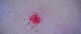
Are red moles dangerous?
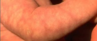
Signs to pay attention to if small red moles appear:
- Color. If it is uneven and begins to change, this is a reason to visit a doctor.
- Inflammation around the formation, that is, redness in the form of a halo around the mole.
- An increase in the size of the mole or its thickening.
- The appearance of cracks and ulcers on the body of the mole.
- Hair loss if it grew from a mole.
- The appearance of itching, burning or tingling in the place where the mole is located.
- A change in the edges of the nevus also indicates that it is time to visit a specialist.
An accurate diagnosis can be made by a dermatologist or oncologist, and you can and should turn to them for help. Nevi are not dangerous as long as they are in a stable condition, in which case you can wait to see a doctor.
When to see a doctor?
Nevi located in the lip area or on their surface need to be closely monitored. Moles are not protected by clothing and are constantly exposed to sunlight. Pigment formations are often injured during shaving or hair removal, eating, etc. Regular damage to the nevus increases the risk of developing melanoma several times.
Signs indicating malignant degeneration include:
- nevus growth - an increase in size by more than 4 mm in a short time;
- color change - sudden fading or darkening, inclusions, uneven coloring;
- lack of skin pattern - the mole is flaky or has a glossy surface;
- asymmetry - asymmetrical halves when visually dividing the nevus;
- dynamics of the process - pain, tingling, itching, hair loss, blurring of boundaries, bleeding, etc.
If you identify at least one alarming sign, you must contact a dermato-oncologist. With a 95% chance, skin cancer can be defeated at the initial stage. During the diagnosis, the doctor will determine the type of formation, establish the process of degeneration, and select the optimal treatment regimen. If necessary, he will prescribe fluorescent microscopy and a blood test to determine the level of tumor markers.
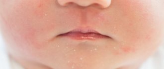
Red moles in children
For most people, the bulk of moles appear in the first 20 years of life, then the formation of moles (nevi) subsides. They also tend to sometimes disappear on their own, and this happens due to the fact that the blood vessels supplying the mole dry out or become clogged. One in 100 newborns are already born with small red moles on their body, and they are considered more prone to change and develop into melanoma. This red neoplasm that rises slightly above the surface of the skin is called a Spitz nevus.
Children can also be born with large blue spots on their backs; they are called Mongolian spots and are most often found in children with Asian roots. Any moles are often injured, especially in childhood. This can cause complications, so it is advisable to sometimes see a doctor, and also try to protect children from such injuries.
Types of moles on the lips
In medicine, nevi are classified into vascular, which arise due to the rapid growth of capillaries, and pigmented, which appear due to excess melanin.
Moles on the lip can vary significantly in appearance: shape, size and color. Based on this, the following types of moles are distinguished:
- Flat - small formations located in the upper part of the epidermis. They are practically invisible and do not create any discomfort.
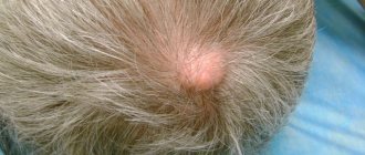
- Convex - their appearance occurs in the deep layers of the skin. These moles usually contain hair follicles.
- Vascular - have a pronounced blue or purple color and a compacted structure.
- Hemangioma - this neoplasm looks like a nodule or wart.
- Age spots are most often congenital. They can reach large sizes and pose the greatest risk of forming a malignant tumor.
Do red moles need to be removed?
Not only is it possible, but it is also necessary. If the hemangioma increases in size, changes shape or color, this is, of course, a direct indication to see an oncologist as soon as possible for further examination, and removal if necessary. Of course, not using artisanal or folk methods, but with the help of modern specialists. If the mole behaves quite calmly and does not change shape or color, then most likely there is no need to be afraid and you can postpone removing the moles.
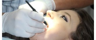
Is it possible to remove all moles with laser?
During the consultation, when a patient requests removal of a mole, the doctor carefully examines the mole and selects the most suitable removal method. If the mole has no lesions and dermatoscopy or siascopy does not reveal signs of atypia, removal for medical purposes is not required.
In this case, the removal of a mole is carried out only when the patient wishes it for aesthetic or comfort reasons, and in each case the method of removing a specific mole is selected individually. However, even unchanged moles are recommended to be removed if they are often injured by clothing, jewelry, shaving, and so on.
Moles are usually removed with a laser after administration of local analgesics. Laser mole removal is a quick procedure and the wound heals faster than surgical removal as there are no stitches, so a better cosmetic result is achieved. In addition, the likelihood of postoperative complications is much lower.
Diagnostics
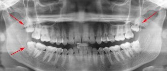
In medicine, there is a general method for diagnosing skin tumors. These include an initial examination and recording of all patient complaints (history). If a malignant tumor is suspected, an MRI or radiography is prescribed. All data obtained is analyzed and conclusions are drawn, on the basis of which a preliminary diagnosis is made. The main and final point in making a diagnosis is the histological method (biopsy). It is a microscopic sampling of tissue from the body of a mole, and is done to identify the pathology of cells in the neoplasm.
“Will you take a test from the mole the day before?”
The only analysis that can reliably determine the nature of the formation is histological examination. The formation is removed entirely and sent for pathomorphological examination. No “plucking” is performed in advance before removing moles.
It is possible to perform a cytological examination in advance if there is discharge, ulceration or trauma to the formation. This study allows you to establish a preliminary diagnosis and is performed when it is difficult to make a preliminary diagnosis. Often, an external examination or dermatoscopy is sufficient to make a diagnosis. For example, in relation to fibroepithelial polyps (papillomas), keratomas, fibromas, viral warts, a fairly large group of nevi (intradermal, warty, non-pigmented, etc.).
Methods for removing red moles
If the mole does not bother you in any way and is not located in high-risk areas, that is, on the arms, feet, or head and back, then it is quite possible not to remove it.
If the hemangioma bothers you, then it can only be removed by specialists, a dermatologist or oncologist. You should not take the risk of removing a nevus from a cosmetologist and exposing the incorrectly removed hemangioma to the risk of growth or the risk of infection.
There are several common procedures for removing them.
- Electrocautery. The nevus is burned with an electric current delivered using a small instrument. The grounding block will be placed somewhere so that the rest of the body is not exposed to electricity.
- Cryosurgery. The mole is frozen using liquid nitrogen. It is destroyed by extreme cold. Liquid nitrogen is sprayed for 10 seconds, the problem is solved in one session. This method is quick and relatively simple. The wound also does not require special care.
- Laser surgery. This procedure uses a concentrated yellow laser beam that emits enough heat to destroy the nevus from the inside. This method is quick and is used on an outpatient basis, meaning it does not require subsequent hospitalization. Depending on how many moles will be removed, one to three procedures may be required. There may be small scars that will disappear within 10 days.
- Surgical method. This is the process of removing a nevus from the upper part of the skin by cutting the lesion and then applying sutures. It is not a frequently used method, and after such an intervention there are scars.
In any case, if you have concerns and questions about why red moles appear, you need to see a doctor, if only to reassure yourself with good tests. Because unexplained questions create stress, which in turn contributes to a decrease in immunity. Be healthy.
I monitor one mole, remove another: how not to miss dangerous neoplasms on your skin
Where do they come from
On the Internet you can find a version that our moles initially grow... due to tanning. They say that a person is born with clear skin, and over time it is because of the sun that moles appear on it.
“In fact, the formation of moles and their number is determined primarily by heredity,” says dermato-oncologist at the Clinic of Skin Diseases, assistant at the Department of Skin Diseases at Sechenov University, candidate of medical sciences Ekaterina Vertieva.
- That is, those who have a lot of new growths on the skin in their family - mom, dad, grandmother or grandfather - will have more moles.
The second factor is the sun. Under the influence of its rays, the number of moles increases.
At the same time, there are several peaks in a person’s life when most moles appear. Such surges are associated with hormonal changes: at a younger age (3 - 6 years), adolescence and from 25 to 35 years. Women may experience additional peaks during pregnancy.
— Is it true that there is a “safe limit” of moles, and if there are more than normal, then a person has an increased risk of skin cancer?
— If there are more than a hundred moles on the skin, then the risk of malignant neoplasms is really higher. But not so much that those with a rich scattering of moles should immediately sound the alarm. The logic is simple: the more tumors there are, in principle, the higher the likelihood that among them there will be nevi with malignant potential (how to protect yourself from danger - see below).
STAY IN TOUCH
What is what
Among the names of growths on the skin you can find the classic, familiar word “mole,” as well as nevus and keratoma. What is the difference?
“ Nevus is the medical name for the same mole,” explains Ekaterina Vertieva. - Essentially, this is a benign skin tumor. A mole consists of pigment cells of melanocytes, which produce the coloring pigment melanin (because of it, our skin darkens when tanning. - Ed.) A mole is a cluster of such melanocyte cells. That is why the appearance of nevi is associated, among other things, with exposure to the sun.
— Keratoma is a benign superficial formation on the skin that can never degenerate into something malignant. Keratomas occur more often in old age. They consist of keratinocyte cells (these are the surface cells of the upper layer of the skin of the epidermis). Externally, a keratome usually looks like a brown formation rising above the surface of the skin. Keratomas often become crusty and flaky. Pieces can fall off, which scares people. In fact, this is a natural desquamation process. There is no need to be scared, the doctor reassures.
On a stem and with hairs - what can you expect from them?
- There are moles that seem to move away from the skin and rise above it - is this dangerous?
- Look: we have superficial layers of skin, the epidermis, and deep layers, the dermis. Common, classic dark moles are superficial, so-called border nevi. They are found in the epidermis. Over time, some superficial nevi absolutely physiologically, that is, safely, naturally grow into the deep layers of the skin, the dermis. And then they begin to rise above the surface of the skin. Nothing wrong with that.
Moreover, over time, by about 50-60 years, dermal nevi completely lose their pigment. They cease to be brown and acquire the color of healthy skin. This is the so-called mole involution. All this is absolutely safe.
- When do hairs grow from a mole? They say this is a good sign, such nevi are definitely safe and will not degenerate into melanoma.
- This is a misconception. Melanoma can easily grow hair. Therefore, if a nevus has suspicious signs (see below), then the hairs are not a reason to calm down and not check the mole.
By the way, doctors categorically do not advise pulling hair out of a nevus; this is unnecessary trauma. You can only cut it carefully.
Meyerson phenomenon
This is the name of the condition when redness appears around the mole, like an inflamed pink corolla, our expert explains. And it reassures: as a rule, it is not dangerous. Most often, the Meyerson phenomenon occurs in patients with a tendency to skin diseases: eczema, atopic dermatitis. If you experience any discomfort or itching, you should consult a doctor. Usually, dermatologists prescribe atopic corticosteroids to relieve such symptoms; with their help, the symptoms are easily removed.
— If you damage a mole and it bleeds, do you need to see a doctor urgently?
— It is necessary, but not necessarily right on the same day or the next day, up to 7 days is quite normal. There is no need to be afraid that after injury, even with bleeding, your mole will definitely degenerate into something bad. Do not believe the stories about a grandmother who picked off a mole, it immediately metastasized into the blood, and death quickly occurred. It is a myth. If a person injures a benign mole, then nothing bad will happen to it. Adverse consequences occur if it was initially melanoma.
— What to do immediately after a mole is damaged, before going to the doctor?
- Treat with a clear antiseptic. Hydrogen peroxide, for example. Just don’t smear it with fucorcin, iodine or brilliant green. Because it will then be difficult for the doctor to examine and assess the condition of your mole due to staining with these substances.
How often to check with a doctor
There are tips on the Internet to show your moles to a doctor every 6 to 12 months.
“If a person has 100 or more nevi and is advised to visit a doctor every six months or a year, then this is still too much,” says Ekaterina Vertieva. - In most cases, moles will not turn into anything bad. But, to be sure of this, it is important to undergo a full examination once by a dermatologist, or even better, an oncodermatologist. Have a specialist carefully check all moles and confirm that none of them are cause for concern. Or he indicated those that could potentially be dangerous, and then told how often they need to be monitored.
If the doctor, after examination, says that all moles are calm, then, as a rule, it is recommended to see the next time in 3 - 4 years. At the same time, no one canceled the self-examination.
THIS WILL BE USEFUL
The golden rule for self-examination
This is the international standard for assessing moles. It can be used as a test at home. The mole is tested according to the ABCDE criteria.
A (asymmetry, asymmetry) - has the mole become asymmetrical?
B (border) - have the borders of the mole become uneven?
C (colour) - has the color of the mole changed: several shades of brown, black, blue, pink have appeared?
D (diameter, diameter) - has the mole grown more than 6 mm in diameter?
E (evolution, evolution) - does the mole change intensively over 4 - 6 months (in shape, color, size)?
Important : if the answers to all questions are “yes,” this is reason to suspect the mole is malignant and immediately consult a doctor. If you have several of the symptoms, then the risk is lower, but you also need to see a doctor.
Delete cannot be saved
“There are three reasons for removing moles,” says Dr. Vertieva. — Cosmetic removal — if a person simply doesn’t like the way a mole looks. We also recommend removing benign moles if they are located in places where they are often injured. For example, for men in the beard area, for women in the bra clasp area. The third option is if we see suspicious symptoms. In these cases, the doctor conducts an examination using a dermatoscope (a device similar to a microscope. - Ed.). If a mole is causing concern, removal is done for medical reasons.
— What are the current methods for removing moles?
- Laser, radio wave method, electrocoagulation (cauterization of the nevus with high-frequency current) are used - these methods destroy the mole. As well as surgical excision.
If the patient has a suspicious nevus, then surgical removal is necessary. Only in this case, the tumor tissue is fully preserved, and histologists will be able to give a reliable conclusion as to whether there are malignant cells in the mole.
In other cases, when the mole is definitely calm, the least traumatic method is the radio wave method or laser (coagulation is a rougher method). After removing the nevus, a neat wound remains; it must be treated with a solution of potassium permanganate or fucorcin. A crust forms, which falls off after an average of two weeks. You can wet the wound site with water from 3 to 4 days.
I strongly do not recommend self-medicating at home and using celandine to remove tumors. At best, this technique leads to a chemical burn, and sometimes to more serious consequences in the case of malignant potential of the formation.
QUESTION ON THE TOPIC
Is it possible to sunbathe if you have a lot of moles?
“Yes, this is not a contraindication for tanning,” our expert rejoices. — The main thing is to follow safety rules: stay in the sun before 11 a.m. and after 4 p.m. And use sunscreen.
BREAKTHROUGH AND PROSPECTS
“Now there is a real revolution in the treatment of melanoma, the most aggressive skin cancer,” says Ekaterina Vertieva. — Previously, chemotherapy was used, which was very toxic and at the same time extended the life of patients by only a couple of months. Since the beginning of the 2010s, targeted drugs have been used. They do not hit all cells of the body, like chemotherapy, but selectively hit specific targets. These drugs are available in Russia under compulsory medical insurance and prolong the life of our patients very significantly. The main trend now is the study of new targets for the fight against melanoma. And also immunotherapy (the method for which the Nobel Prize in Medicine was awarded this year. - Ed.).
CONGRATULATIONS!
The oldest department of Sechenov University is 150 years old
On May 27, the Department of Skin and Venereal Diseases named after. V.A. Rakhmanov Sechenov University - 150 years. Already in the first years of its work, the department was recognized as the best in Europe. Over a century and a half history, vast experience in teaching the science of dermatovenereology has been accumulated here. Today, about 2,500 skin and venereal diseases are known, say department employees. The most common are psoriasis, atopic dermatitis, eczema and fungal infections; In recent decades, there has been a steady increase in the incidence of allergic dermatoses.
The head of the department, Professor Olga Olisova, lists modern promising scientific developments of the department's employees: improving the early diagnosis of lymphoproliferative skin diseases (T-cell lymphomas) using genetic markers; development of hardware methods for diagnosing melanocytic skin neoplasia; studying the pathogenetic connections of HPV with the development of skin tumors using molecular genetic detection methods; development of diagnostic markers for severe bullous dermatoses and orphan skin diseases (using the example of mastocytosis).
BY THE WAY
At the Department of Skin and Venereal Diseases of Sechenovka, a unique plaster museum has been created, containing more than 3,000 wax plaster casts with images of various skin and venereal diseases. In terms of the quality and quantity of exhibits, the dummy museum is a national treasure that has no equal not only in Russia, but throughout the world.