In the process of evolution of living beings, including humans and microorganisms, their mutual adaptation occurred. As a result, a group of microorganisms was formed that constantly live on the external surfaces and internal organs of humans.
Each person, being surrounded by natural sources of microflora, communicating with other people and entering into various relationships with them, as a result of direct and indirect contacts “exchanges” microflora with them.
Microorganisms enter the human body with water, food, from various objects, and from the air. The composition of this “random” group depends not only on environmental factors, but also on the specifics of work, everyday life, individual physiology and metabolism of each person (oily or dry skin, sweating, etc.).
Appreciating with surprise the diversity and abundance of microbes in the environment, and primarily on his body, A. Leeuwenhoek noted that there are more of them in his own mouth than there are people in the entire United Kingdom.
In recent years, information has emerged that the microflora of the human body, with all its abundance and diversity, also has strictly individual characteristics for each individual. This individual specificity of microflora is so high that it can be compared with the specificity of fingerprints. This, in principle, makes it possible to identify a person’s personality based on the “prints” of a person’s microflora.
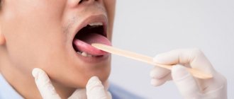
The microflora of the oral cavity is extremely diverse. Suitable temperature, alkaline reaction of saliva, leftover food are favorable conditions for the development of a wide variety of microorganisms. Almost all people in the oral cavity are inhabited by micrococci, streptococci, staphylococci, spore and non-spore bacilli, vibrios, spirochetes, spirilla, acidophilus, yeast, actinomycetes, etc.
Plaque and deposits in carious teeth and tonsil folds are especially rich in microbes.
In sick people and bacteria carriers, hemolytic streptococci, diphtheria bacillus, meningococci, tuberculosis bacillus, etc. can be found in the oral cavity.
The human respiratory organs do not have a permanent microflora. Its composition depends entirely on the content of microbes in the inhaled air. The microflora of the gastrointestinal tract, especially the large intestine, is very abundant. To illustrate, it can be stated that an adult person excretes many hundreds of billions of microorganisms from the intestines every day. The intestines are constantly inhabited by coli bacteria, some cocci, tetanus bacillus, bacillus perfringens, etc.
A lot of different microorganisms are on the hands, where they get from all other parts of the body and from the environment. The detection of typical inhabitants of the gastrointestinal tract, in particular E. coli, on the hands indicates an unsatisfactory sanitary and hygienic regime of work and life, and a low level of personal hygiene.
Cleanliness of hands, body, and maintenance of normal health are necessary for trade and catering workers who have constant contact with food products.
MAIN BIOTOPE OF THE ORAL CAVITY METHODS OF THEIR STUDY
The oral cavity, as an ecological niche, can be divided into several smaller biotopes, different from each other in terms of living conditions:
∨ oral mucosa,
∨ ducts of the salivary glands with saliva in them,
∨ gingival fluid and gingival groove area,
∨ oral fluid,
∨ dental plaque.
The physicochemical characteristics of each biotope—environmental pH, viscosity, temperature, the presence of organic compounds and food residues, partial pressure of gases—provide significant differences in the composition of the microbiocenosis of each of the listed biotopes.
The mucous membrane of the oral cavity is the most extensive biotope in area and diverse in living conditions. Therefore, the microflora of the mucous membrane varies significantly in different areas. On the surface of the mucous membrane, predominantly gram-negative anaerobic and facultative anaerobic flora, as well as microaerophiles, vegetate. In the sublingual region, on the inner surface of the cheeks, in the folds and crypts of the oral mucosa, obligate anaerobic species usually predominate: veillonella, peptostreptococci, lactobacilli, as well as S. mitis
.
S. salivarius
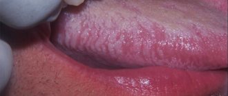
typically colonizes the dorsum of the tongue.
On the mucous membrane of the hard and soft palate, palatine arches, tonsils, a variety of streptococci, corynebacteria, neisseria, hemophila and pseudomonads, as well as yeast-like fungi and nocardia are found in large numbers.
Quantitative study of the microflora of the mucous membrane is carried out microscopically - using imprints on glass, or bacteriologically - using the method of imprints on agar blocks, followed by counting the number of colonies grown per 1 cm of the plate. Normally, this figure ranges from 2x10² to 2x10⁴.
Salivary gland ducts and saliva . Saliva in the ducts of the glands of a healthy person is practically sterile due to the high bactericidal activity of enzymes, lysozyme, secretory immunoglobulins and other factors of specific and nonspecific protection. There may be a small amount of bacteria, mainly belonging to obligate anaerobic species (Veylonella).
Studying the microflora of saliva from the duct is very difficult. For sterile examination of saliva, various cannulas are currently used, fixed in the area of the excretory duct of the salivary gland.
Gingival fluid and gingival groove. Gingival fluid is a transudate that is secreted in the area of the gingival groove and is almost immediately contaminated by the microflora of the gum mucosa and oral fluid. This biotope is dominated by filamentous and convoluted obligate anaerobic species of bacteria: fusobacteria, leptotrichia, actinomycetes, spirilla, anaerobibrio, campylobacter and spirochetes. This is the main habitat of representatives of the genera Bacteroides
,
Porphyrom
o
has
and
Prevotella
. Protozoa, yeast-like fungi and mycoplasmas are also found here.
The concentration of the listed microorganisms in the gingival fluid increases sharply with the formation of a pathological gingival pocket during periodontitis (periodontal pocket). Retention of food, detritus in the pocket, and impaired fluid circulation lead to a sharp drop in redox potential and create optimal conditions for the reproduction of obligate anaerobic flora, including Porphyrom
o
has gingivalis
and
Prevotella melaninogeni
ca. The toxic factors of these microbes play a decisive role in the progression of the inflammatory process in the periodontium. For microbiological examination of the contents of gingival fluid, the method of quantitative sampling with micropipettes, cannulas, and capillaries is usually used. The concentration of bacteria in a healthy person in the gingival fluid is no more than 10 × 105 cells per ml, and with the development of gingivitis or periodontitis - tens and hundreds of millions of cells per ml.
Oral fluid is the most important biotope of the oral cavity. Through it, interaction occurs between other parts of the oral microbiocenosis and various regulatory influences on the part of the macroorganism are realized. The basis of oral fluid is saliva, secreted from the ducts of the salivary glands, which is populated by a variety of microflora.
Microbes constantly enter the oral fluid, multiplying on the oral mucosa, in the gingival groove, pockets, folds and in dental plaque. The oral fluid contains significant quantities of Veillonella and microaerophilic streptococci S. salivarius
, facultative anaerobic streptococci, aerococci, mycoplasmas, as well as mobile forms - vibrios, selenomonas, spirochetes, spirilla.
To study the microflora of oral fluid, a quantitative cultural study is used. The normal concentration of bacteria ranges from several tens of millions to billions of cells per ml.
Quantitatively, about half of all residents of the oral cavity are streptococci; they are detected in all samples of material taken from any part of the oral cavity. In saliva samples there are approximately 2 times more of them than in material from plaque or gingival groove.
Based on their effect on red blood cells and the ability to cause their hemolysis when growing on blood agar, all streptococci are divided into: beta-hemolytic, alpha-greening and gamma-non-hemolytic.
The most significant group of oral streptococci should be considered microaerophilic alpha-hemolytic (“greening”) streptococci and gamma-non-hemolytic streptococci, which include most anaerobic peptostreptococci. However, it should be noted that from 40 to 90°/o strains belonging to the species S. mulleri
, widely distributed in the oral cavity, may be beta-hemolytic.
This species is considered to be quite virulent; it is often isolated during pathological processes. Of the alpha-greening species, the most virulent are S. intermedius
, which is part of the group of periodontopathogenic species, as well as individual strains of
S. sanguis
, capable of causing bacteremia and septic processes (septic endocarditis) with the slightest dental interventions (tooth extraction, curettage).
Different types of streptococci predominate in different biotopes due to different adhesive activity. So, S. mutans
and
S. sanguis
have a high ability to adhere to tooth enamel and dominate dental plaque.
S. salivarius
inhabits predominantly the back of the tongue, from where it is washed off with saliva, making up a significant part of the microflora of the oral fluid.
Streptococci S. mitis
,
S. mille
gi and
S. sanguis
have high adhesive properties to epithelial cells of the oral mucosa.
Lactobacilli constitute an important, although relatively small, group of residents of the oral cavity. The number of lactobacilli in the saliva of adults ranges from values close to zero to 105 cells per 1 ml. On average, their number is about 7 × 10⁴ cells in 1 ml of saliva.
Lactobacilli have rather low adhesive properties to the mucosal epithelium and, especially, to tooth enamel, however, they are represented in all niches of the oral cavity. Very often, lactobacilli are fixed on various tissues due to coaggregation with various other symbiont microbes, in particular with peptostreptococci and microaerophilic streptococci of the oral cavity. They are retained purely mechanically in the fissures of the teeth and folds of the mucous membrane.
Lactobacilli multiply rapidly when carbohydrate foods enter the oral cavity and abundantly produce lactic and other acids, which allows them to be considered a cariogenic factor. At the same time, lactobacilli play a critical stabilizing role in the formation of oral microbiocenosis, as they synthesize vitamins B and K, necessary for the development of other bacteria and macroorganisms. It is known that vitamin K and its metabolites are powerful stimulators of the growth of bacteroides and fusobacteria.
4. Dental plaque as a typical variant of biofilm. Methods of its research.
Dental plaque is the most complex and multicomponent biotope that forms on the surface of the tooth. Almost all representatives of the microbial flora of the oral cavity are determined in the composition of dental plaque. However, their number varies significantly among different people and at different periods of their lives.
The uniqueness of this biotope lies in the fact that it is largely the result of the vital activity of various microorganisms of the oral biocenosis.
In the formation of dental plaque, there is no doubt the determining role of the macroorganism and environmental factors that influence it throughout life (diet, lifestyle, occupational hazards, etc.).
According to modern concepts, dental plaque is a typical variant of biofilm - a symbiont community of microbial species formed in fluid liquid environments. Quantitative and qualitative disturbances in the composition of the symbionts of a given biotope, disturbances in their interaction with the macroorganism play a decisive role in the emergence of such important nosological forms as dental caries and periodontitis. To study the composition of dental plaque, a technique is used to take the material with a probe or metal spatula, followed by weighing on an analytical balance. Then mechanical grinding of the plaque or its disintegration by ultrasound and quantitative inoculation using anaerobic cultivation techniques are carried out.
References
- Pomazanov, V.V. Microecological status of a person and its definition. — Prospects for the implementation of innovative technologies in medicine and pharmacy, 2021. — pp. 167-184.
- Gostry, A.V., Simonova, A.V., Mikhailova, A.N. and others. Chronic pharyngitis: etiology, pathogenesis, treatment. New approaches to assessing etiopathogenesis. - Archives of Internal Medicine, 2021. - No. 1 (45). — P.32-43.
- Man, W., Clerc, M., Pieters, W. et al. Loss of microbial topography between oral and nasopharyngeal microbiota and development of respiratory infections early in life. – American journal of respiratory and critical care medicine, 2021. – Vol. 200(6). — P. 760-770.
Resident microorganisms
The ecosystem of the oral mucosa includes permanent symbiont microorganisms and non-permanent opportunistic ones. Symbionts - take part in metabolism, digestion, form the immune status and general nonspecific resistance. Opportunistic microflora is the causative agent of diseases and enters the active stage under favorable conditions - against the background of immunodeficiency states, during injuries, operations, stress and illness. Microorganisms multiply, reach a critical mass and provoke disease.